

Methyltetrazine as a small live-cell compatible bioorthogonal handle for imaging enzyme activities in situ
- PMID: 36349224
- PMCID: PMC9627743
- DOI: 10.1039/d2cb00120a
Methyltetrazine as a small live-cell compatible bioorthogonal handle for imaging enzyme activities in situ
Abstract
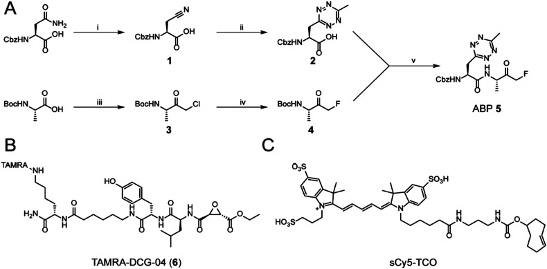
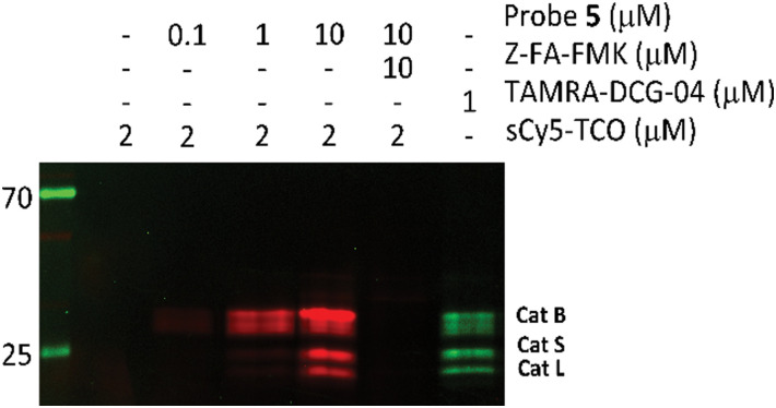
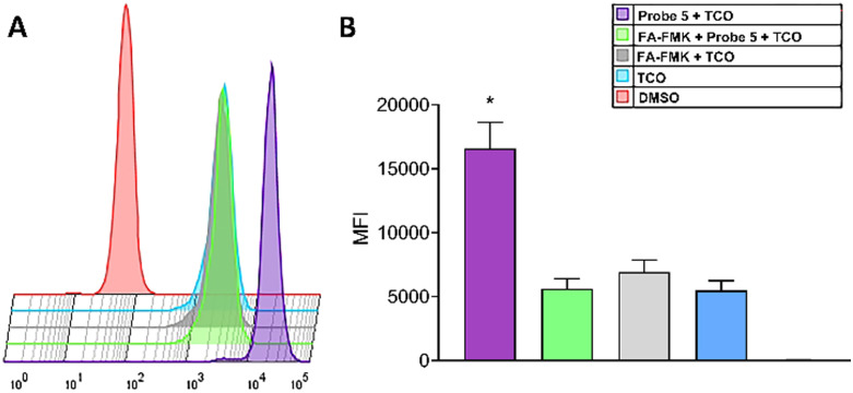
Bioorthogonal chemistry combines well with activity-based protein profiling, as it allows for the introduction of detection tags without significantly influencing the physiochemical and biological functions of the probe. In this work, we introduced methyltetrazinylalanine (MeTz-Ala), a close mimic of phenylalanine, into a dipeptide fluoromethylketone cysteine protease inhibitor. Following covalent and irreversible inhibition, the tetrazine allows vizualisation of the captured cathepsin activity by means of inverse electron demand Diels Alder ligation in cell lysates and live cells, demonstrating that tetrazines can be used as live cell compatible, minimal bioorthogonal tags in activity-based protein profiling.
This journal is © The Royal Society of Chemistry.
Conflict of interest statement
There are no conflicts to declare.
Figures


Similar articles
-
A Minimal, Unstrained S-Allyl Handle for Pre-Targeting Diels-Alder Bioorthogonal Labeling in Live Cells.Angew Chem Int Ed Engl. 2016 Nov 14;55(47):14683-14687. doi: 10.1002/anie.201608438. Epub 2016 Oct 20. Angew Chem Int Ed Engl. 2016. PMID: 27763724 Free PMC article.
-
Inverse Electron-Demand Diels-Alder Bioorthogonal Reactions.Top Curr Chem (Cham). 2016 Feb;374(1):3. doi: 10.1007/s41061-015-0005-z. Epub 2015 Dec 22. Top Curr Chem (Cham). 2016. PMID: 27572986 Review.
-
Development of a novel antibody-tetrazine conjugate for bioorthogonal pretargeting.Org Biomol Chem. 2016 Aug 21;14(31):7544-51. doi: 10.1039/c6ob01411a. Epub 2016 Jul 19. Org Biomol Chem. 2016. PMID: 27431745
-
Site-Specific Protein Labeling Utilizing Lipoic Acid Ligase (LplA) and Bioorthogonal Inverse Electron Demand Diels-Alder Reaction.Methods Mol Biol. 2018;1728:365-387. doi: 10.1007/978-1-4939-7574-7_23. Methods Mol Biol. 2018. PMID: 29405010
-
Bioorthogonal Reactions in Activity-Based Protein Profiling.Molecules. 2020 Dec 18;25(24):5994. doi: 10.3390/molecules25245994. Molecules. 2020. PMID: 33352858 Free PMC article. Review.
Cited by
-
Degradable and Multifunctional PEG-Based Hydrogels Formed by iEDDA Click Chemistry with Stable Click-Induced Supramolecular Interactions.Macromolecules. 2024 Feb 16;57(4):1556-1568. doi: 10.1021/acs.macromol.3c01855. eCollection 2024 Feb 27. Macromolecules. 2024. PMID: 38435678 Free PMC article.
References
-
- Chakrabarty S., Kahler J. P., van de Plassche M. A. T., Vanhoutte R. and Verhelst S. H. L., in Activity-Based Protein Profiling, ed. B. F. Cravatt, K.-L. Hsu and E. Weerapana, Springer International Publishing, Cham, 2019, pp. 253–281 - PubMed
LinkOut - more resources
Full Text Sources