. 2022 Sep 28;13(42):12389-12395.
doi: 10.1039/d2sc03612f. eCollection 2022 Nov 2.

Deciphering and reprogramming the cyclization regioselectivity in bifurcation of indole alkaloid biosynthesis

Affiliations

Deciphering and reprogramming the cyclization regioselectivity in bifurcation of indole alkaloid biosynthesis

Zhuo Wang et al. Chem Sci. .

Abstract

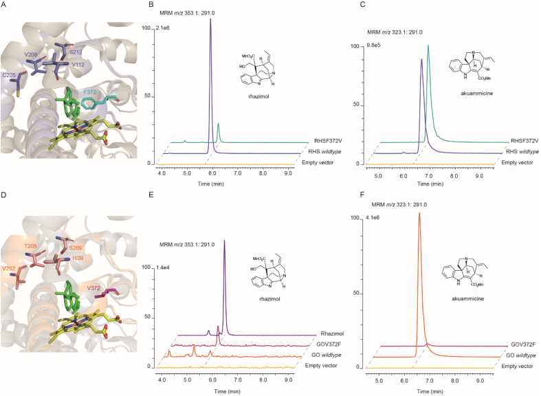
The metabolism of monoterpene indole alkaloids (MIAs) is an outstanding example of how plants shape chemical diversity from a single precursor. Here we report the discovery of novel enzymes from the Alstonia scholaris tree, a cytochrome P450, an NADPH dependent oxidoreductase and a BAHD acyltransferase that together synthesize the indole alkaloid akuammiline with a unique methanoquinolizidine cage structure. The two paralogous cytochrome P450 enzymes rhazimal synthase (AsRHS) and geissoschizine oxidase (AsGO) catalyse the cyclization of the common precursor geissoschizine and they direct the MIA metabolism towards to the two structurally distinct and medicinally important MIA classes of akuammilan and strychnos alkaloids, respectively. To understand the pathway divergence, we investigated the catalytic mechanism of the two P450 enzymes by homology modelling and reciprocal mutations. Upon conducting mutant enzyme assays, we identified a single amino acid residue that mediates the space in active sites, switches the enzymatic reaction outcome and impacts the cyclization regioselectivity. Our results represent a significant advance in MIA metabolism, paving the way for discovery of downstream genes in akuammilan alkaloid biosynthesis and facilitating future synthetic biology applications. We anticipate that our work presents, for the first time, insights at the molecular level for plant P450 catalytic activity with a significant key role in the diversification of alkaloid metabolism, and provides the basis for designing new drugs.


Conflict of interest statement

The authors declare no conflict of interest.

Figures

Fig. 1
Fig. 1. Schematic overview of monoterpene indole alkaloid (MIA) metabolic pathways in the Apocynaceae family. Strictosidine and its reduced aglycon geissoschizine generated by the activity of strictosidine glucosidase (SGD) and alcohol dehydrogenase geissoschizine synthase (GS), are the precursors of different important classes of MIAs. P450 geissoschizine oxidase (GO, CYP71D1V1) catalyzes the formation of a bond between C2 and C16 (highlighted in orange) followed by the breaking of the C2–C3 bond, the formation of a new C3–C7 bond (highlighted in orange) and breaking of the C16–C17 bond and the loss of the formaldehyde group for the synthesis of a strychnos scaffold of akuammicine. P450 sarpagan bridge enzyme (SBE, CYP71AY4) catalyzes the formation of a bond between C5 and C16 (highlighted in magenta) for the synthesis of a sarpagan scaffold of polyneuridine aldehyde. The paralog P450 enzyme alstonine synthase (AS, CYP71AY1) catalyzes the aromatization of a tetrahydroalstonine piperidine ring. Until the present work the enzymes that catalyze the formation of the bond between C7 and C16 (highlighted in purple) for the synthesis of the methanoquinolizidine scaffold and later steps in metabolism of akuammilan alkaloids were unknown. Newly discovered functionally characterized enzymes in this study are shown in red, while enzymes with known activity that are identified in this study are shown in light blue. Dashed arrows represent unknown (multiple) enzymatic steps.
Fig. 2
Fig. 2. Discovery of akuammiline biosynthetic pathway genes. (A) The methanoquinolizidine structure and the biosynthetic pathway of akuammiline. (B) LCMS chromatograms at m/z 351 (highlighted in red) and 353 showing the in vitro catalytic activity of AsRHS using geissoschizine as the substrate and quenched either by MeOH or NaBH4. (C) LCMS chromatograms at m/z 353 showing the in vitro catalytic activity of AsRHR1 and AsRHR2 by incubation together with geissoschizine and AsRHS. (D) LCMS chromatograms at m/z 395 showing the in vitro catalytic activity of AKS1 and AKS2 using synthetic rhazimol as the substrate. Denatured enzymes were used for negative controls, the peak at m/z 353 with a retention time of 5.9 min corresponds to the substrate (geissoschizine). RHS: rhazimal synthase; RHR: rhazimal reductase; AKS: akuammiline synthase.
Fig. 3
Fig. 3. Molecular modelling and mutagenesis on active sites of AsRHS and AsGO. (A and D) Overlay of the homology model structures of AsRHS and AsGO and the template X-ray structure 7CB9 of SmCYP76AH1 showing the P450 active site, with the heme (in yellow) and substrate milliradian (in green) from the template structure. The structural elements (SRSs and amino acids) from AsRHS and AsGO are depicted in purple and orange, respectively. The divergent amino acids selected for reciprocal mutations are shown in sticks. The key amino acid residues with side chains occupying the space between the heme and substrate that direct the enzyme assay outcome are highlighted in cyan (AsRHS F372) and magenta (AsGO V372). (B and C) Characterization of rhazimal synthase (B) and geissoschizine oxidase (C) activities by in vitro enzyme assays of the AsRHS wildtype, AsRHSF372V and the empty vector (control) using geissoschizine as the substrate and stopped by NaBH4; assays analyzed by LCMS using MRM chromatographic traces m/z 353.1 : m/z 291.0 for rhazimol and m/z 323.1 : m/z 291.0 for akuammicine. (E and F) Characterization of rhazimal synthase (E) and geissoschizine oxidase (F) activities by in vitro enzyme assays of the AsGO wildtype, AsGOV372F and the empty vector (control) using geissoschizine as the substrate and stopped by NaBH4; assays analyzed by LCMS using MRM chromatographic traces m/z 353.1 : m/z 291.0 for rhazimol and m/z 323.1 : m/z 291.0 for akuammicine. The intensity of LCMS chromatograms in each panel was normalized to the intensity of the higher peak.
Fig. 4
Fig. 4. Proposed mechanism for catalytic activity of RHS transforming geissoschizine (3) to akuammilan alkaloid rhazimal (4) and the formation of a methanoquinolizidine scaffold. The plausible mechanism for formation of rhazimal (4) (methanoquinolizidine scaffold) through the oxidation/hydroxylation of geissoschizine on C7 followed by nucleophilic attack by C16. The newly formed bond C7–C16 is highlighted in blue.

References

    1. Ramírez A. García-Rubio S. Curr. Med. Chem. 2003;10:1891–1915. doi: 10.2174/0929867033457016. - DOI - PubMed
    1. Gaich T. Eckermann R. Synthesis. 2013;45:2813–2823. doi: 10.1055/s-0033-1339711. - DOI
    1. Smith J. M. Moreno J. Boal B. W. Garg N. K. Angew. Chem., Int. Ed. Engl. 2015;54:400–412. - PubMed
    1. Adams G. L. and Smith 3rd A. B., The Alkaloids: Chemistry and Biology, 2016, vol. 76, pp. 171–257 - PubMed
    1. Kearney S. E. Zahoránszky-Kőhalmi G. Brimacombe K. R. Henderson M. J. Lynch C. Zhao T. Wan K. K. Itkin Z. Dillon C. Shen M. Cheff D. M. Lee T. D. Bougie D. Cheng K. Coussens N. P. Dorjsuren D. Eastman R. T. Huang R. Iannotti M. J. Karavadhi S. Klumpp-Thomas C. Roth J. S. Sakamuru S. Sun W. Titus S. A. Yasgar A. Zhang Y.-Q. Zhao J. Andrade R. B. Brown M. K. Burns N. Z. Cha J. K. Mevers E. E. Clardy J. Clement J. A. Crooks P. A. Cuny G. D. Ganor J. Moreno J. Morrill L. A. Picazo E. Susick R. B. Garg N. K. Goess B. C. Grossman R. B. Hughes C. C. Johnston J. N. Joullie M. M. Kinghorn A. D. Kingston D. G. I. Krische M. J. Kwon O. Maimone T. J. Majumdar S. Maloney K. N. Mohamed E. Murphy B. T. Nagorny P. Olson D. E. Overman L. E. Brown L. E. Snyder J. K. Porco J. A. Rivas F. Ross S. A. Sarpong R. Sharma I. Shaw J. T. Xu Z. Shen B. Shi W. Stephenson C. R. J. Verano A. L. Tan D. S. Tang Y. ACS Cent. Sci. 2018;4:1727–1741. doi: 10.1021/acscentsci.8b00747. - DOI - PMC - PubMed
    2. Taylor R. E. Thomson R. J. Vosburg D. A. Wu J. Wuest W. M. Zakarian A. Zhang Y. Ren T. Zuo Z. Inglese J. Michael S. Simeonov A. Zheng W. Shinn P. Jadhav A. Boxer M. B. Hall M. D. Xia M. Guha R. Rohde J. M. ACS Cent. Sci. 2018;4:1727–1741. doi: 10.1021/acscentsci.8b00747. - DOI - PMC - PubMed