. 2022 Oct 26:2022:9795682.
doi: 10.34133/2022/9795682. eCollection 2022.

Adhesive and Self-Healing Polyurethanes with Tunable Multifunctionality

Affiliations

Adhesive and Self-Healing Polyurethanes with Tunable Multifunctionality

Lei Zhou et al. Research (Wash D C). .

Abstract

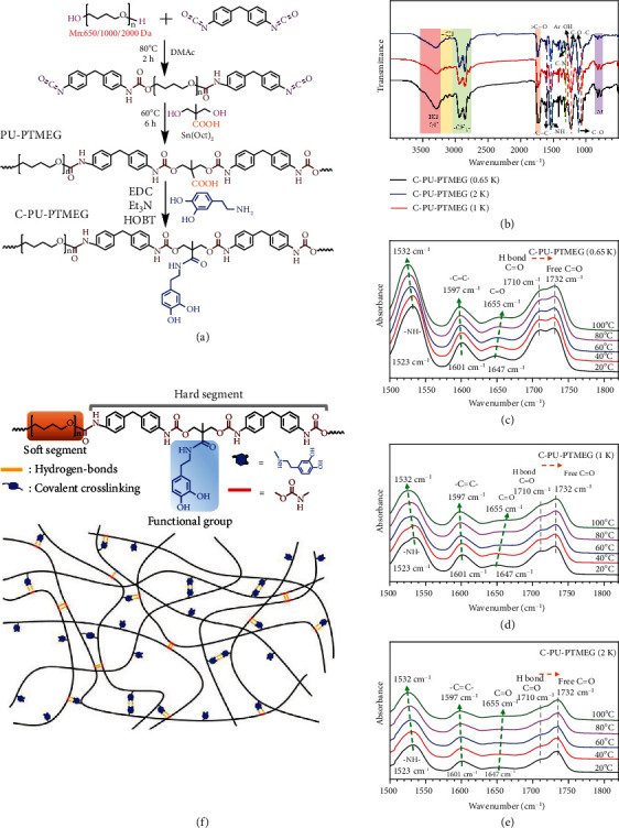
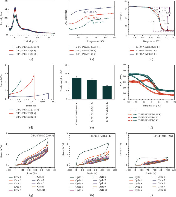
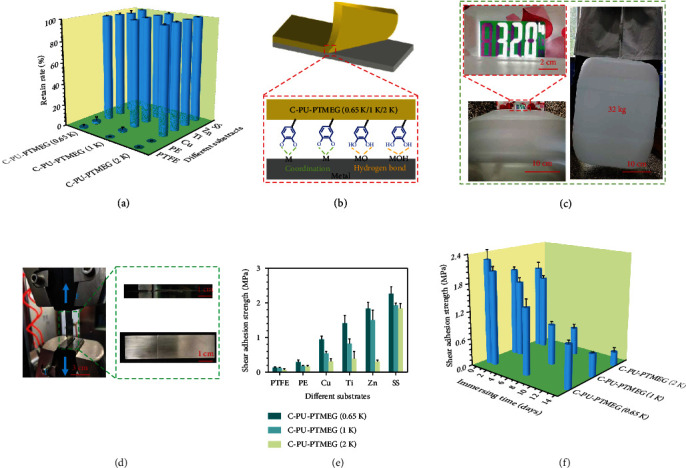
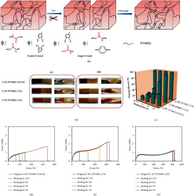
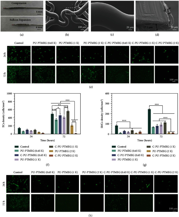
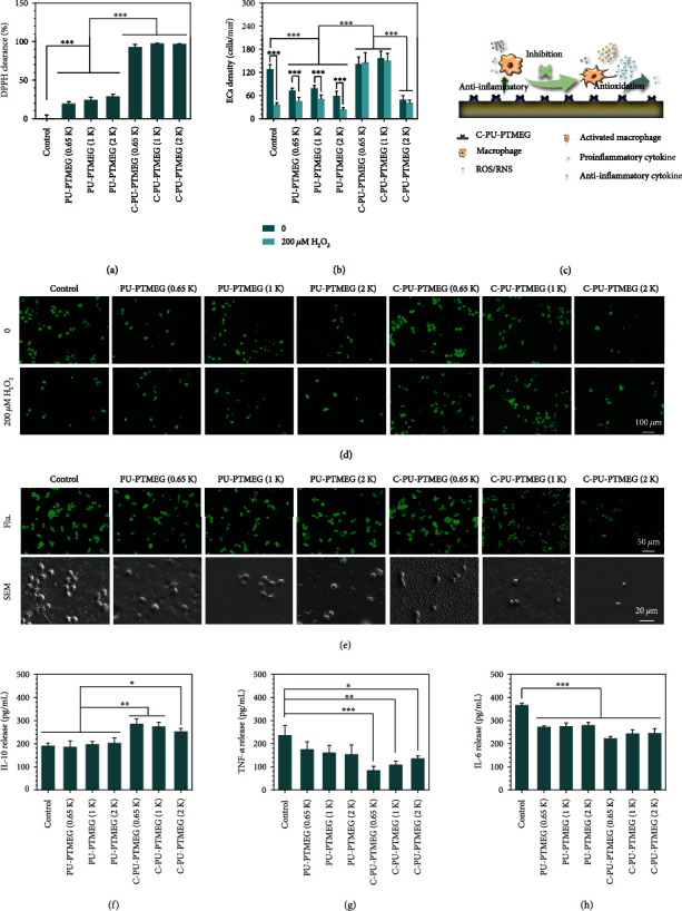
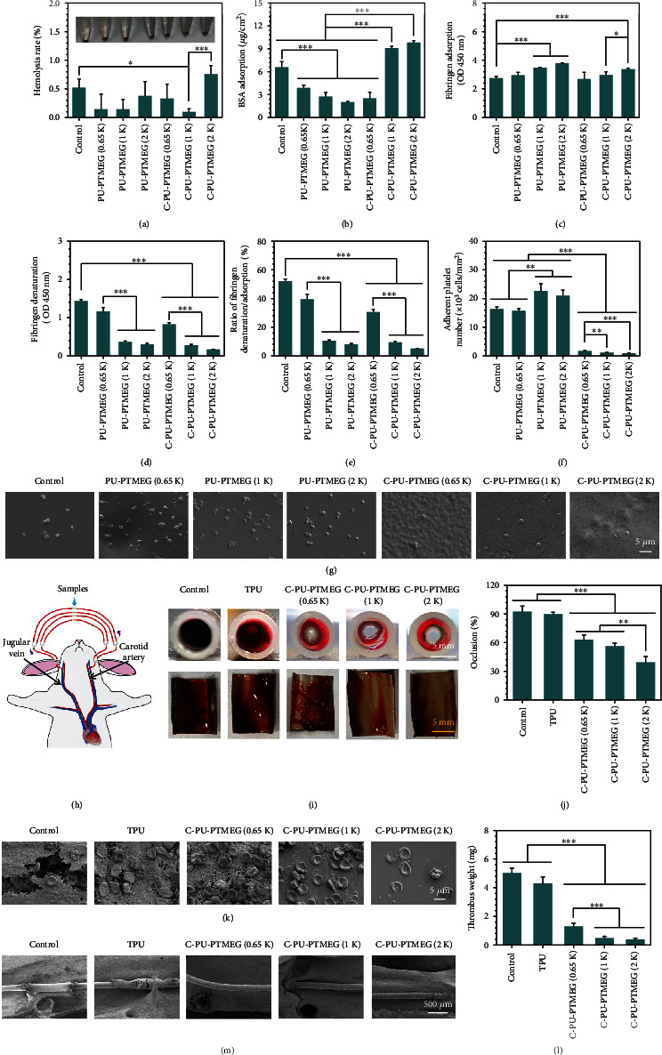
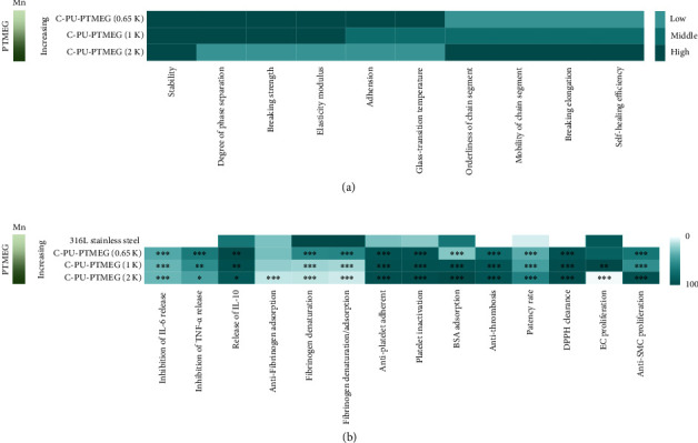
Many polyurethanes (PUs) are blood-contacting materials due to their good mechanical properties, fatigue resistance, cytocompatibility, biosafety, and relatively good hemocompatibility. Further functionalization of the PUs using chemical synthetic methods is especially attractive for expanding their applications. Herein, a series of catechol functionalized PU (C-PU-PTMEG) elastomers containing variable molecular weight of polytetramethylene ether glycol (PTMEG) soft segment are reported by stepwise polymerization and further introduction of catechol. Tailoring the molecular weight of PTMEG fragment enables a regulable catechol content, mobility of the chain segment, hydrogen bond and microphase separation of the C-PU-PTMEG elastomers, thus offering tunability of mechanical strength (such as breaking strength from 1.3 MPa to 5.7 MPa), adhesion, self-healing efficiency (from 14.9% to 96.7% within 2 hours), anticoagulant, antioxidation, anti-inflammatory properties and cellular growth behavior. As cardiovascular stent coatings, the C-PU-PTMEGs demonstrate enough flexibility to withstand deformation during the balloon dilation procedure. Of special importance is that the C-PU-PTMEG-coated surfaces show the ability to rapidly scavenge free radicals to maintain normal growth of endothelial cells, inhibit smooth muscle cell proliferation, mediate inflammatory response, and reduce thrombus formation. With the universality of surface adhesion and tunable multifunctionality, these novel C-PU-PTMEG elastomers should find potential usage in artificial heart valves and surface engineering of stents.


Conflict of interest statement

We declare that there are no competing financial and personal relationships in this work.

Figures

Figure 1
Figure 1
Chemical structure and FT-IR spectra of C-PU-PTMEGs. (a) Synthesis schematic and structures of PU-PTMEGs and C-PU-PTMEGs. (b) FT-IR spectra of C-PU-PTMEGs. Variable-temperature FI-TR spectra of (c) C-PU-PTMEG (0.65 K), (d) C-PU-PTMEG (1 K), and (e) C-PU-PTMEG (2 K) with an increasing temperature in the range of 1500 to 1850 cm−1. (f) Molecular structure of C-PU-PTMEGs and a schematic description of hydrogen bond moiety.
Figure 2
Figure 2
Characterization of C-PU-PTMEG polymers with different molecular weights of the PTMEG soft segment (0.65, 1, and 2 kDa). (a) XRD spectra. (b) DSC spectra in the range of -76 to 120°C. (c) TG within the scope of 25 to 600°C at a rate of 10°C min−1. (d) Tensile test and (e) its elastic modulus. (f) Temperature sweep of storage modulus and loss modulus. The successive loading-unloading cycles of (g) C-PU-PTMEG (0.65 K), (h) C-PU-PTMEG (1 K), and (i) C-PU-PTMEG (2 K) for 10 times at a tensile rate of 100 mm min−1.
Figure 3
Figure 3
Adhesion tests. (a) The degree of retention of C-PU-PTMEGs with different molecular weight of the PTMEG component (0.65, 1, and 2 kDa) on the various material surfaces after scribing by special exact cutter and further adhering with 3 M tape for 5 min. (b) Schematic diagram of the adhesion mechanism of C-PU-PTMEGs on the surface of metallic materials. (c) Joint bonding of two pieces of 316L SS with a C-PU-PTMEG (1 K) film (40 μm thick) for one day withstand a weight of 32 kg. (d) Photograph of the joints prepared with C-PU-PTMEGs and different materials being stretched by a universal stretching machine at a speed of 1 mm/min. (e) Shear adhesion strength of the three C-PU-PTMEGs on various surfaces in dry environment. (f) Shear adhesion strength of C-PU-PTMEGs in a wet environment by bonding of two pieces of 316L SS.
Figure 4
Figure 4
Self-healing of the C-PU-PTMEGs. (a) Schematic illustration of self-healing process through a variety of hydrogen bond interactions and a dynamics of molecular chain segments for C-PU-PTMEGs. (b) Digital photographs of C-PU-PTMEGs healing in air and PBS for 5 h at 37°C temperature after being cut into two pieces. (c) Dependency of self-healing efficiency of C-PU-PTMEGs on time at 37°C. Tensile stress-strain curve of the original and self-healing (d) C-PU-PTMEG (0.65 K), (e) C-PU-PTMEG (1 K), and (f) C-PU-PTMEG (2 K) after different healing times at a 37°C temperature in PBS.
Figure 5
Figure 5
C-PU-PTMEG (1 K) as a coating of vascular stents and the cellular growth behavior of endothelial (HUVEC) and smooth muscle (HUASMC) cells on the various C-PU-PTMEG polymers. (a) Digital photograph of a vascular stent with C-PU-PTMEG (1 K) coating after compression and balloon expansion. (b) SEM morphology of the vascular stent with C-PU-PTMEG (1 K) coating after balloon expansion and (c) high magnification of the marked area. (d) Cross-sectional image of the stent with C-PU-PTMEG (1 K) coating. (e) Fluorescence images by cell tracker (CellTracker™ Green BODIPY®, Thermo Fisher Scientific, Inc.) and (f) proliferation of HUVECs on the PU surfaces. (g) Proliferation and (h) fluorescence images of HUASMCs on the PU surfaces. Statistical significance was regarded as follows: P < 0.05,  ∗∗P < 0.01, and∗∗∗P < 0.001.
Figure 6
Figure 6
Antioxidant and anti-inflammatory evaluation. (a) The free radical clearing activity of the PU films by DPPH assay. (b) HUVECs proliferation on PU films in an oxidative stress condition mediated by 200 μM H2O2 concentrations after culturing for one day. (c) Schematic presentation of the C-PU-PTMEGs for eliminating excess ROS/NOS and inhibiting inflammation and macrophage activation. (d) the fluorescent morphology of HUVECs by cell tracker on PU films in an oxidative stress condition mediated by 200 μM H2O2 concentrations after culturing for one day. (e) Fluorescent and SEM images of macrophages culturing on the PU films after one day. Expression of anti-inflammatory factors (f) IL-10 and proinflammatory factors (g) TNF-α and (h) IL-6 in macrophages. Statistical significance was regarded as follows: P < 0.05, ∗∗P < 0.01, and ∗∗∗P < 0.001.
Figure 7
Figure 7
Hemocompatibility of C-PU-PTMEGs. (a) Hemolysis rate of various samples incubated by whole blood. (b) BSA adsorption on various films. (c) Fibrinogen adsorption and (d) denaturation on various films. (e) The ratio of the absorbance for fibrinogen denaturation and adsorption. (f) Platelet count and (g) the SEM image of various samples. Platelets from a healthy human volunteer platelet-rich plasma and incubated for 30 min on various samples. (h) Scheme of the ex vivo whole blood dynamic circulation system. (i) Cross-sectional photographs of the catheter and photographs of the sample after spreading to expose the thrombus. (j) Occlusion rate of the catheters containing various samples after one-hour circulation. (k) SEM images of the surface on samples going through the blood circulation. (l) Dry weight of the thrombi on the sample surface. (m) SME images of the samples in the rat abdominal aorta after implanting for 15 days. Statistical significance was regarded as follows: P < 0.05,  ∗∗P < 0.01, and∗∗∗P < 0.001.
Figure 8
Figure 8
Heat maps of properties and functions for C-PU-PTMEGs. (a) Heat map of the physicochemical properties and function. (b) Heat map of C-PU-PTMEG-biological properties and function. Statistical significance was regarded as follows: P < 0.05,  ∗∗P < 0.01, and∗∗∗P < 0.001.

References

    1. Wendels S., Avérous L. Biobased polyurethanes for biomedical applications. Bioactive Materials . 2021;6(4):1083–1106. doi: 10.1016/j.bioactmat.2020.10.002. - DOI - PMC - PubMed
    1. Adipurnama I., Yang M.-C., Ciach T., Butruk-Raszeja B. Surface modification and endothelialization of polyurethane for vascular tissue engineering applications: a review. Biomaterials Science . 2017;5(1):22–37. doi: 10.1039/C6BM00618C. - DOI - PubMed
    1. Zia F., Zia K. M., Zuber M., Tabasum S., Rehman S. Heparin based polyurethanes: a state-of-the-art review. International Journal of Biological Macromolecules . 2016;84:101–111. doi: 10.1016/j.ijbiomac.2015.12.004. - DOI - PubMed
    1. Bezuidenhout D., Williams D. F., Zilla P. Polymeric heart valves for surgical implantation, catheter-based technologies and heart assist devices. Biomaterials . 2015;36:6–25. doi: 10.1016/j.biomaterials.2014.09.013. - DOI - PubMed
    1. Zhu Z., Gao Q., Long Z., et al. Polydopamine/poly(sulfobetaine methacrylate) co-deposition coatings triggered by CuSO4/H2O2 on implants for improved surface hemocompatibility and antibacterial activity. Bioactive Materials . 2021;6(8):2546–2556. doi: 10.1016/j.bioactmat.2021.01.025. - DOI - PMC - PubMed

LinkOut - more resources