

CerebNet: A fast and reliable deep-learning pipeline for detailed cerebellum sub-segmentation
- PMID: 36349595
- PMCID: PMC9771831
- DOI: 10.1016/j.neuroimage.2022.119703
CerebNet: A fast and reliable deep-learning pipeline for detailed cerebellum sub-segmentation
Abstract
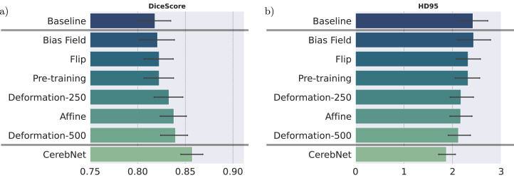
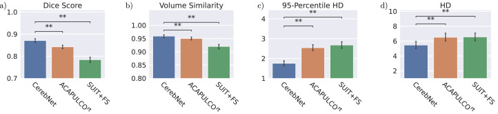
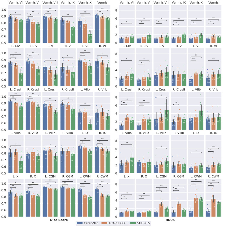
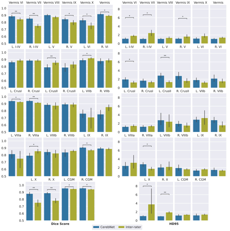
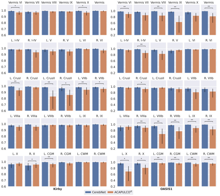
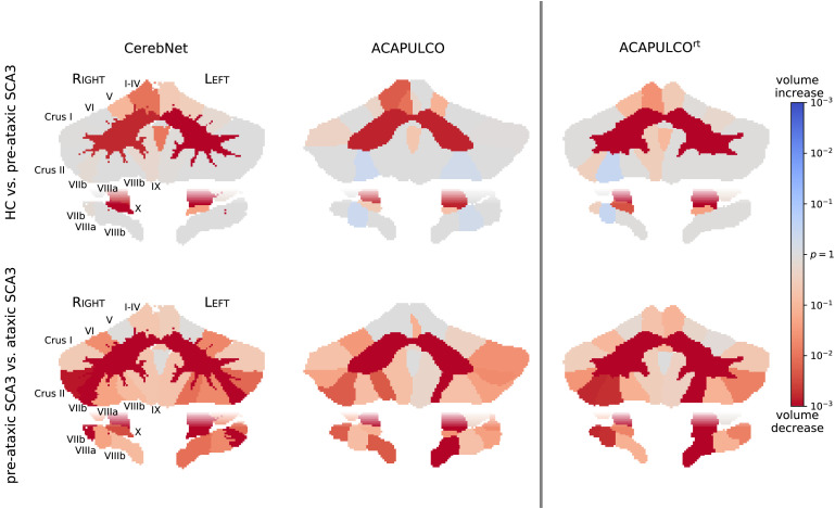
Quantifying the volume of the cerebellum and its lobes is of profound interest in various neurodegenerative and acquired diseases. Especially for the most common spinocerebellar ataxias (SCA), for which the first antisense oligonculeotide-base gene silencing trial has recently started, there is an urgent need for quantitative, sensitive imaging markers at pre-symptomatic stages for stratification and treatment assessment. This work introduces CerebNet, a fully automated, extensively validated, deep learning method for the lobular segmentation of the cerebellum, including the separation of gray and white matter. For training, validation, and testing, T1-weighted images from 30 participants were manually annotated into cerebellar lobules and vermal sub-segments, as well as cerebellar white matter. CerebNet combines FastSurferCNN, a UNet-based 2.5D segmentation network, with extensive data augmentation, e.g. realistic non-linear deformations to increase the anatomical variety, eliminating additional preprocessing steps, such as spatial normalization or bias field correction. CerebNet demonstrates a high accuracy (on average 0.87 Dice and 1.742mm Robust Hausdorff Distance across all structures) outperforming state-of-the-art approaches. Furthermore, it shows high test-retest reliability (average ICC >0.97 on OASIS and Kirby) as well as high sensitivity to disease effects, including the pre-ataxic stage of spinocerebellar ataxia type 3 (SCA3). CerebNet is compatible with FreeSurfer and FastSurfer and can analyze a 3D volume within seconds on a consumer GPU in an end-to-end fashion, thus providing an efficient and validated solution for assessing cerebellum sub-structure volumes. We make CerebNet available as source-code (https://github.com/Deep-MI/FastSurfer).
Keywords: CerebNet; Cerebellum; Computational neuroimaging; Deep learning.
Copyright © 2022 The Author(s). Published by Elsevier Inc. All rights reserved.
Figures


References
-
- Bogovic J.A., Bazin P.L., Ying S.H., Prince J.L. Lecture Notes in Computer Science (including subseries Lecture Notes in Artificial Intelligence and Lecture Notes in Bioinformatics) Vol. 7917 LNCS. 2013. Automated segmentation of the cerebellar lobules using boundary specific classification and evolution; pp. 62–73. - PMC - PubMed
-
- Buckner R., Head D., Parker J., Fotenos A., Marcus D., Morris J., Snyder A. A unified approach for morphometric and functional data analysis in young, old, and demented adults using automated atlas-based head size normalization: reliability and validation against manual measurement of total intracranial volume. NeuroImage. 2004;23:724–738. doi: 10.1016/j.neuroimage.2004.06.018. - DOI - PubMed
Publication types
MeSH terms
Grants and funding
- R01 AG021910/AG/NIA NIH HHS/United States
- R01 NS080816/NS/NINDS NIH HHS/United States
- S10 OD017974/OD/NIH HHS/United States
- P41 EB027061/EB/NIBIB NIH HHS/United States
- U24 RR021382/RR/NCRR NIH HHS/United States
- P30 NS076408/NS/NINDS NIH HHS/United States
- R01 AG064027/AG/NIA NIH HHS/United States
- P30 AG066444/AG/NIA NIH HHS/United States
- R56 MH121426/MH/NIMH NIH HHS/United States
- U54 MH091657/MH/NIMH NIH HHS/United States
- P01 AG003991/AG/NIA NIH HHS/United States
- P50 AG005681/AG/NIA NIH HHS/United States
- P41 EB030006/EB/NIBIB NIH HHS/United States
- P01 AG026276/AG/NIA NIH HHS/United States
- U01 AG024904/AG/NIA NIH HHS/United States
LinkOut - more resources
Full Text Sources
