. 2023 Jan;1519(1):173-185.
doi: 10.1111/nyas.14927. Epub 2022 Nov 9.

Effects of melodic intonation therapy in patients with chronic nonfluent aphasia

Affiliations

Effects of melodic intonation therapy in patients with chronic nonfluent aphasia

Sarah Marchina et al. Ann N Y Acad Sci. 2023 Jan.

Abstract

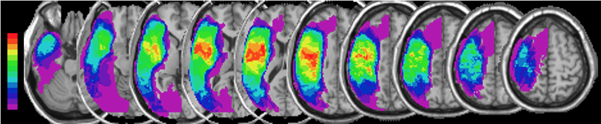
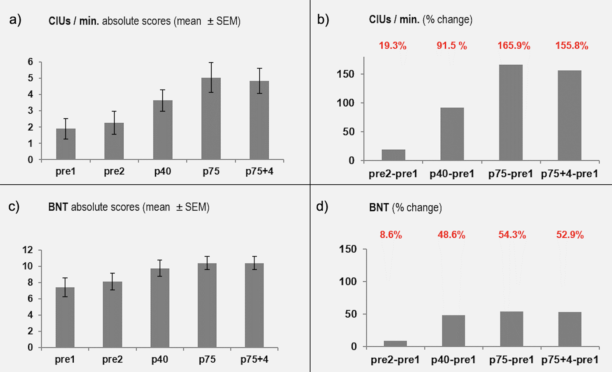
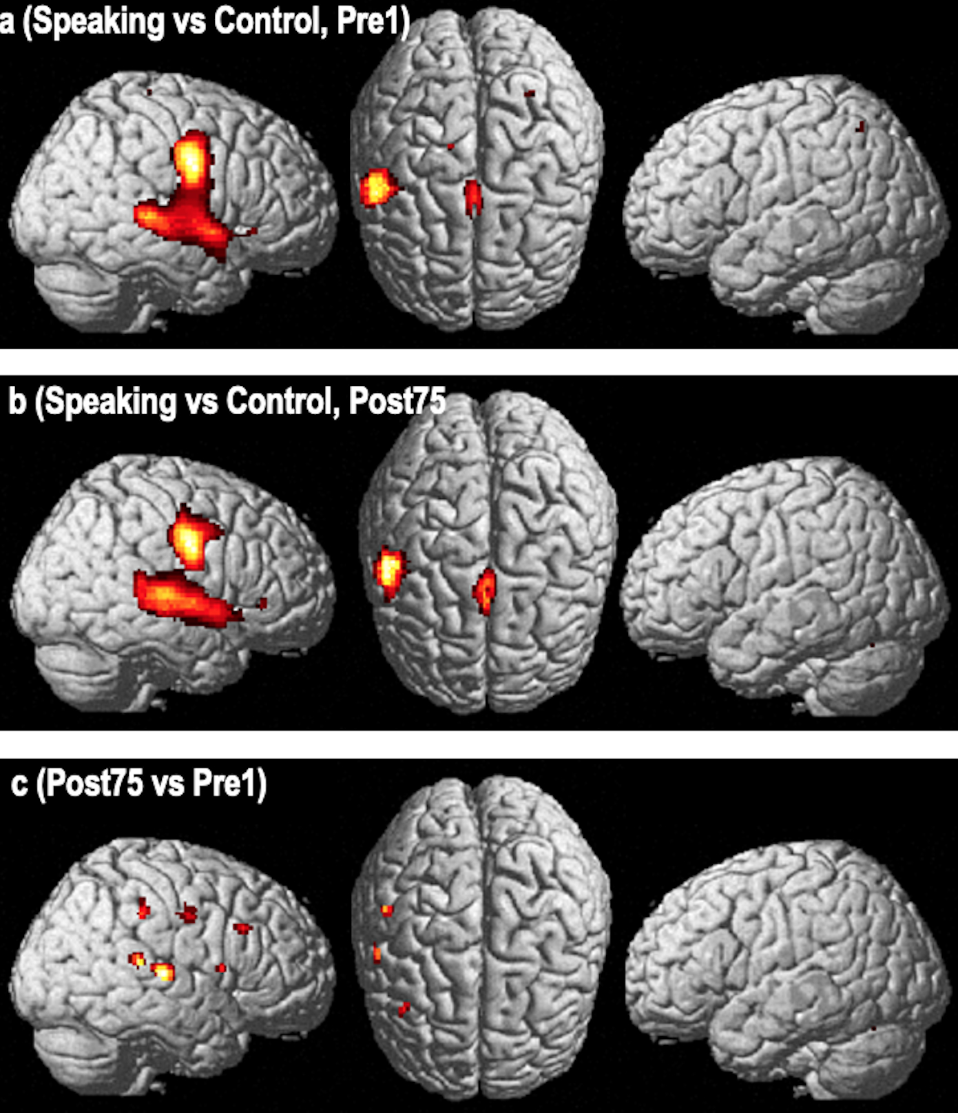
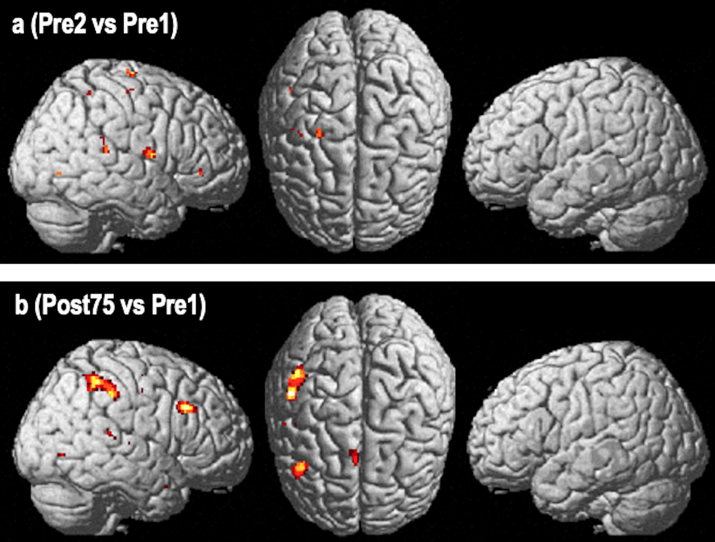
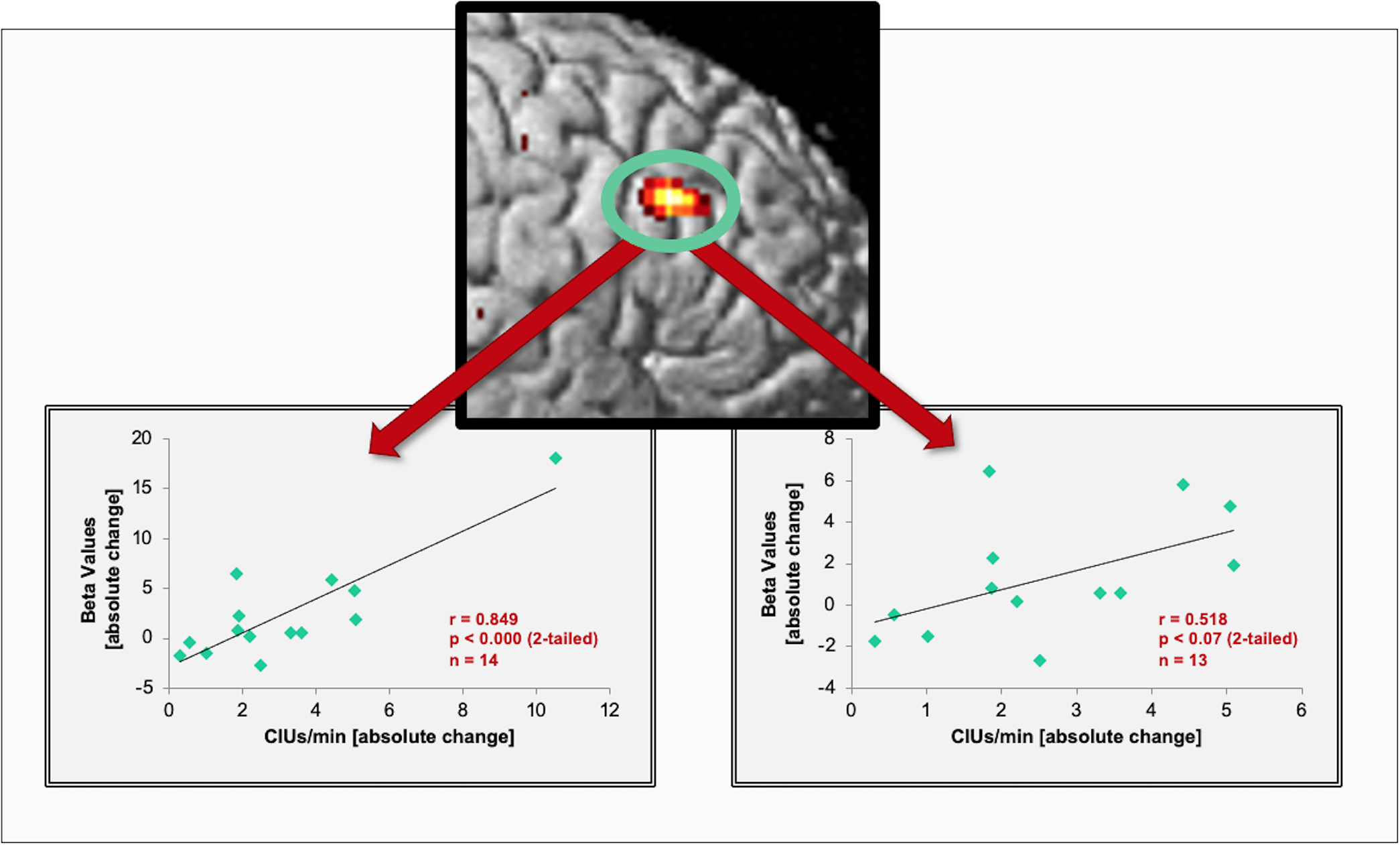
Patients with large left-hemisphere lesions and post-stroke aphasia often remain nonfluent. Melodic intonation therapy (MIT) may be an effective alternative to traditional speech therapy for facilitating recovery of fluency in those patients. In an open-label, proof-of-concept study, 14 subjects with nonfluent aphasia with large left-hemisphere lesions (171 ± 76 cc) underwent two speech/language assessments before, one at the midpoint, and two after the end of 75 sessions (1.5 h/session) of MIT. Functional MR imaging was done before and after therapy asking subjects to vocalize the same set of 10 bi-syllabic words. We found significant improvements in speech output after a period of intensive MIT (75 sessions for a total of 112.5 h) compared to two pre-therapy assessments. Therapy-induced gains were maintained 4 weeks post-treatment. Imaging changes were seen in a right-hemisphere network that included the posterior superior temporal and inferior frontal gyri, inferior pre- and postcentral gyri, pre-supplementary motor area, and supramarginal gyrus. Functional changes in the posterior right inferior frontal gyri significantly correlated with changes in a measure of fluency. Intense training of intonation-supported auditory-motor coupling and engaging feedforward/feedback control regions in the unaffected hemisphere improves speech-motor functions in subjects with nonfluent aphasia and large left-hemisphere lesions.

Keywords: MRI; aphasia; melodic intonation therapy; neuroplasticity; neurorehabilitation; stroke recovery.


Conflict of interest statement

COMPETING INTERESTS

None

Figures

FIGURE 1
FIGURE 1
Composite lesion map. After spatial normalization to MNI space, the individual lesion maps of all 14 patients were overlaid and superimposed on a canonical T1-weighted image. The color spectrum bar indicates the number of patients with lesioned tissue in a given voxel or region (e.g., purple represents the lesioned voxels/regions of a single patient; bright red indicates that all 14 patients have lesioned tissue in that voxel or region).
FIGURE 2
FIGURE 2
Behavioral outcomes over time. Each pair of graphs shows the absolute performance (Figure 2a and 2c) and percent change (Figure 2b and 2d) in Correct Information Units (CIUs/min) and the Boston Naming Test (BNT). Percent change is always compared to the first baseline (pre1). Assessments were done after 40 and 75 treatment sessions as well as four weeks after the last treatment sessions to assess maintenance effects.
FIGURE 3
FIGURE 3
Functional imaging changes using a fixed effects analysis. Activation pattern of the “Over Speaking vs Silence Control Condition” contrast in a group of 14 nonfluent aphasia patients pre- (Fig. 3a) and post-treatment (Fig. 3b) with Melodic Intonation Therapy (Figure 3a and 3b are p<0.05, FWE whole brain corrected, voxel extent threshold=10). The third row (Fig. 3c) is the direct voxel-by-voxel contrast of the pre- and post-therapy studies revealing a pattern of activation changes involving the middle to posterior superior temporal, inferior parietal lobule (supramarginal gyrus), inferior precentral gyrus, and the inferior frontal gyrus (p<0.05, FDR corrected, voxel extent threshold=10).
FIGURE 4
FIGURE 4
Functional imaging changes using a random effects analysis. Results of image analysis are derived from a random effects analysis using a full factorial model. The Post>Pre-therapy contrast of the Overt Speaking vs Silence Control condition. Fig. 4a – shows prominent changes in a right hemisphere network involving the inferior parietal region (supramarginal gyrus) and the inferior frontal region (Broca’s homotop) with smaller changes in the pre-SMA and the posterior superior temporal gyrus region (p<0.05, uncorr.). The comparison of both baselines (pre2>pre1). Fig. 4b - reveals a scatter of small changes in various regions, but most importantly, no overlap with the post>pre-therapy comparison (p<0.05, uncorrected).
FIGURE 5
FIGURE 5
Correlations between speech output improvements and imaging changes. The scatter plot (left) shows a significant correlation between the strength of activation changes (beta-values) in the posterior IFG and improvements in patients’ propositional speech as measured by CIUs/min. The graph on the right shows the resulting non-significant, but similarly positive trend after excluding the subject with the highest correlation score.

References

    1. Kertesz A, Harlock W, & Coates R (1979). Computer tomographic localization, lesion size, and prognosis in aphasia and nonverbal impairment. Brain Lang, 8(1), 34–50. - PubMed
    1. Pedersen PM, Vinter K, & Olsen TS (2004). Aphasia after stroke: type, severity and prognosis. The Copenhagen aphasia study. Cerebrovasc Dis, 17(1), 35–43. - PubMed
    1. Pedersen PM, Jorgensen HS, Nakayama H, Raaschou HO, & Olsen TS (1995). Aphasia in acute stroke: incidence, determinants, and recovery. Ann Neurol, 38, 659–666. - PubMed
    1. Rosen HJ, Petersen SE, Linenweber MR, Snyder AZ, White DA, Chapman L, Dromerick AW, Fiez JA, & Corbetta MD (2000). Neural correlates of recovery from aphasia after damage to left inferior frontal cortex. Neurology, 55(12), 1883–1894. - PubMed
    1. Wade DT, Hewer RL, David RM, & Enderby PM (1986). Aphasia after stroke: natural history and associated deficits. J Neurol Neurosurg Psychiatry, 49(1), 11–16. - PMC - PubMed

Publication types