. 2022 Dec 1;128(6):1483-1500.
doi: 10.1152/jn.00397.2022. Epub 2022 Nov 9.

Chronic, episodic nicotine exposure alters GABAergic synaptic transmission to hypoglossal motor neurons and genioglossus muscle function at a critical developmental age

Affiliations

Chronic, episodic nicotine exposure alters GABAergic synaptic transmission to hypoglossal motor neurons and genioglossus muscle function at a critical developmental age

Lila Buls Wollman et al. J Neurophysiol. .

Abstract

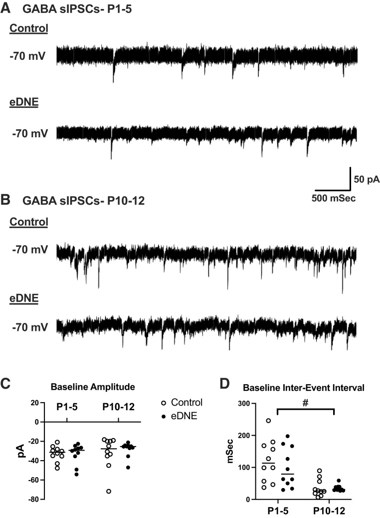
Regulation of GABAergic signaling through nicotinic acetylcholine receptor (nAChR) activation is critical for neuronal development. Here, we test the hypothesis that chronic episodic developmental nicotine exposure (eDNE) disrupts GABAergic signaling, leading to dysfunction of hypoglossal motor neurons (XIIMNs), which innervate the tongue muscles. We studied control and eDNE pups at two developmentally vulnerable age ranges: postnatal days (P)1-5 and P10-12. The amplitude and frequency of spontaneous and miniature inhibitory postsynaptic currents (sIPSCs, mIPSCs) at baseline were not altered by eDNE at either age. In contrast, eDNE increased GABAAR-α1 receptor expression on XIIMNs and, in the older group, the postsynaptic response to muscimol (GABAA receptor agonist). Activation of nAChRs with exogenous nicotine increased the frequency of GABAergic sIPSCs in control and eDNE neurons at P1-5. By P10-12, acute nicotine increased sIPSC frequency in eDNE but not control neurons. In vivo experiments showed that the breathing-related activation of tongue muscles, which are innervated by XIIMNs, is reduced at P10-12. This effect was partially mitigated by subcutaneous muscimol, but only in the eDNE pups. Taken together, these data indicate that eDNE alters GABAergic transmission to XIIMNs at a critical developmental age, and this is expressed as reduced breathing-related drive to XIIMNs in vivo.NEW & NOTEWORTHY Here, we provide a thorough assessment of the effects of nicotine exposure on GABAergic synaptic transmission, from the cellular to the systems level. This work makes significant advances in our understanding of the impact of nicotine exposure during development on GABAergic neurotransmission within the respiratory network and the potential role this plays in the excitatory/inhibitory imbalance that is thought to be an important mechanism underlying neonatal breathing disorders, including sudden infant death syndrome.

Keywords: GABA; development; hypoglossal; nicotine.


Conflict of interest statement

No conflicts of interest, financial or otherwise, are declared by the authors.

Figures

None
Graphical abstract
Figure 1.
Figure 1.
Weight, age, resting membrane potential, holding current, and input resistance. There were no differences in age (A) or weight (B) between control and episodic developmental nicotine exposure (eDNE) pups at either age [postnatal days(P)1–5: n = 20 control and n = 18 eDNE; P10–P12: n = 9 control and n = 14 eDNE]. There was also no difference in resting membrane potential (C), holding current (D), or input resistance (E) of hypoglossal motor neurons (XIIMNs) from control and eDNE pups at either age (n =30 control and n =30 eDNE neurons in each age group). All data presented as means ± SD. Comparisons were made between control and eDNE across ages with a 2-way ANOVA, with group means compared by Tukey’s post hoc test. #Significant difference between age groups.
Figure 2.
Figure 2.
Amplitude and interevent interval (IEI) of GABAergic spontaneous inhibitory postsynaptic currents (sIPSCs) at baseline. A: representative traces of pharmacologically isolated GABAergic sIPSCs in hypoglossal motor neurons (XIIMNs) from a postnatal day (P)3 control (top) and a P3 episodic developmental nicotine exposure (eDNE, bottom) animal. B: representative traces of pharmacologically isolated GABAergic sIPSCs in XIIMNs from a P10 control (top) and a P11 eDNE (bottom) animal. C: there was no difference in baseline amplitude of sIPSCs between control and eDNE at either age (2-way ANOVA, n = 10 control and eDNE: P1–5, P > 0.9999; P10–12, P = 0.9797), nor was there a difference in sIPSC amplitude between age groups (control, P > 0.9999; eDNE, P = 0.9151). D: there was no difference in the IEI of sIPSCs between control and eDNE at either age (2-way ANOVA, n = 10 control and eDNE: P1–5, P = 0.7428; P10–12, P > 0.9999); however, in both control and eDNE neurons, IEI was significantly shorter at P10–12 than at P1–5 (control, P = 0.0017; eDNE, P = 0.0227). Horizontal lines indicate mean values. #Significant difference between age groups.
Figure 3.
Figure 3.
Amplitude and interevent interval (IEI) of GABAergic miniature inhibitory postsynaptic currents (mIPSCs) at baseline. A: representative traces of pharmacologically isolated GABAergic mIPSCs in hypoglossal motor neurons (XIIMNs) from a postnatal day (P)2 control (top) and a P3 episodic developmental nicotine exposure (eDNE, bottom) animal. B: representative traces of pharmacologically isolated GABAergic mIPSCs in XIIMNs from a P11 control (top) and a P12 eDNE (bottom) animal. Asterisks highlight individual currents. C: there was no difference in baseline amplitude of mIPSCs between control and eDNE at either age (2-way ANOVA, n = 10 control and eDNE: P1–5, P = 0.9944; P10–12, P = 0.9114), nor was there a difference in mIPSC amplitude between age groups (control, P = 0.2786; eDNE, P = 0.1332). D: there was no difference in the IEI of mIPSCs between control and eDNE at either age (2-way ANOVA, n = 10 control and eDNE: P1–5, P = 0.9885; P10–12, P = 0.9551); however, in both control and eDNE neurons, IEI was significantly shorter at P10–12 than at P1–5 (control, P = 0.1219; eDNE P = 0.1774). Horizontal lines indicate mean values. #Significant difference between age groups.
Figure 4.
Figure 4.
Activation of postsynaptic GABAA receptors. A: representative traces of the whole cell GABAA receptor-mediated inward current in hypoglossal motor neurons (XIIMNs) from a postnatal (P)4 control (left) and a P4 episodic developmental nicotine exposure (eDNE, right) animal. B: representative traces of the whole cell GABAA receptor-mediated inward current in XIIMNs from a P10 control (left) and a P10 eDNE (right) animal. C: at P1–5, activation of GABAA receptors with bath application of 0.5 µM muscimol resulted in a similar peak inward current in control and eDNE neurons. In the P10–12 age group, bath-applied muscimol resulted in a larger peak inward current in eDNE neurons compared with control (2-way ANOVA, n = 10 control and eDNE: P1–5, P = 0.8052; P10–12, P = 0.0373). #Significant difference between control and eDNE.
Figure 5.
Figure 5.
GABAA receptor (GABAAR)-α1 immunohistochemistry. A: example images of tissue samples stained for the GABAAR-α1 subunit in a postnatal (P)3 control (left) and a P3 episodic developmental nicotine exposure (eDNE, right) preparation. B: example images of tissue samples stained for the GABAAR-α1 subunit in a P10 control (left) and a P11 eDNE (right) preparation. C: in both age groups, GABAAR-α1 receptor subunit immunostaining was increased in eDNE compared with control (2-way ANOVA, P1–5: n = 255 control and n = 257 eDNE, P < 0.0001; P10–12: n = 114 control and n = 178 eDNE, P < 0.0001). In both control (P < 0.0001) and eDNE (P < 0.0001) groups, receptor expression decreased with age. #Significant differences.
Figure 6.
Figure 6.
Amplitude and interevent interval (IEI) of GABAergic spontaneous inhibitory postsynaptic currents (sIPSCs) at baseline and during acute nicotine application in postnatal days (P)1–5 neurons. A: representative traces of pharmacologically isolated GABAergic sIPSCs in hypoglossal motor neurons (XIIMNs) from a P3 control animal at baseline (top) and during acute nicotine application (bottom). B: representative traces of pharmacologically isolated GABAergic sIPSCs in XIIMNs from a P3 episodic developmental nicotine exposure (eDNE) animal at baseline (top) and during acute nicotine application (bottom). C: acute nicotine application did not alter the amplitude of sIPSCs in either control or eDNE neurons (2-way ANOVA, n = 10 control, P = 0.2863 and n = 10 eDNE, P = 0.2319). D: in both control neurons and eDNE neurons, acute nicotine application caused a decrease in sIPSC IEI (2-way ANOVA, n = 10 control, P = 0.0391; n = 10 eDNE, P = 0.0372). Horizontal lines within the symbols indicate mean values. #Significant difference between baseline and acute nicotine application.
Figure 7.
Figure 7.
Amplitude and interevent interval (IEI) of GABAergic spontaneous inhibitory postsynaptic currents (sIPSCs) at baseline and during acute nicotine application in postnatal days (P)10–12 neurons. A: representative traces of pharmacologically isolated GABAergic sIPSCs in hypoglossal motor neurons (XIIMNs) from a P10 control animal at baseline (top) and during acute nicotine application (bottom). B: representative traces of pharmacologically isolated GABAergic sIPSCs in XIIMNs from a P10 episodic developmental nicotine exposure (eDNE) animal at baseline (top) and during acute nicotine application (bottom). C: acute nicotine application did not alter the amplitude of sIPSCs in either control or eDNE neurons (2-way ANOVA, n = 10 control, P = 0.9974 and n = 10 eDNE, P = 0.7698). D: in control neurons, acute nicotine application did not alter IEI of sIPSCs (2-way ANOVA, n = 10 control, P = 0.5914); however, in eDNE neurons acute nicotine application caused a decrease in sIPSC IEI compared with baseline (n = 10 eDNE, P = 0.0198). Horizontal lines indicate mean values. #Significant difference between baseline and acute nicotine application.
Fig. 8.
Fig. 8.
In vivo experimental preparation and average genioglossus muscle (GG) latency in postnatal days (P)10–12 pups after saline or muscimol injection. A: schematic of the in vivo experimental preparation, which includes concurrent diaphragm (Dia) and GG electromyographic (EMG) recordings in lightly anesthetized rats. For details, see methods. High-frequency spikes in the diaphragm EMG are ECG artifacts. B: example traces from a P10 control animal (left) and a P11 episodic developmental nicotine exposure (eDNE) animal (right) showing the raw and the rectified and integrated GG and diaphragm EMG activities during a 15-s nasal occlusion (indicated by bold square), which was applied 30 min after a subcutaneous saline injection (sham). The time between the onset of the nasal occlusion and the first discernable GG muscle EMG burst (arrow) is defined as onset latency. C: in P10–12 rats after saline injection, onset latency of GG EMG bursts was significantly longer in eDNE pups compared with control (saccharin) pups (P = 0.0101). Muscimol injection had no effect on latency in control pups (P = 0.3589); however, in eDNE pups latency was decreased after muscimol injection (P = 0.0321) (1-way ANOVA, n = 12 control and n = 12 eDNE pups). All data represented as means ± SD. #Significant differences between groups.

Similar articles

Cited by

References

    1. Olsen RW, Tobin AJ. Molecular biology of GABAA receptors. FASEB J 4: 1469–1480, 1990. doi:10.1096/fasebj.4.5.2155149. - DOI - PubMed
    1. Macdonald RL, Olsen RW. GABAA receptor channels. Annu Rev Neurosci 17: 569–602, 1994. doi:10.1146/annurev.ne.17.030194.003033. - DOI - PubMed
    1. Goetz T, Arslan A, Wisden W, Wulff P. GABAA receptors: structure and function in the basal ganglia. Prog Brain Res 160: 21–41, 2007. doi:10.1016/S0079-6123(06)60003-4. - DOI - PMC - PubMed
    1. Ben-Ari Y. Excitatory actions of GABA during development: the nature of the nurture. Nat Rev Neurosci 3: 728–739, 2002. doi:10.1038/nrn920. - DOI - PubMed
    1. Owens DF, Liu X, Kriegstein AR. Changing properties of GABAA receptor-mediated signaling during early neocortical development. J Neurophysiol 82: 570–583, 1999. doi:10.1152/jn.1999.82.2.570. - DOI - PubMed

Publication types

LinkOut - more resources