. 2023 Jan 18;83(2):301-315.
doi: 10.1158/0008-5472.CAN-22-1433.

Immunotherapeutic Targeting and PET Imaging of DLL3 in Small-Cell Neuroendocrine Prostate Cancer

Affiliations

Immunotherapeutic Targeting and PET Imaging of DLL3 in Small-Cell Neuroendocrine Prostate Cancer

Jonathan Chou et al. Cancer Res. .

Abstract

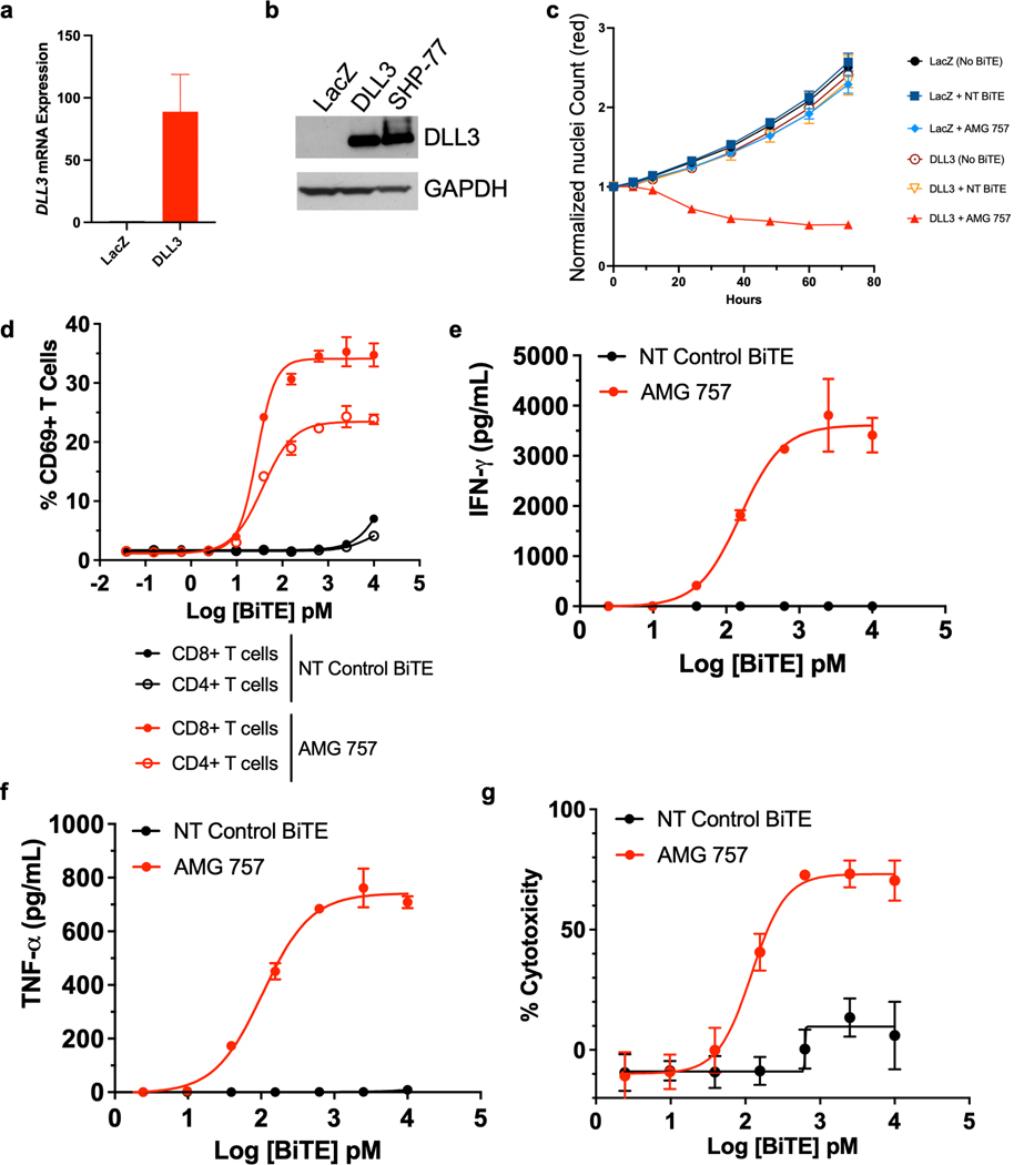
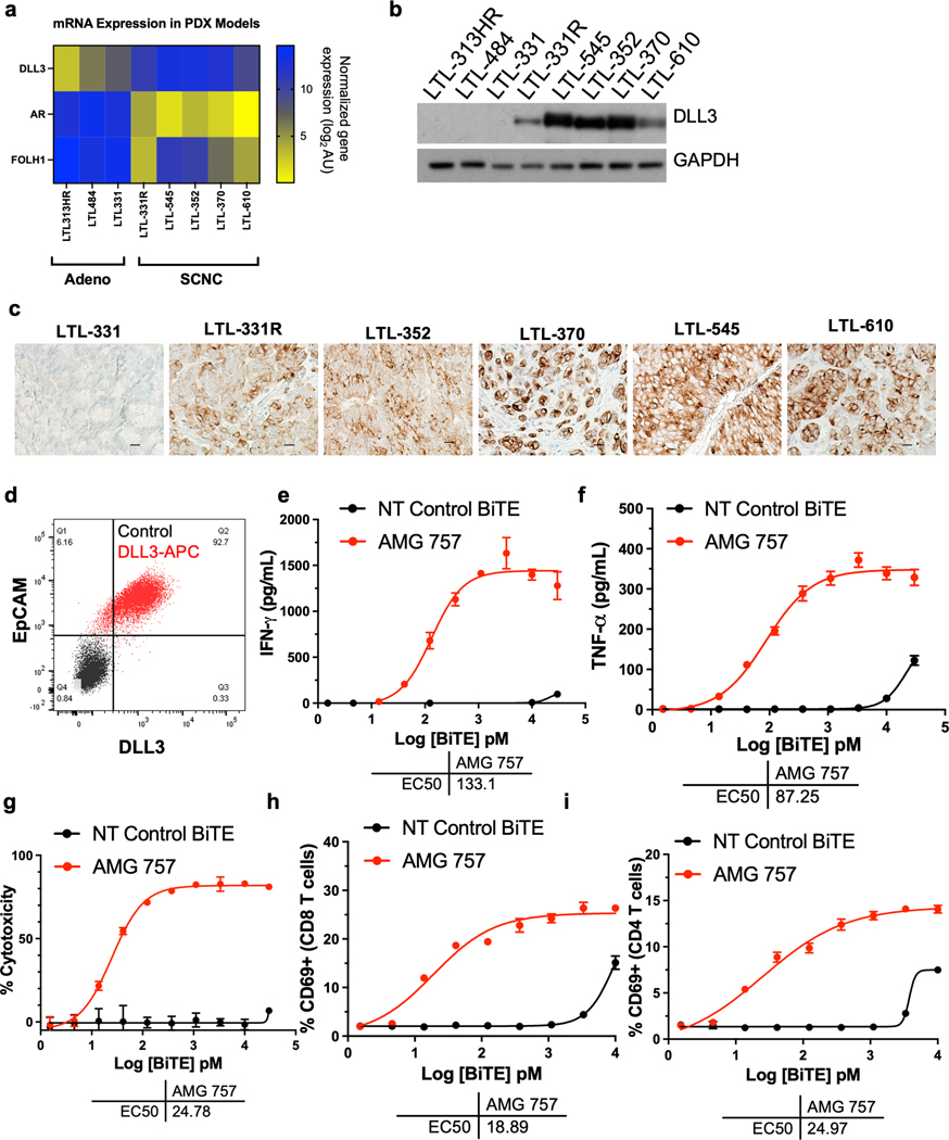
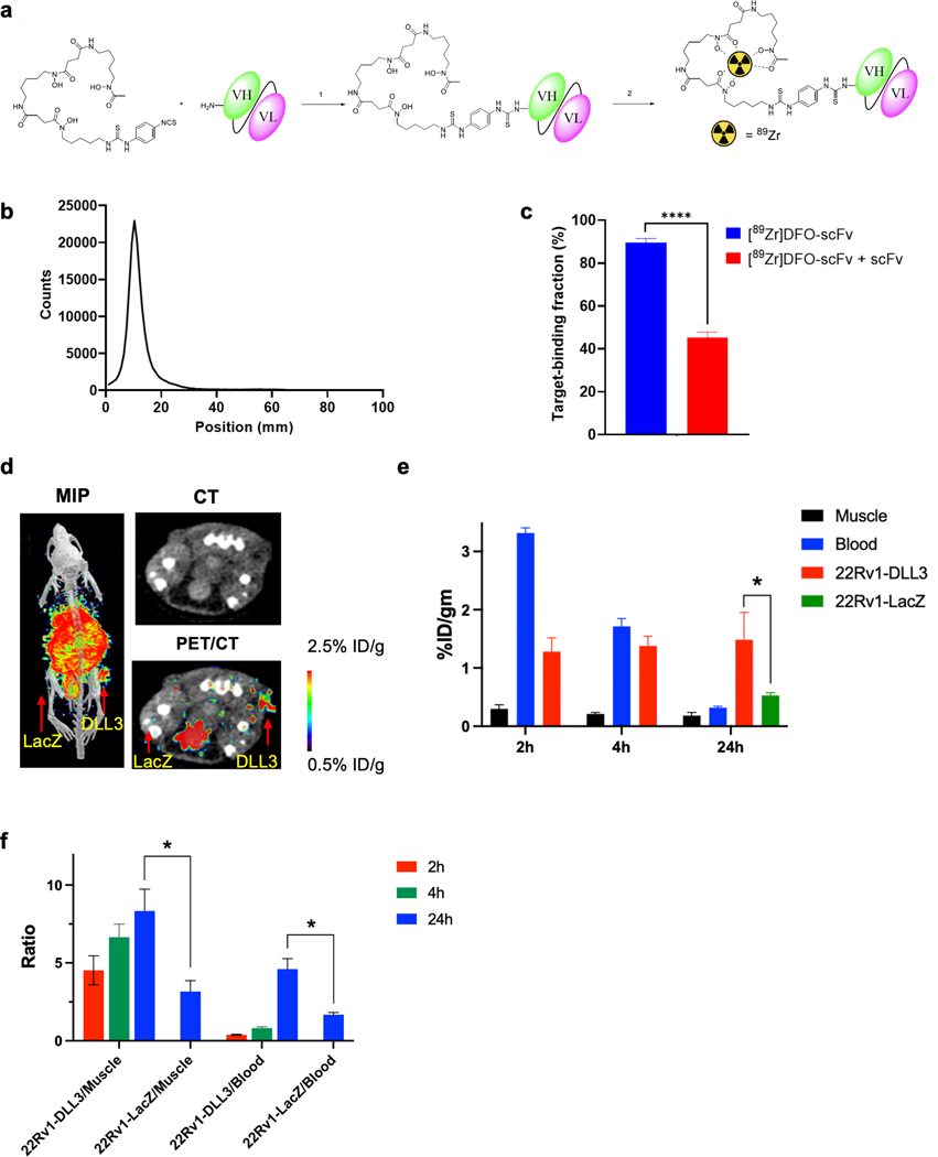
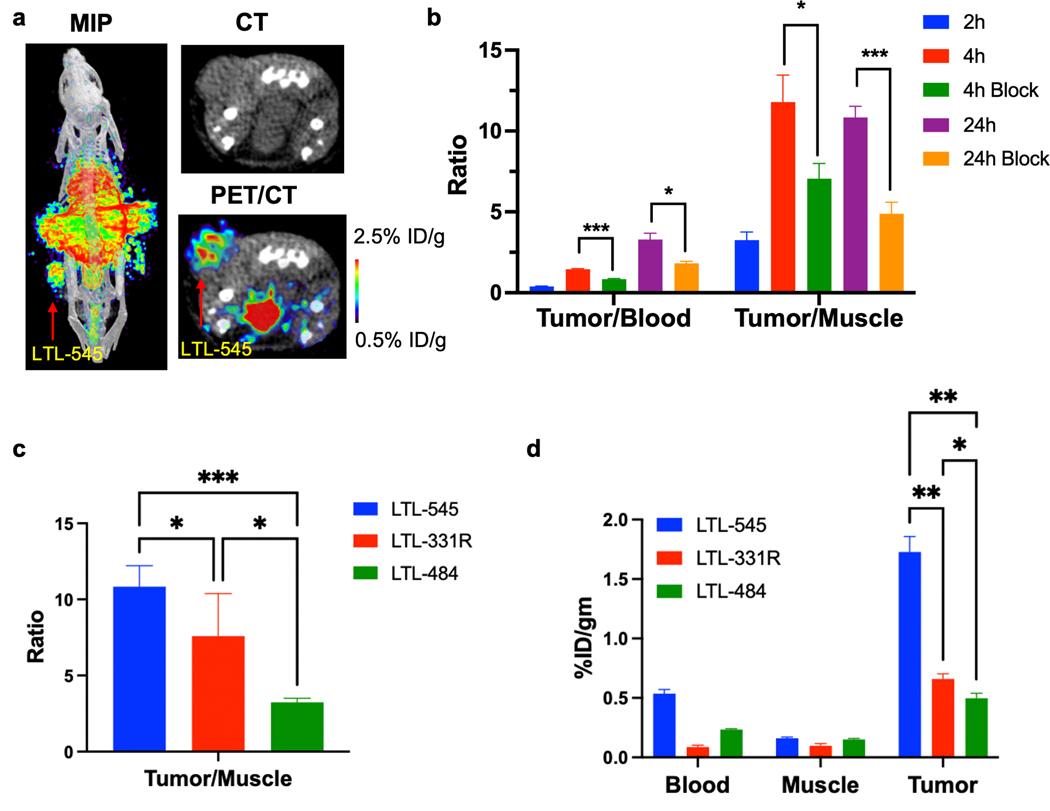
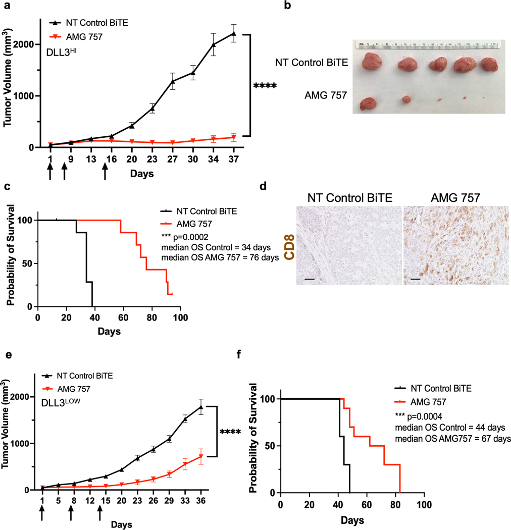
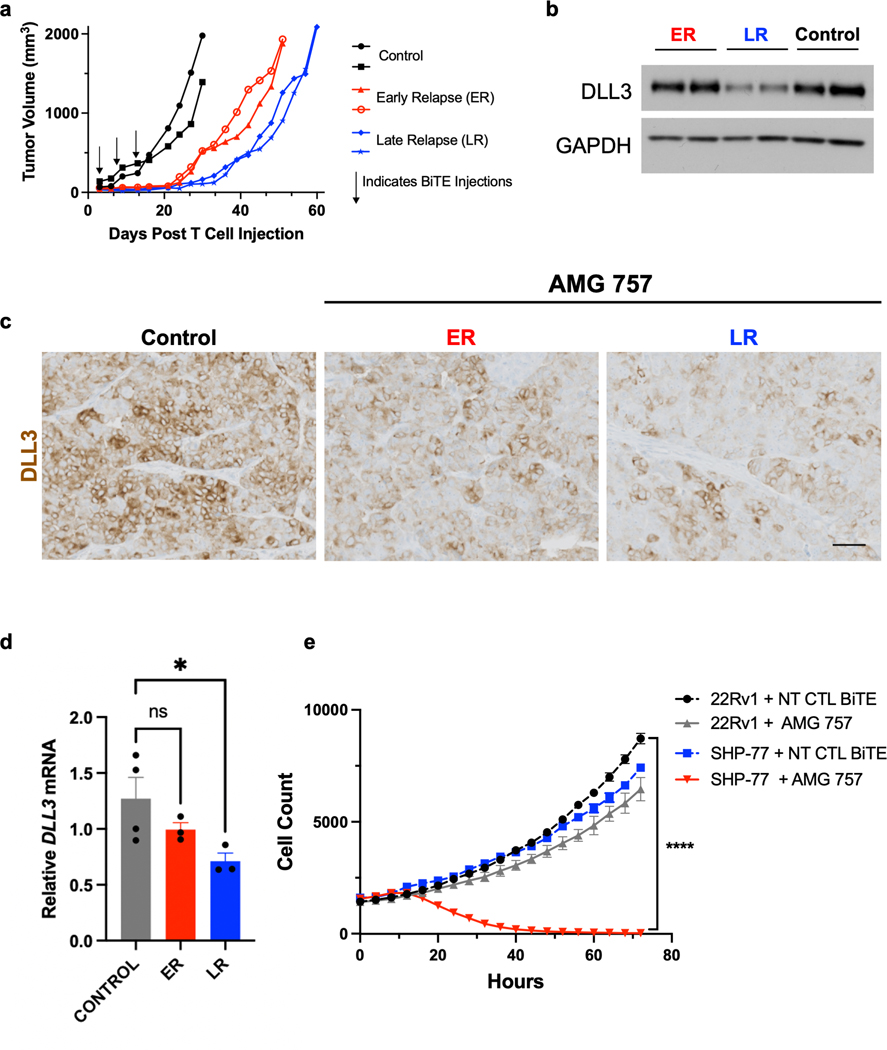
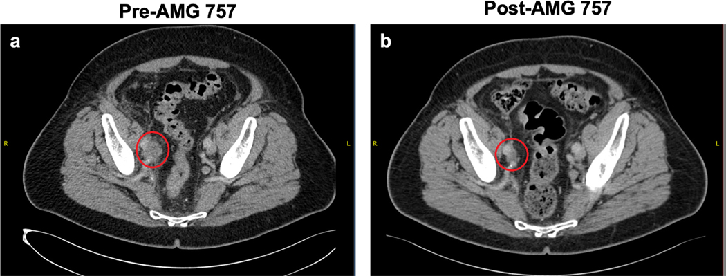
Effective treatments for de novo and treatment-emergent small-cell/neuroendocrine (t-SCNC) prostate cancer represent an unmet need for this disease. Using metastatic biopsies from patients with advanced cancer, we demonstrate that delta-like ligand 3 (DLL3) is expressed in de novo and t-SCNC and is associated with reduced survival. We develop a PET agent, [89Zr]-DFO-DLL3-scFv, that detects DLL3 levels in mouse SCNC models. In multiple patient-derived xenograft models, AMG 757 (tarlatamab), a half-life-extended bispecific T-cell engager (BiTE) immunotherapy that redirects CD3-positive T cells to kill DLL3-expressing cells, exhibited potent and durable antitumor activity. Late relapsing tumors after AMG 757 treatment exhibited lower DLL3 levels, suggesting antigen loss as a resistance mechanism, particularly in tumors with heterogeneous DLL3 expression. These findings have been translated into an ongoing clinical trial of AMG 757 in de novo and t-SCNC, with a confirmed objective partial response in a patient with histologically confirmed SCNC. Overall, these results identify DLL3 as a therapeutic target in SCNC and demonstrate that DLL3-targeted BiTE immunotherapy has significant antitumor activity in this aggressive prostate cancer subtype.

Significance: The preclinical and clinical evaluation of DLL3-directed immunotherapy, AMG 757, and development of a PET radiotracer for noninvasive DLL3 detection demonstrate the potential of targeting DLL3 in SCNC prostate cancer.


Figures

Figure 1.
Figure 1.. DLL3 Expression by Histologic Subtype of mCRPC.
a. DLL3 expression by H-score across samples with de novo and t-SCNC (n = 12) versus adenocarcinoma (n = 12) histologic differentiation. b. H&E and IHC stains for DLL3 in de novo and t-SCNC biopsies. Scale bar, 50 μM. c. Overall survival from date of biopsy for patients harboring mCRPC biopsies that were positive vs. negative for DLL3 expression. (HR = hazard ratio; CI = confidence interval). The number at risk for each group is indicated and censored data are indicated by the gray tick marks.
Figure 2.
Figure 2.. AMG 757 Activity Against DLL3-expressing Prostate Cancer Cells in vitro.
a. Relative DLL3 mRNA expression in 22RV1 prostate cancer cells expressing LacZ (control) or DLL3. b. Western blot showing DLL3 protein expression in 22RV1 cells expressing LacZ (control) or DLL3. SCLC SHP-77 cells were used as a positive control. GAPDH is shown as loading control. c. Growth curves normalized by nuclei count (NucLightRed) in 22RV1-LacZ and 22RV1-DLL3 cells co-cultured with human T cells and treated with a NT control BiTE® molecule or AMG 757. d-g. Co-cultures of NCI-H660 prostate cancer cells with human T cells and AMG 757 (red) or a NT control BiTE molecule (black) were assayed for d. CD69 activation of CD4+ and CD8+ T cells; e-f. IFNγ (e) and TNFα (f) secretion; g. cytotoxicity of NCI-H660 cells.
Figure 3.
Figure 3.. DLL3 Expression and AMG 757 Activity in Prostate PDX Tumor Cells.
a. mRNA expression of DLL3, AR and FOLH1 across 3 prostate cancer adenocarcinoma and 5 prostate cancer SCNC lines, including one t-SCNC line (LTL-331R). mRNA expression is normalized gene expression (log2) arbitrary units (AU). b. Western blot showing DLL3 expression in the PDX cell lines. c. DLL3 expression by IHC in the models indicated. Scale bar, 50 μM. d. Scatter plot showing DLL3 expression in dissociated tumor cells from the LTL-331R PDX line. Cells stained with a nontargeting antibody as a control are shown in black (Control). Cells stained with an anti-EpCAM antibody conjugated to FITC and anti-DLL3 antibody conjugated to APC are shown in red. e-i. Co-cultures of LTL-331R PDX cells with human T cells were incubated with AMG 757 (red) or NT control BiTE molecule (black) and assayed for e. IFNγ; f. TNFα; g. LTL-331R tumor cell cytotoxicity; h. activation of CD8+ T cells; and i. activation of CD8+ T cells. The AMG 757 EC50 is shown for each assay.
Figure 4.
Figure 4.. [89Zr]DFO-DLL3-scFv ImmunoPET Imaging of DLL3+ Tumor Xenografts.
a. Schematic for [89Zr]DFO-scFv generation. b. iTLC analysis of purified [89Zr]DFO-scFv. c. Magnetic bead–based radioligand binding assay for DLL3 target binding fraction in the absence or presence of unlabeled DLL3 scFv. **** p < 0.0001. d. Maximum-intensity projection (MIP) μPET/CT, transaxial CT, and transaxial μPET/CT slices obtained 24h after administration of [89Zr]DFO-scFv in 22RV1-LacZ and 22RV1-DLL3 dual tumor xenografts. e. Biodistribution data of [89Zr]DFO-scFv from selected tissues from 2h to 24h in mice bearing 22Rv1-LacZ and 22RV1-DLL3 dual tumors. f. Tumor/blood and tumor/muscle ratio of [89Zr]DFO-scFv from 2h to 24h in 22Rv1-LacZ and 22RV1-DLL3 dual tumors. *p < 0.05.
Figure 5.
Figure 5.. DLL3 ImmunoPET Imaging Across Multiple PDX Models.
a. Maximum-intensity projection (MIP) μPET/CT, transaxial CT, and transaxial μPET/CT slices obtained 24h after administration of [89Zr]DFO-scFv in mice bearing LTL-545 PDX tumors. b. Tumor/blood and tumor/muscle ratio of [89Zr]DFO-DLL3-scFv (with scFv block) at 2h, 4h, and 24h in mice bearing LTL-545 PDX xenografts. ns= not significant, * p < 0.05, *** p < 0.001. c. Tumor/blood and tumor/muscle ratio of [89Zr]DFO-DLL3-scFv in LTL-545 (DLL3HI), LTL-331R (DLL3LOW) and LTL-484 (DLL3NEGATIVE) models at 24h post-injection. * p< 0.05, *** p < 0.001. d. Biodistribution data of [89Zr]DFO-scFv from selected tissues in LTL-545, LTL-331R and LTL-484 models at 24h post-injection.
Figure 6.
Figure 6.. AMG 757 Antitumor Activity in DLLHI and DLL3LOW PDX Models.
a. Tumor growth curves in mice bearing LTL-545 PDX tumors (DLLHI) treated with AMG 757 (red) or NT Control BiTE® (black). Mice were administered human T cells on Day 0. Arrows indicate days of BiTE® treatment. b. Images of the endpoint tumors from mice in the study shown in (a). c. Kaplan-Meier survival curve of mice in the study shown in (a). Median OS is shown, n=15 mice per group. d. IHC images of CD8+ T cells in LTL-545 tumors taken 5 days after the first AMG 757 or NT control BiTE® treatment in (a). Scale bar, 50 μM. e. Tumor growth curves in mice bearing LTL-331R PDX tumors (DLL3LOW) treated with AMG 757 (red) or NT control BiTE® (black). f. Kaplan-Meier survival curve of mice from the study shown in (e). Median OS is shown, n = 10 mice per group. No mice were censored for panels c and f.
Figure 7.
Figure 7.. DLL3 Expression in Relapsed LTL-331R Tumors.
a. Individual tumor growth curves in mice bearing LTL-331R PDX tumors treated with AMG 757 or NT Control (CTL) BiTE®. Mice that received AMG 757 showed early relapse (ER, red) or late relapse (LR, blue). b. Western blot for DLL3 expression in control or relapsed tumor samples. c. IHC showing DLL3 expression in control or relapsed tumor samples. Scale bar, 100 μM. d. DLL3 mRNA expression in control or relapsed tumor samples by qPCR. * p<0.05. e. 22RV1 (labeled with NucLightGreen) and SHP-77 (labeled with NucLightRed) cells were mixed at a 1:1 ratio, and co-cultured with T cells. For each condition, the number of 22RV1 and SHP-77 cells in the presence of either NT CTL BiTE® or AMG 757 is shown and plotted over time. **** p<0.0001 for cell count at the final time point, between SHP-77 + AMG 757 vs all other conditions.
Figure 8.
Figure 8.. Confirmed Objective Response in a Patient with Histologically-Confirmed SCNC Treated with AMG 757.
a. Computed tomography (CT) scan before AMG 757 therapy showing the patient’s right pelvic soft tissue mass, indicated by the red circle, and significant right-sided hydronephrosis. b. CT scan of the patient’s pelvis after 15 weeks on AMG 757 therapy shows a partial response by RECIST criteria (greater than 30% reduction in size) and near resolution of the right-sided hydronephrosis.

References

    1. Aggarwal R, Huang J, Alumkal JJ, Zhang L, Feng FY, Thomas GV, et al. Clinical and Genomic Characterization of Treatment-Emergent Small-Cell Neuroendocrine Prostate Cancer: A Multi-institutional Prospective Study. J Clin Oncol 2018;36(24):2492–503 doi 10.1200/JCO.2017.77.6880. - DOI - PMC - PubMed
    1. Spetsieris N, Boukovala M, Patsakis G, Alafis I, Efstathiou E. Neuroendocrine and Aggressive-Variant Prostate Cancer. Cancers (Basel) 2020;12(12) doi 10.3390/cancers12123792. - DOI - PMC - PubMed
    1. Aparicio AM, Harzstark AL, Corn PG, Wen S, Araujo JC, Tu SM, et al. Platinum-based chemotherapy for variant castrate-resistant prostate cancer. Clin Cancer Res 2013;19(13):3621–30 doi 10.1158/1078-0432.CCR-12-3791. - DOI - PMC - PubMed
    1. Chapman G, Sparrow DB, Kremmer E, Dunwoodie SL. Notch inhibition by the ligand DELTA-LIKE 3 defines the mechanism of abnormal vertebral segmentation in spondylocostal dysostosis. Hum Mol Genet 2011;20(5):905–16 doi 10.1093/hmg/ddq529. - DOI - PubMed
    1. Dunwoodie SL, Clements M, Sparrow DB, Sa X, Conlon RA, Beddington RS. Axial skeletal defects caused by mutation in the spondylocostal dysplasia/pudgy gene Dll3 are associated with disruption of the segmentation clock within the presomitic mesoderm. Development 2002;129(7):1795–806. - PubMed

Publication types

MeSH terms