Case Reports
. 2022 Nov 10;387(19):1795-1803.
doi: 10.1056/NEJMcpc2115857.

Case 34-2022: A 57-Year-Old Woman with Covid-19 and Delusions

Affiliations
Case Reports

Case 34-2022: A 57-Year-Old Woman with Covid-19 and Delusions

Gregory L Fricchione et al. N Engl J Med. .
No abstract available


Figures

Figure 1
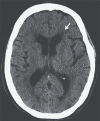
Figure 1. CT of the Head.
An axial image, obtained without the administration of intravenous contrast material, shows no evidence of an acute territorial infarct, intracranial mass, or hemorrhage. There is moderate confluent hypoattenuation involving the supratentorial white matter (arrow).
Figure 2
Figure 2. MRI of the Head.
An axial fluid-attenuated inversion recovery image (Panel A) shows moderate confluent hypoattenuation involving the supratentorial white matter (arrow), a finding consistent with small-vessel change, which is advanced given the patient’s age. There is equivalent prominence of the ventricles and sulci (arrowheads), a finding consistent with mild generalized parenchymal volume loss, which is also advanced given the patient’s age. A coronal T2-weighted image (Panel B) shows that the hippocampi are normal with respect to size, signal, and morphologic features (arrows). An axial T1-weighted image (Panel C), obtained after the administration of intravenous contrast material, shows no abnormal enhancement of the brain parenchyma. An axial diffusion-weighted image (Panel D) shows no restricted diffusion. An axial gradient–echo image (Panel E) shows no susceptibility signal. There is no evidence of a cause or consequence of seizure.
Figure 3
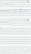
Figure 3. Electroencephalograms.
A bifrontal electroencephalographic (EEG) montage obtained after the patient had a seizure (Panel A) shows bilateral slowing without epileptiform discharges; there is a sharp frontal wave that may be an artifact (arrows). A follow-up EEG obtained 2 weeks later (Panel B) shows more slowing.

References

    1. Boyer EW, Shannon M. The serotonin syndrome. N Engl J Med 2005;352:1112-1120. - PubMed
    1. Hall RC, Popkin MK, Stickney SK, Gardner ER. Presentation of the steroid psychoses. J Nerv Ment Dis 1979;167:229-236. - PubMed
    1. Dono F, Nucera B, Lanzone J, et al. Status epilepticus and COVID-19: a systematic review. Epilepsy Behav 2021;118:107887-107887. - PMC - PubMed
    1. Dalmau J, Graus F. Antibody-mediated encephalitis. N Engl J Med 2018;378:840-851. - PubMed
    1. Pilotto A, Masciocchi S, Volonghi I, et al. The clinical spectrum of encephalitis in COVID-19 disease: the ENCOVID multicentre study. June 20, 2020. (http://medrxiv.org/lookup/doi/10.1101/2020.06.19.20133991). preprint.

Publication types