. 2022 Nov 8;41(6):111593.
doi: 10.1016/j.celrep.2022.111593.

Tuft-cell-intrinsic and -extrinsic mediators of norovirus tropism regulate viral immunity

Affiliations

Tuft-cell-intrinsic and -extrinsic mediators of norovirus tropism regulate viral immunity

Madison S Strine et al. Cell Rep. .

Abstract

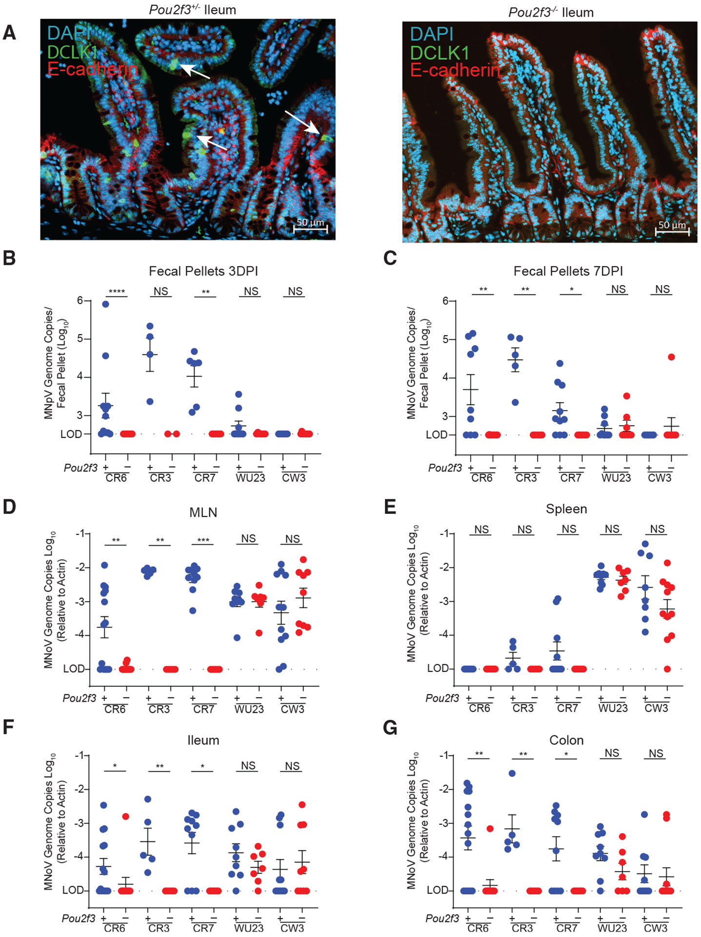
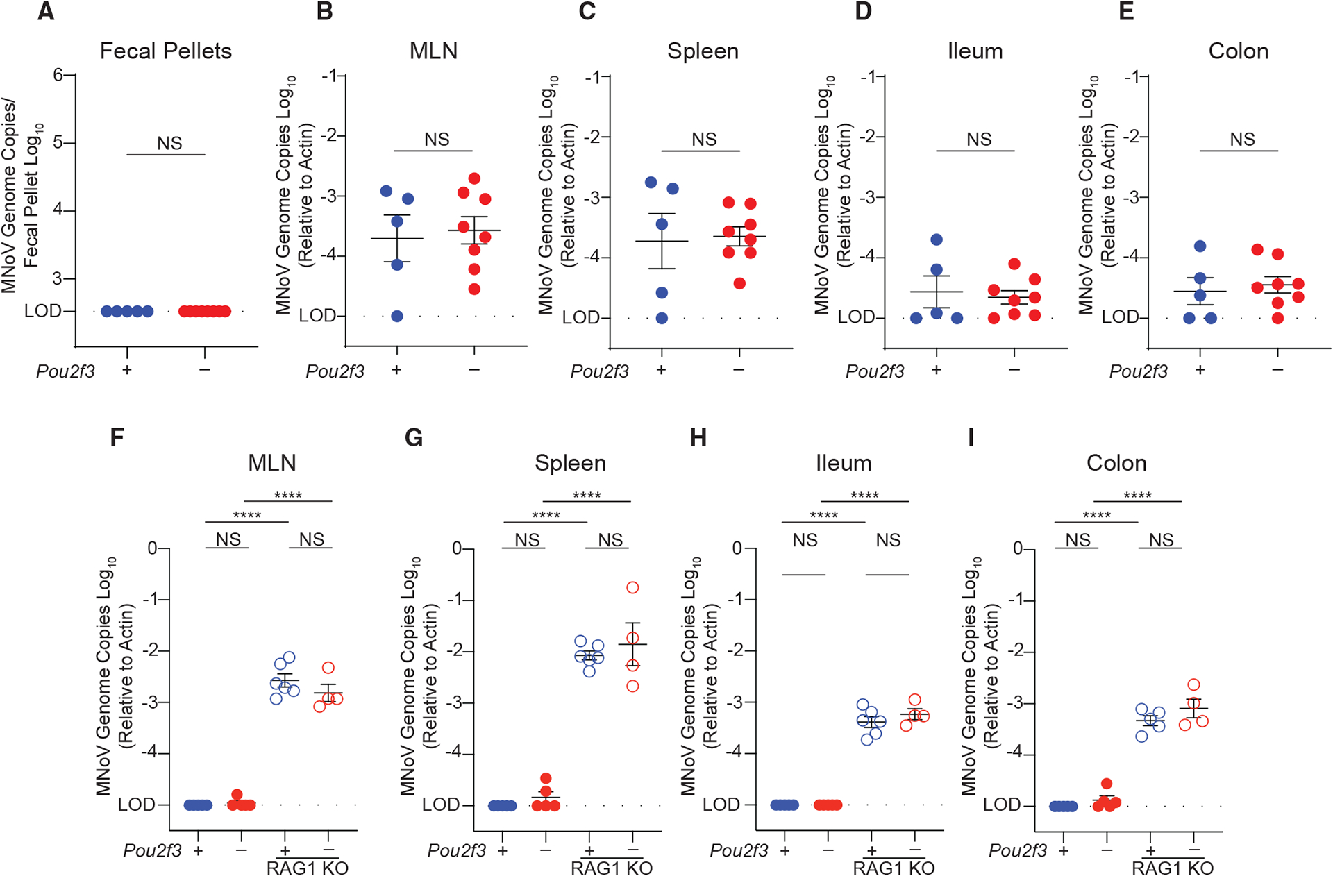
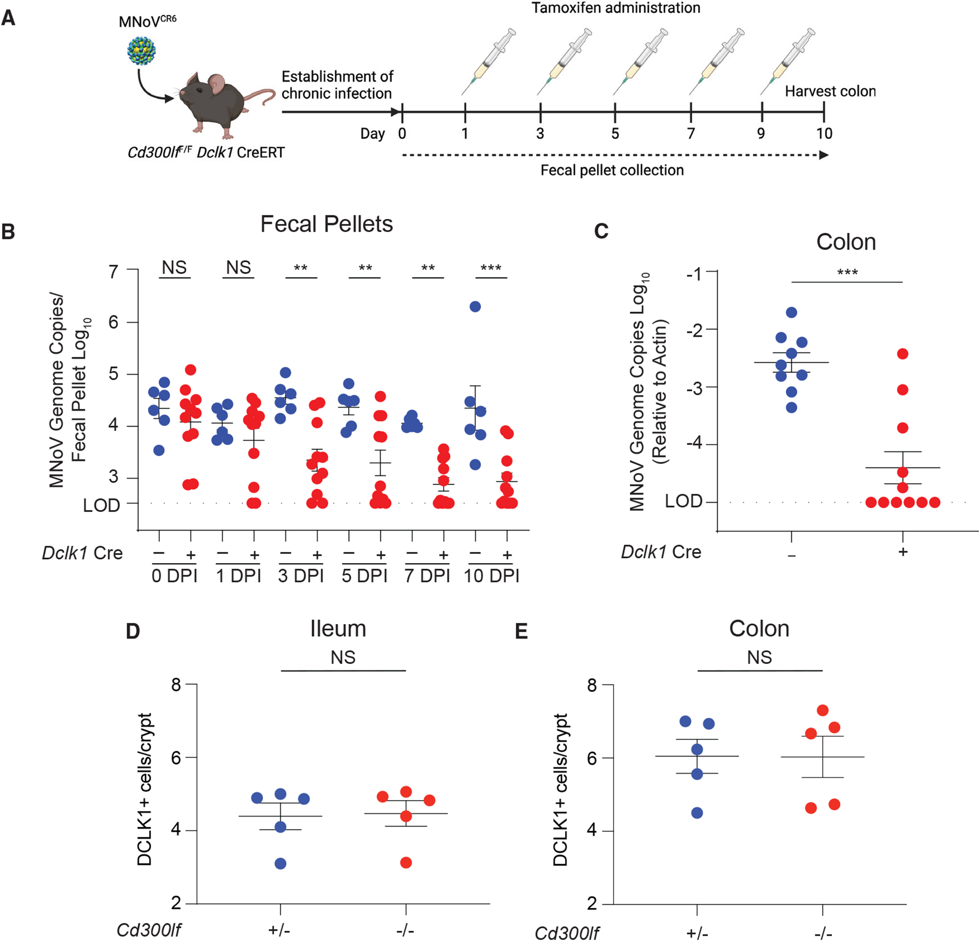
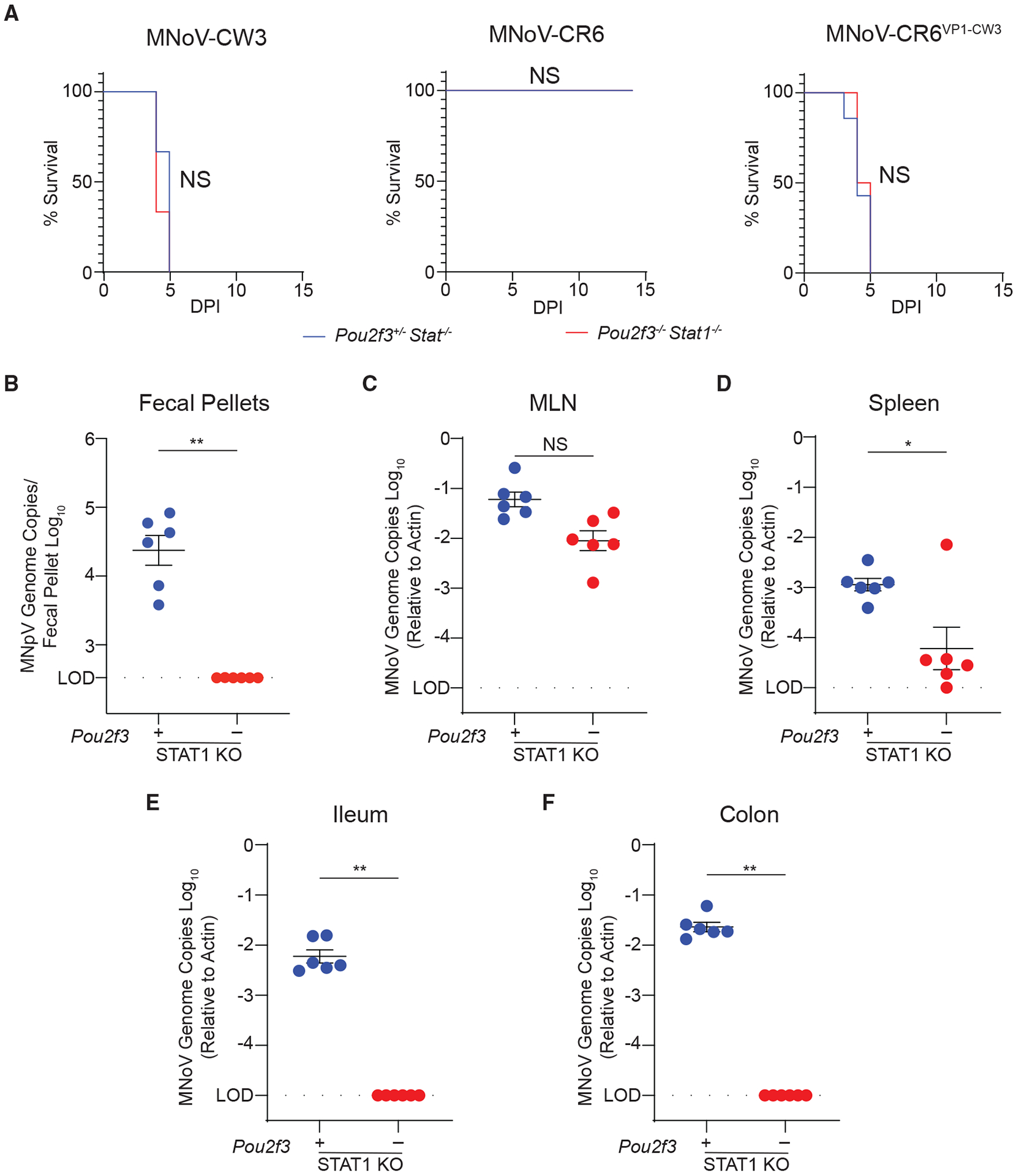
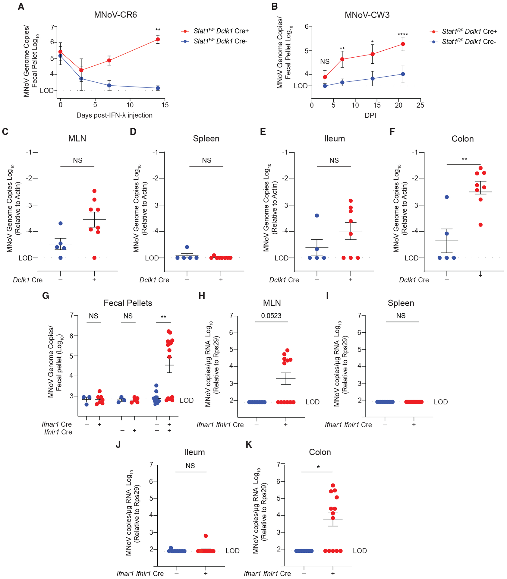
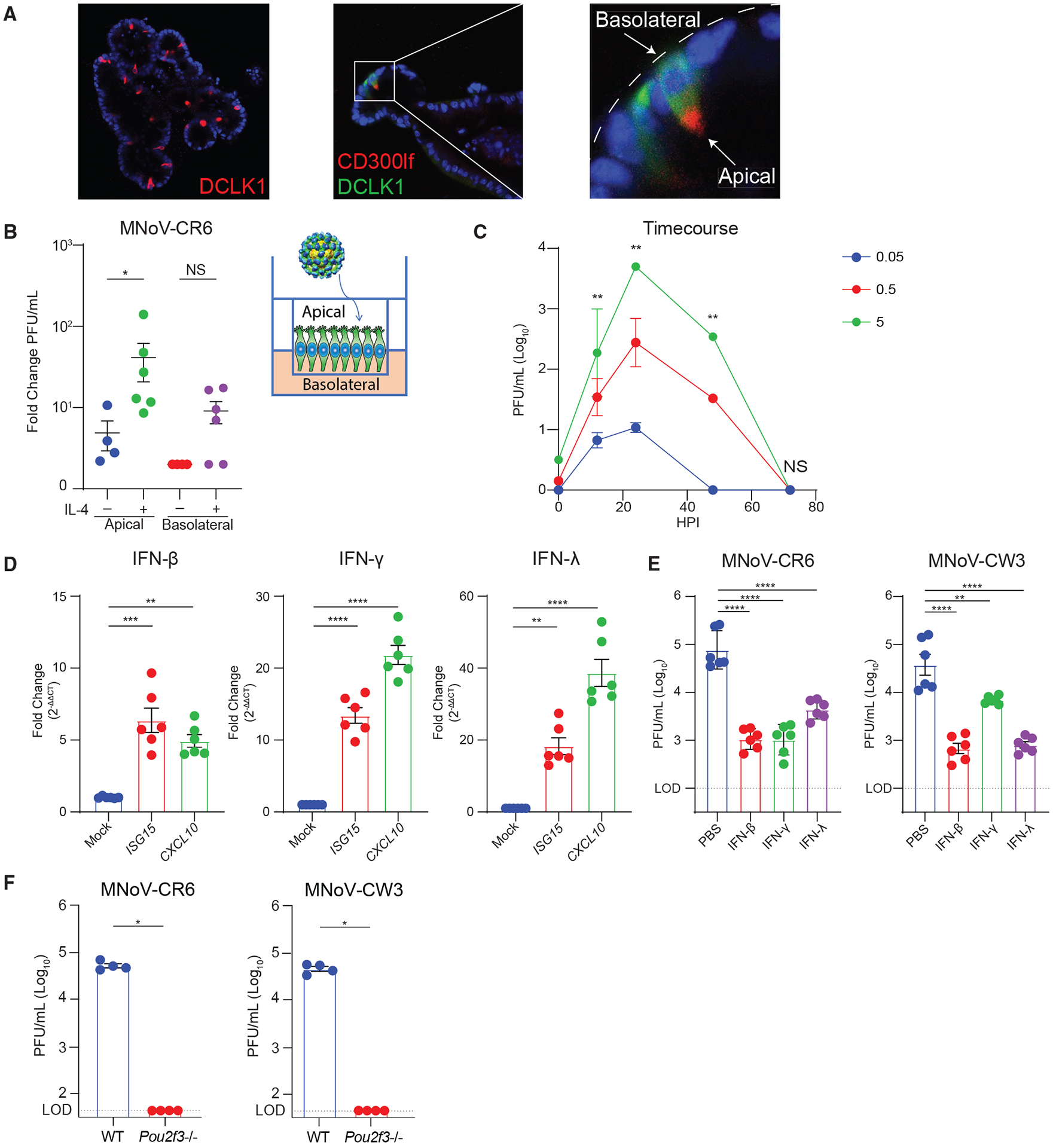
Murine norovirus (MNoV) is a model for human norovirus and for interrogating mechanisms of viral tropism and persistence. We previously demonstrated that the persistent strain MNoVCR6 infects tuft cells, which are dispensable for the non-persistent strain MNoVCW3. We now show that diverse MNoV strains require tuft cells for chronic enteric infection. We also demonstrate that interferon-λ (IFN-λ) acts directly on tuft cells to cure chronic MNoVCR6 infection and that type I and III IFNs signal together via STAT1 in tuft cells to restrict MNoVCW3 tropism. We then develop an enteroid model and find that MNoVCR6 and MNoVCW3 similarly infect tuft cells with equal IFN susceptibility, suggesting that IFN derived from non-epithelial cells signals on tuft cells in trans to restrict MNoVCW3 tropism. Thus, tuft cell tropism enables MNoV persistence and is determined by tuft cell-intrinsic factors (viral receptor expression) and -extrinsic factors (immunomodulatory signaling by non-epithelial cells).

Keywords: CP: Immunology; CP: Microbiology; immune evasion; interferon; mucosal immunity; norovirus; tuft cells; viral persistence; viral tropism.


Conflict of interest statement

Declaration of interests The authors declare no competing interests.

Figures

Figure 1.
Figure 1.. Tuft cells are essential for enteric infection by diverse persistent MNoV strains
(A) Pou2f3+/− (left) and Pou2f3−/− (right) mice were generated and stained with DAPI (blue), E-cadherin (red), and DCLK1 (green) for tuft cells in the mouse ileum. Images are representative of one of at least three independent experiments. (B–G) Pou2f3+/− and Pou2f3−/− mice were challenged perorally (PO) with 106 plaque-forming units (PFUs) of MNoV strains CR6, CR3, CR7, and Wu23. MNoV strain CW3 data are also shown and were previously published elsewhere (Grau et al., 2020). Fecal samples were collected at 3 and 7 days post-infection, and mice were sacrificed at 7 days post-infection (dpi). MNoV genome copies were quantified by qPCR in the (B and C) feces, (D) MLN, (E) spleen, (F) distal ileum, and (G) proximal colon. Infection experiments were performed at least two independent times, and qPCR was performed in triplicate. Data in (B)–(G) are pooled from two to four independent experiments with at least three mice per group and were analyzed by Mann-Whitney test. Shown are means ± SEM. Statistical significance annotated as follows: NS, not significant; *p < 0.05; **p < 0.01; ***p < 0.001; ****p < 0.0001; LOD, limit of detection.
Figure 2.
Figure 2.. Tuft cells are not essential for extraintestinal infection by persistent MNoV strain CR6
Pou2f3+/−, Pou2f3−/−, Pou2f3+/− Rag1−/−, and Pou2f3−/−Rag1−/− mice were challenged with 106 PFUs MNoVCR6 by intraperitoneal (i.p.) injection. Mice were sacrificed at (A–E) 7 dpi or (F–I) the chronic time point of 21 dpi. MNoV genome copies were quantified by qPCR in the (A) feces, (B and F) MLN, (C and G) spleen, (D and H) distal ileum, and (E and I) proximal colon. Experiments were performed at least two independent times with at least two mice per group, and qPCR was performed in triplicate. Data were analyzed by Mann-Whitney test (A–E) and ordinary one-way ANOVA (F–I). Shown are means ± SEM. Statistical significance annotated as follows: NS, not significant; *p < 0.05; **p < 0.01; ***p < 0.001; ****p < 0.0001; LOD, limit of detection.
Figure 3.
Figure 3.. Short-lived tuft cells support persistent MNoV infection
(A) Schematic depicting the infection and tamoxifen treatment used on Cd300lfF/F Dclk1 CreERT mice. Mice were infected PO with 106 PFUs MNoVCR6 for 15 to 33 days to establish chronic infection. Mice were then dosed with tamoxifen via i.p. injection every other day for 9 days to ablate CD300lf expression on newly differentiated tuft cells. (B) Fecal samples were collected, and MNoVCR6 fecal shedding was quantified on days 1, 3, 6, 7, and 10. (C) MNoVCR6 genome copies were assessed in the proximal colon at 10 days post-tamoxifen treatment. Genome copies were measured by qPCR and normalized to actin in tissues. Experiments were performed two independent times with at least five mice per group, and qPCR was performed in triplicate. (D and E) Tuft cell abundance was quantified along the crypt-villus axis in the ileum (D) and colon (E) of Cd300lf+/− and Cd300lf−/− mice. Data were analyzed by Mann-Whitney test (B, D, and E) and ordinary one-way ANOVA (C). Shown are means ± SEM. Statistical significance annotated as follows: NS, not significant; *p < 0.05; **p < 0.01; ***p < 0.001; LOD, limit of detection.
Figure 4.
Figure 4.. Tuft cells are essential for enteric infection in Stat1-deficient mice
(A) Pou2f3+/− Stat1−/− or Pou2f3−/− Stat1−/− mice were infected PO with 106 PFUs MNoV strains CR6 or CW3 or the chimeric strain CR6-VP1-CW3. Mouse survival was tracked for up to 7 days. (B–F) Mice challenged with MNoVCR6 were sacrificed and viral load was assessed by qPCR at 7 dpi for MNoV genome copies in the (B) feces, (C) MLN, (D) spleen, (E) distal ileum, and (F) proximal colon. Experiments were performed at least two independent times with at least three mice per group and data were analyzed by Mann-Whitney. Kaplan-Meier curves were generated for survival experiments. Shown are means ± SEM. Statistical significance annotated as follows: NS, not significant; *p < 0.05; **p < 0.01; LOD, limit of detection.
Figure 5.
Figure 5.. Tuft-cell-intrinsic innate immune signaling restricts MNoV infection
(A) Stat1F/F Dclk1 Cre mice were infected with 106 PFUs PO MNoVCR6 to establish chronic infection. Mice were treated with IFN-λ for 2 weeks. Fecal pellets were collected longitudinally at days 0, 3, 7, and 14 post-IFN-λ. (B–F) Stat1F/F Dclk1 Cre mice were infected with 106 PFUs PO MNoVCW3. Fecal pellets were collected at (B) 4, 7, 14, and 21 dpi and (C) MLN, (D) spleen, (E) ileum, and (F) colon at 21 dpi, mice were sacrificed, and tissues were harvested. (G–K) Ifnar1F/F Villin Cre, Ifnlr1F/F Villin Cre , and Ifnar F/F Ifnlr1F/F Villin Cre mice were infected with 106 PFUs PO MNoVCW3, and (G) fecal pellets, (H) MLN, (I) spleen, (J) ileum, and (K) colon were harvested at 14 dpi. Viral loads were assessed by qPCR for MNoV genome copies. Experiments were performed two independent times, and data were analyzed by Mann Whitney or two-way ANOVA. Shown are means ± SEM. Statistical significance annotated as follows: NS, not significant; *p < 0.05; **p < 0.01; ***p < 0.001; ****p < 0.0001; LOD, limit of detection.
Figure 6.
Figure 6.. Development of an enteroid culture system reveals MNoVCR6 and MNoVCW3 can infect tuft cells in vitro
Intestinal epithelial stem cells (IECs) were isolated from crypts of wild-type mice. IECs were differentiated and treated with recombinant IL-4 (rIL-4) to induce tuft cell differentiation. (A) Immunofluorescence staining for the tuft cell markers DCLK1 and CD300lf, showing apical distribution of CD300lf facing the interior lumen of the 2D enteroid. Images are representative of one of at least three independent experiments. (B) Enteroids were polarized and differentiated in monolayers on Transwell inserts to expose the apical surface of tuft cells. Monolayers were infected with MNoVCR6 (MOI = 5) at the apical surface ± rIL-4 administered to the apical or basolateral surface. Data are shown as fold change (increase) in viral titer from 1 to 24 hours post-infection (hpi). (C) Growth kinetics of MNoVCR6 (MOI = 0.05, 0.5, or 5) demonstrating efficient viral replication in the polarized tuft cell enteroid culture system. (D) Enteroids are susceptible to ISG induction, shown by upregulation of ISG15 and CXCL10 after treatment with 1,000 ng/mL IFN-β1, IFN-γ, or IFN-λ for 24 h. (E) Enteroids were pre-treated for 24 h with 1,000 ng/mL IFN-β1, IFN-γ, or IFN-λ and infected with MNoVCR6 or MNoVCW3. (F) Tuft cells are required for infection by MNoVCR6 and MNoVCW3 in enteroids. (D–F) Viral replication was quantified by plaque assay on BV2 cells at (D) 12, 24, 48, and 72 or (E and F) 24 hpi of enteroid culture. Data are pooled from two to four independent experiments and analyzed by Mann-Whitney (C and F) and one-way ANOVA (D and E). Shown are means ± SEM. Statistical significance annotated as follows: NS, not significant; *p < 0.05; **p < 0.01; ***p < 0.001; ****p < 0.0001; LOD, limit of detection.

Similar articles

Cited by

References

    1. Agudo J, Park E, Rose S, Alibo E, Sweeney R, Dhainaut M, Kobayashi K, Sachidanandam R, Baccarini A, Merad M, and Brown B (2018). Quiescent tissue stem cells evade immune surveillance. Immunity 48, 271–285.e5. 10.1016/j.immuni.2018.02.001. - DOI - PMC - PubMed
    1. Arias A, Bailey D, Chaudhry Y, and Goodfellow I (2012). Development of a reverse-genetics system for murine norovirus 3: long-term persistence occurs in the caecum and colon. J. Gen. Virol 93, 1432–1441. 10.1099/vir.0.042176-0. - DOI - PMC - PubMed
    1. Atmar R, Opekun A, Gilger M, Estes M, Crawford S, Neill F, and Graham D (2008). Norwalk virus shedding after experimental human infection. Emerg. Infect. Dis 14, 1553–1557. 10.3201/eid1410.080117. - DOI - PMC - PubMed
    1. Baldridge M, Lee S, Brown J, McAllister N, Urbanek K, Dermody T, Nice N, and Virgin H (2017). Expression of Ifnlr1 on intestinal epithelial cells is critical to the antiviral effects of interferon lambda against norovirus and reovirus. J. Virol 91, e02079. 10.1128/JVI.02079-16. - DOI - PMC - PubMed
    1. Baldridge M, Nice T, McCune B, Yokoyama C, Kambal A, Wheadon M, Diamond M, Ivanova Y, Artyomov M, and Virgin H (2015). Commensal microbes and interferon-λ determine persistence of enteric murine norovirus infection. Science 347, 266–269. 10.1126/science.1258025. - DOI - PMC - PubMed

Publication types