. 2022 Nov 8;41(6):111610.
doi: 10.1016/j.celrep.2022.111610.

Sex differences in resilience to ferroptosis underlie sexual dimorphism in kidney injury and repair

Affiliations

Sex differences in resilience to ferroptosis underlie sexual dimorphism in kidney injury and repair

Shintaro Ide et al. Cell Rep. .

Abstract

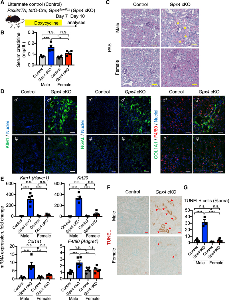
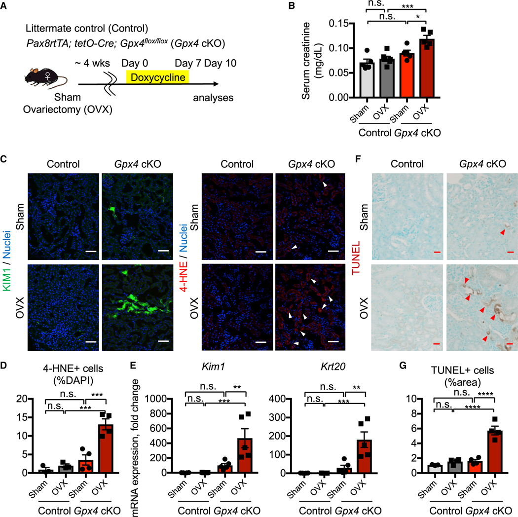
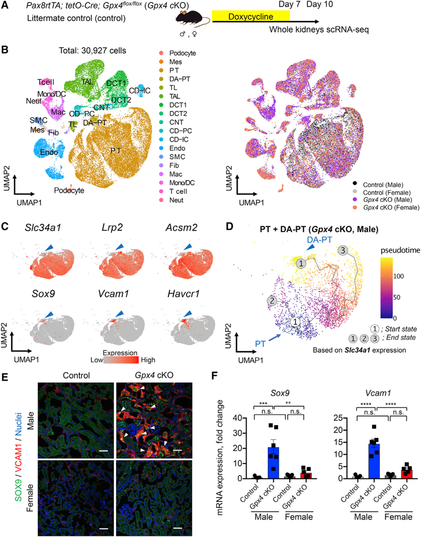
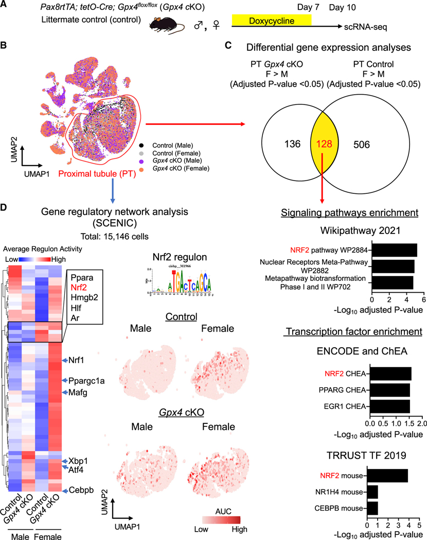
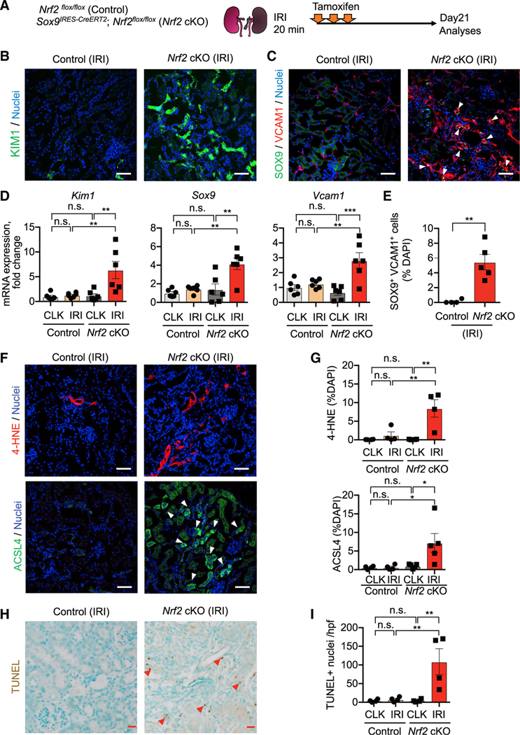
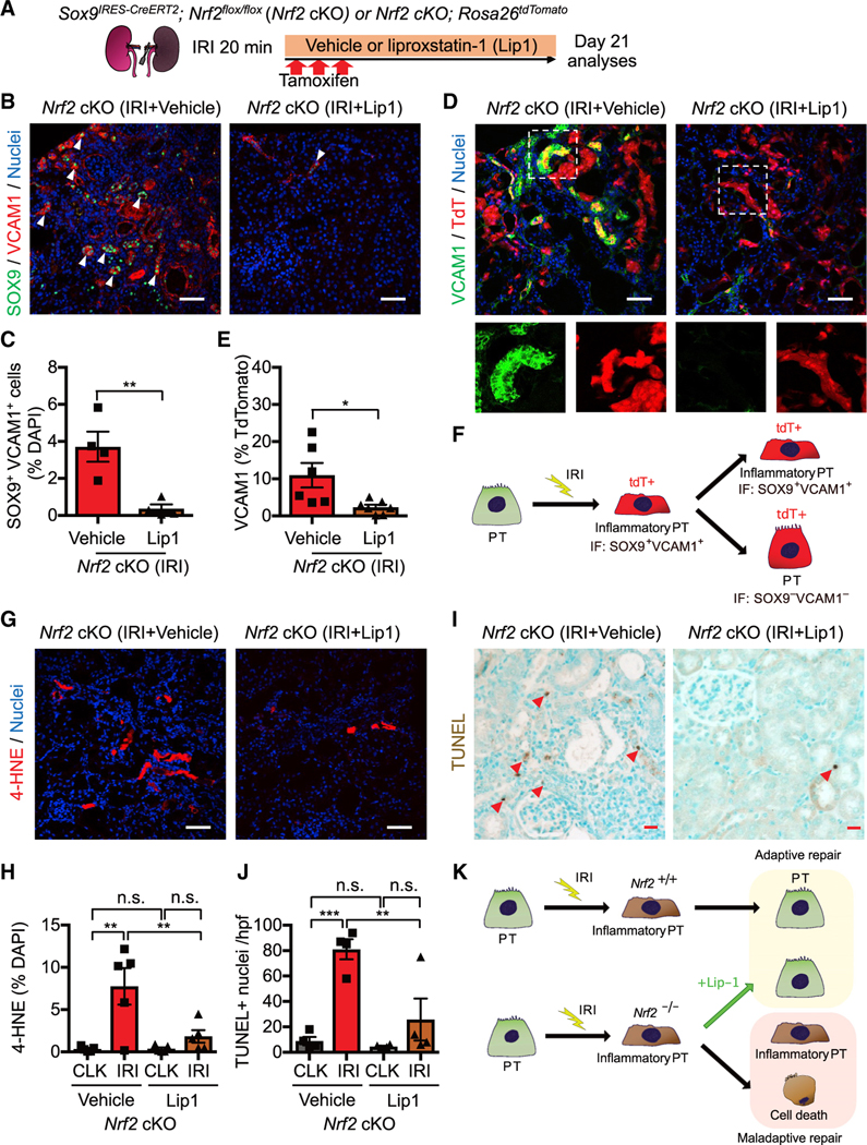
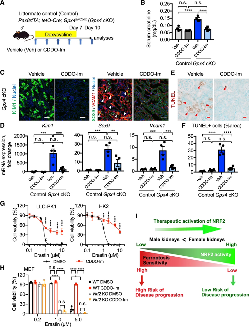
In both humans and mice, repair of acute kidney injury is worse in males than in females. Here, we provide evidence that this sexual dimorphism results from sex differences in ferroptosis, an iron-dependent, lipid-peroxidation-driven regulated cell death. Using genetic and single-cell transcriptomic approaches in mice, we report that female sex confers striking protection against ferroptosis, which was experimentally induced in proximal tubular (PT) cells by deleting glutathione peroxidase 4 (Gpx4). Single-cell transcriptomic analyses further identify the NFE2-related factor 2 (NRF2) antioxidant protective pathway as a female resilience mechanism against ferroptosis. Genetic inhibition and pharmacological activation studies show that NRF2 controls PT cell fate and plasticity by regulating ferroptosis. Importantly, pharmacological NRF2 activation protects male PT cells from ferroptosis and improves cellular plasticity as in females. Our data highlight NRF2 as a potential therapeutic target to prevent failed renal repair after acute kidney injury in both sexes by modulating cellular plasticity.

Keywords: CP: Molecular biology; GPX4; NRF2; acute kidney injury; cell fate; cellular plasticity; ferroptosis; glutathione Peroxidase 4; kidney injury and repair; sexual dimorphism; single-cell transcriptomics.


Conflict of interest statement

Declaration of interests The authors declare no competing interests.

Figures

Figure 1.
Figure 1.. Female kidneys are protected from ferroptosis
(A) Experimental workflow for genetic deletion of Gpx4, encoding a canonical anti-ferroptosis enzyme, in renal tubular epithelial cells. Doxycycline was given to control and Gpx4 cKO mice for 7 days, and kidneys were harvested on day 10. (B) Serum creatinine levels in control versus Gpx4 cKO male and female mice. n = 4–5. (C) Representative images of periodic-acid Schiff (PAS)-stained kidneys. *, hyaline casts. n = 4. (D) Immunostaining for tubular injury markers (KIM1 and NGAL), macrophages (F4/80), and fibrosis (COL1A1). For quantification, see Figure S1F. n = 4–5. (E) Real-time PCR analyses of gene expression. n = 5–6. (F and G) TUNEL staining for evaluating cell death. Quantification of TUNEL+ area is shown in (G) n = 3–4. Arrowheads, TUNEL+ cells. cKO, conditional knockout. One-way ANOVA with post hoc multiple comparisons test. n.s., not significant. *p < 0.05; **p < 0.01; ***p < 0.001; ****p < 0.0001. Scale bars: 50 μm in (C) and (D) and 20 μm in (F). Data are represented as mean ± SEM. See also Figure S1.
Figure 2.
Figure 2.. Intact ovarian function underlies female ferroptosis resilience
(A) Experimental workflow for testing ovarian function in female ferroptosis resistance by ovariectomy (OVX). Animals were allowed to recover for 4 weeks after OVX and then treated with doxycycline for 7 days to delete Gpx4. Kidneys were harvested on day 10. Control, Gpx4-intact control genotype; Sham, sham-operated mice. (B) Serum creatinine levels. n = 5–6. (C) Immunostaining for KIM1 (n = 4) and 4-HNE, a toxic lipid peroxide product (n = 3–4). Arrowheads, 4-HNEhigh cells. For quantification of KIM1, see Figure S2B. (D) Quantification of 4-HNE+ cells in (C). n = 3–4. (E) Real-time PCR analyses of indicated gene expression. n = 4–5. (F and G) TUNEL staining for evaluating cell death. Quantification of TUNEL+ area is shown in (G) n = 3–4. Arrowheads, TUNEL+ cells. One-way ANOVA with post hoc multiple comparisons test. n.s., not significant. *p < 0.05; **p < 0.01; ***p < 0.001; ****p < 0.0001. Scale bars: 50 μm in (C) and 20 μm in (F). Data are represented as mean ± SEM. See also Figure S2.
Figure 3.
Figure 3.. scRNA-seq identifies inflammatory PT cells in male Gpx4-deficient kidneys
(A) Experimental workflow for single-cell RNA sequencing (scRNA-seq). Mice were fed with doxycycline-containing water for 7 days, and kidneys were harvested on day 10 to generate scRNA-seq datasets. (B) Integrated single-cell transcriptome map. Unsupervised clustering identified all major renal cell types in the UMAP plot. PT, proximal tubule; DA-PT, damage-associated PT; TL, thin limb; TAL, thick ascending limb; DCT, distal convoluted tubule; CNT, connecting tubule; CD, collecting duct; PC, principal cells; IC, intercalated cells; Mes, mesangial cells; Endo, endothelial cells; SMC, smooth muscle cells; Fib, fibroblasts; Mac, macrophages; Mono/DC, monocytes and dendritic cells; Neut, neutrophils. (C) UMAP plots showing the expression of indicated genes in PT and DA-PT clusters. Differentiated/mature PT cell markers: Slc34a1 (sodium-dependent phosphate transporter 2a [NaPi2a]), Lrp2 (megalin), and acyl-coenzyme A synthetase (Acsm2), and damage-induced genes: Sry-box 9 (Sox9), vascular adhesion molecule 1 (Vcam1), and Havcr1 (KIM1). Arrowheads: DA-PT. (D) Pseudotime trajectory analysis of proximal tubular cells (PT and DA-PT) from male Gpx4 cKO mice. A region occupied with cells with high Slc34a1 expression was set as a starting state. (E) Immunostaining for SOX9 and VCAM1. n = 3. Arrowheads: SOX9+/VCAM1+ cells. (F) Real-time PCR analyses of indicated gene expression. n = 5–6. One-way ANOVA with post hoc multiple comparisons test. n.s., not significant. **p < 0.01; ***p < 0.001; ****p < 0.0001. Scale bars: 50 μm in (E). Data are represented as mean ± SEM. See also Figure S3 and Data S1.
Figure 4.
Figure 4.. scRNA-seq identifies NRF2 as a mechanism for female ferroptosis resilience
(A and B) Experimental workflow for identifying regulatory nodes underlying sex differences of ferroptosis sensitivity. A total of 15,146 PT cells from all conditions was used for downstream analyses. (C) Differential gene expression analyses identify 128 genes that are highly expressed in female PT cells compared with male counterparts both at Gpx4-intact control and Gpx4-knockout conditions. The 128 genes were subjected to Enrichr analysis. Overrepresented signaling pathways and enriched transcription factors are shown. (D) Single-cell regulatory network inference and clustering (SCENIC) identifies potential nodes that regulate PT cell states in the ferroptotic process. Heatmap of regulons derived from SCENIC is shown. Regulon activity for NFE2-related factor 2 (NRF2; also known as Nfe2l2) antioxidant transcription factor is shown on UMAP of PT cells (red dots represent cells with high NRF2 activity). AUC, enrichment score for the activity of each regulon. See also Figures S4–S8 and Data S2.
Figure 5.
Figure 5.. Nrf2 deletion prevents successful renal repair after IRI
(A) Experimental workflow for testing Nrf2 function in regulating PT cell fate after IRI. Nrf2 cKO mice and their littermate controls (control) were subjected to the same ischemic stress (20 min) and tamoxifen treatment. Nrf2 is deleted in Sox9-lineage cells after IRI with tamoxifen administration. Kidneys were harvested on day 21 post-IRI. (B and C) Immunostaining for indicated proteins. Representative images are shown. Arrowheads, SOX9+/VCAM1+ cells. See Figure S8F for KIM1 quantification. (D) Real-time PCR analyses of indicated gene expression. n = 6. (E) Quantification of SOX9+/VCAM1+ cells (arrowheads in C). n = 4–5. (F and G) Immunostaining for 4-HNE and ACSL4. Quantifications are shown in (G). n = 4–5. Arrowheads, ACSL4+ cells. (H and I) TUNEL staining for evaluating cell death. Quantification of TUNEL+ cells is shown in (I). n = 4. Arrowheads, TUNEL+ cells. One-way ANOVA with post hoc multiple comparisons test for (D), (G), and (I) and t test for (E). n.s., not significant. *p < 0.05; **p < 0.01; ***p < 0.001; ****p < 0.0001. Scale bars: 50 μm in (B), (C), and (F) and 20 μm in (H). Data are represented as mean ± SEM.
Figure 6.
Figure 6.. NRF2 governs PT cell fate and plasticity by mitigating ferroptotic stress
(A) Experimental workflow for testing NRF2 function in regulating ferroptotic stress and ferroptosis. Nrf2 cKO mice were subjected to ischemic stress (20 min) and tamoxifen treatment. The same volume of liproxstatin-1 (Lip-1) or vehicle was intraperitoneally injected daily into the mice. Kidneys were harvested on day 21 post-IRI. (B and C) Immunostaining for SOX9 and VCAM1. Representative images are shown. Arrowheads, SOX9+/VCAM1+ cells. Quantification is shown in (C). n = 4. (D and E) Immunostaining for VCAM1 and fate mapping using tdTomato fluorescence. Sox9-lineage cells express tdTomato. Insets: individual fluorescence channels of the dotted box area. Quantification of VCAM1+ cells in Sox9-lineage cells is shown in (E). n = 6–7. (F) Schematic model for PT cell state changes after IRI. (G and H) Immunostaining for 4-HNE and its quantification. n = 5. (I and J) TUNEL staining for evaluating cell death. Quantification of TUNEL+ cells is shown in (J). n = 4. Arrowheads, TUNEL+ cells. (K) Schematic model. Nrf2 regulates PT cell fate by mitigating ferroptotic stress. Student’s t test for (C) and (E) and one-way ANOVA with post hoc multiple comparisons test for (H) and (J). n.s., not significant. *p < 0.05; **p < 0.01; ***p < 0.001; ****p < 0.0001. Scale bars: 50 μm in (B), (D), and (G) and 20 μm in (I). Data are represented as mean ± SEM.
Figure 7.
Figure 7.. Pharmacological NRF2 activation prevents ferroptosis in Gpx4-deficient kidneys
(A) Experimental workflow for testing NRF2 function in regulating ferroptotic stress and ferroptosis using Gpx4 cKO mice. Gpx4 cKO mice were administered either vehicle or CDDO-Im (NRF2 inducer) on alternate days. Doxycycline was given for 7 days, and mice were harvested on day 10. (B) Serum creatinine levels in vehicle- versus CDDO-Im-treated Gpx4 cKO mice. n = 4. (C) Immunostaining for KIM1, SOX9, and VCAM1. See Figure S10D for KIM1 quantification. Representative images are shown. Arrowheads, SOX9+/VCAM1+ cells. n = 4. (D) Real-time PCR analyses of indicated gene expression. n = 4. (E and F) TUNEL staining for evaluating cell death. Quantification of TUNEL+ area is shown in (F). n = 4. Arrowheads, TUNEL+ cells. (G) Cellular viability assays using pig (LLC-PK1) and human (HK2) proximal tubular cell lines. (H) Cellular viability assays using wild-type (WT) and Nrf2 knockout mouse embryonic fibroblasts (MEFs). Erastin (system Xc inhibitor) was used for inducing ferroptosis in these cells. One-way ANOVA (B, D, and F) and two-way ANOVA (G and H) with post hoc multiple comparisons test. n.s., not significant. *p < 0.05; **p < 0.01; ***p < 0.001; ****p < 0.0001. Scale bars: 50 μm in (C) and 20 μm in (E). Data are represented as mean ± SEM in (B, D, and F) and mean ± SD in (G and H). (I) Proposed model. NRF2 acts as a rheostat for modulating ferroptosis sensitivity of proximal tubular cells.

Similar articles

Cited by

References

    1. Aibar S, Gonzá lez-Blas CB, Moerman T, Huynh-Thu VA, Imrichova H, Hulselmans G, Rambow F, Marine JC, Geurts P, Aerts J, et al. (2017). SCENIC: single-cell regulatory network inference and clustering. Nat. Methods 14, 1083–1086. 10.1038/nmeth.4463. - DOI - PMC - PubMed
    1. Aufhauser DD Jr., Wang Z, Murken DR, Bhatti TR, Wang Y, Ge G, Redfield RR 3rd, Abt PL, Wang L, Svoronos N, et al. (2016). Improved renal ischemia tolerance in females influences kidney transplantation outcomes. J. Clin. Invest 126, 1968–1977. 10.1172/JCI84712. - DOI - PMC - PubMed
    1. Bagshaw SM, Laupland KB, Doig CJ, Mortis G, Fick GH, Mucenski M, Godinez-Luna T, Svenson LW, and Rosenal T. (2005). Prognosis for long-term survival and renal recovery in critically ill patients with severe acute renal failure: a population-based study. Crit. Care 9, R700–R709. 10.1186/cc3879. - DOI - PMC - PubMed
    1. Bairey Merz CN, Dember LM, Ingelfinger JR, Vinson A, Neugarten J, Sandberg KL, Sullivan JC, Maric-Bilkan C, Rankin TL, Kimmel PL, et al. (2019). Sex and the kidneys: current understanding and research opportunities. Nat. Rev. Nephrol 15, 776–783. 10.1038/s41581-019-0208-6. - DOI - PMC - PubMed
    1. Bellanti F, Matteo M, Rollo T, De Rosario F, Greco P, Vendemiale G, and Serviddio G. (2013). Sex hormones modulate circulating antioxidant enzymes: impact of estrogen therapy. Redox Biol. 1, 340–346. 10.1016/j.redox.2013.05.003. - DOI - PMC - PubMed

Publication types

Substances