. 2022 Nov 8;41(6):111628.
doi: 10.1016/j.celrep.2022.111628.

An epitope-enriched immunogen expands responses to a conserved viral site

Affiliations

An epitope-enriched immunogen expands responses to a conserved viral site

Timothy M Caradonna et al. Cell Rep. .

Abstract

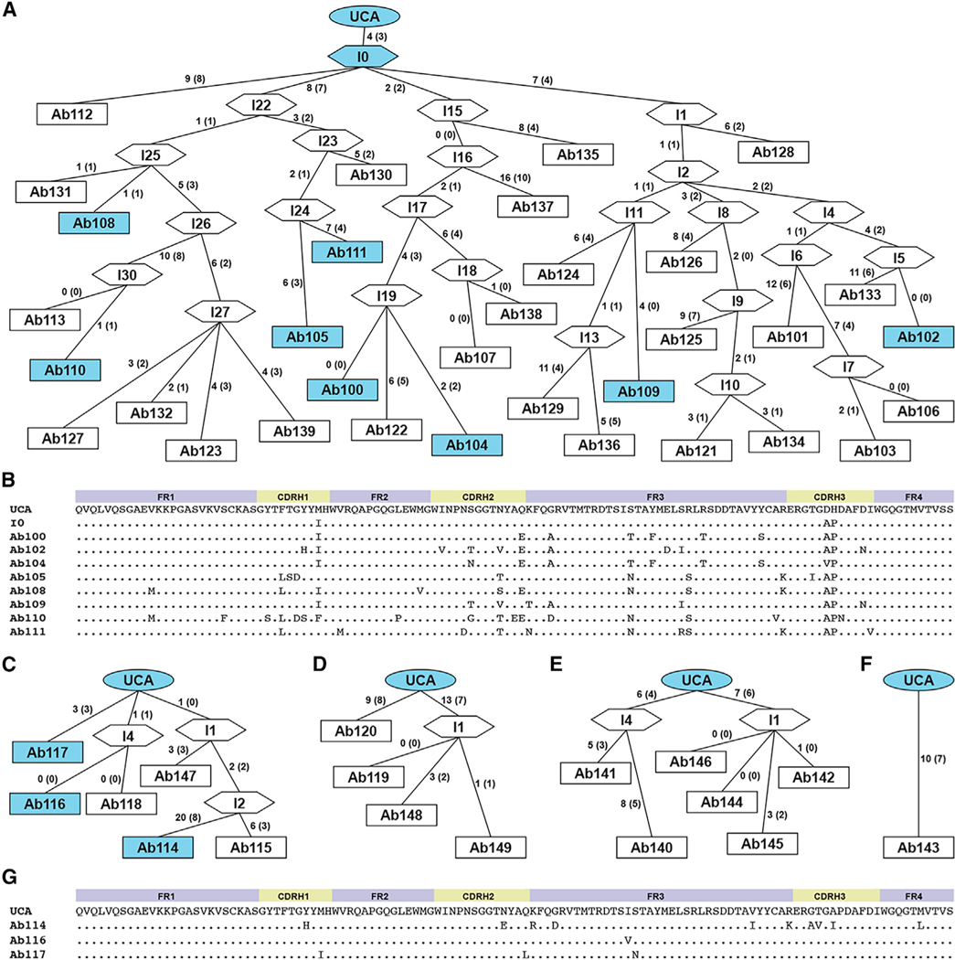
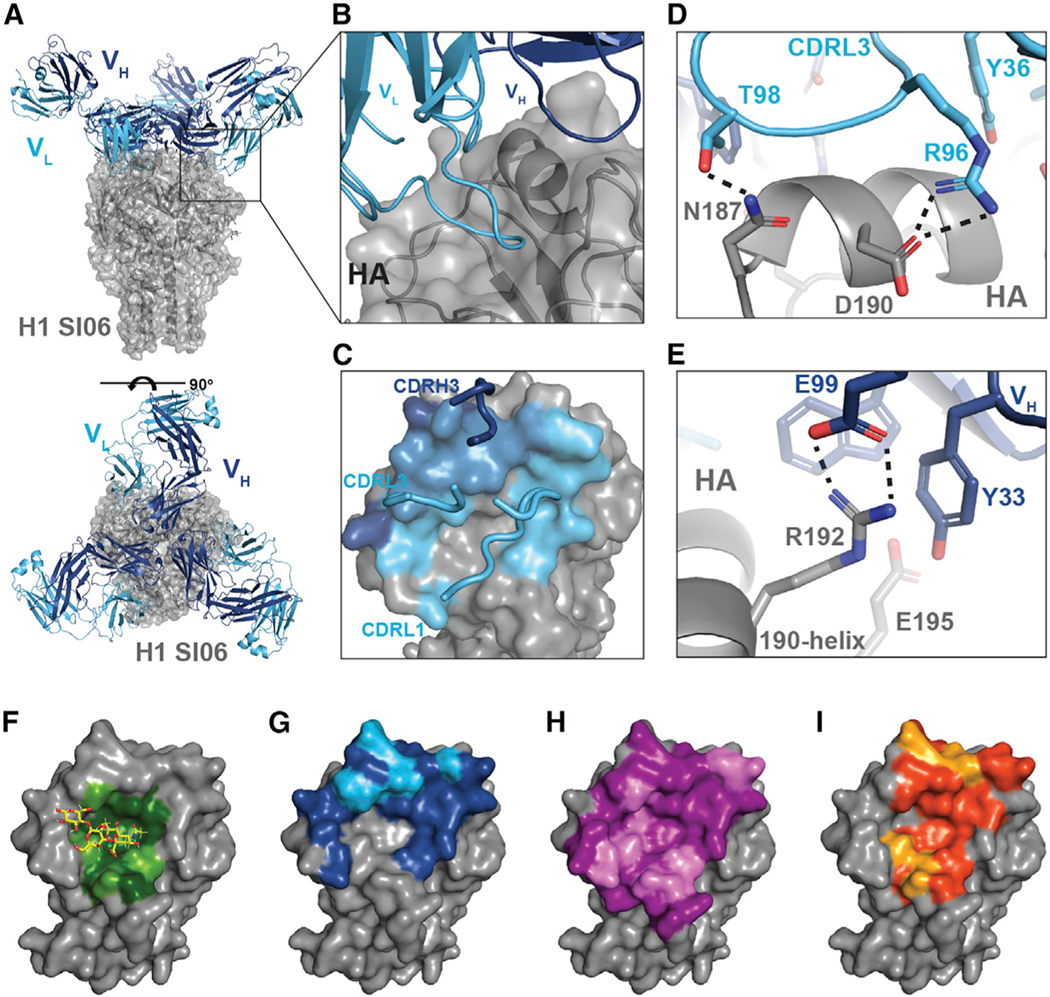
Pathogens evade host humoral responses by accumulating mutations in surface antigens. While variable, there are conserved regions that cannot mutate without compromising fitness. Antibodies targeting these conserved epitopes are often broadly protective but remain minor components of the repertoire. Rational immunogen design leverages a structural understanding of viral antigens to modulate humoral responses to favor these responses. Here, we report an epitope-enriched immunogen presenting a higher copy number of the influenza hemagglutinin (HA) receptor-binding site (RBS) epitope relative to other B cell epitopes. Immunization in a partially humanized murine model imprinted with an H1 influenza shows H1-specific serum and >99% H1-specific B cells being RBS-directed. Single B cell analyses show a genetically restricted response that structural analysis defines as RBS-directed antibodies engaging the RBS with germline-encoded contacts. These data show how epitope enrichment expands B cell responses toward conserved epitopes and advances immunogen design approaches for next-generation viral vaccines.

Keywords: CP: Immunology; adaptive immunity; epitope enrichment; hemagglutinin; immunogen design; influenza virus; protein engineering.


Conflict of interest statement

Declaration of interests T.M.C., M.K., and A.G.S. have filed a provisional patent for the described rsHAtCh immunogen.

Figures

Figure 1.
Figure 1.. Design and characterization of rsHAtCh
(A) Structure of the hemagglutinin RBS epitope and sequence alignment of the four primary RBS boundaries across various influenza A subtypes. “.” denotes amino acid identity with H1 SI-06, and “_” denotes a skipped residue. (B) Matrix showing percentage of amino acid identified between H1 SI06 head and representative rsHA heads from different subtypes of influenza A. (C) Size-exclusion chromatography traces showing relative efficiency of cystine bond formation for the six possible interfaces between rsH3, rsH4, and rsH14 head heterodimers. Dimeric (D) and monomeric (M) peaks labeled. “***” denotes high efficiency, “**” denotes ~50% efficiency, and “*” denotes low efficiency. (D) Schematic and sequences of an NC2-derived heterotrimerization tag used to selectively express heterotrimeric rsHAtCh. Yellow bar represents an intratag disulfide bond. (E) Assembly of a polycistronic “plug and play” DNA cassette composed of each component of rsHAtCh containing tPA (signal sequence), rsHA (insert), 3C (protease cleavage site), NC2 strand (heterotrimerization tag), and orthogonal affinity chromatography tag (AT1–3; His-tag, FLAG tag, or SBP tag, respectively), each separated by 2A peptides. “**” denotes a double stop codon. (F) SDS-PAGE analysis of purified rsHAtCh under non-reducing (1) and reducing (2) conditions. Trimer (T) and component monomer (M) bands shown. (G) BLI analysis of purified rsHAtCh and H1 SI-06 head, screened against a panel of H2227, H2526, CH67, 641 I-9, K03.12, C05, and HRV41.1 (negative control Ab), at 10 and 5 μM conditions. (H) Design process for generating epitope-enriched immunogens. Related to Figure S1.
Figure 2.
Figure 2.. Serum and IgG+ B cell analysis of rsHAtCh immunization
(A) Schematic representation of immunization regimen in IGHV1–2 HC2 mice. Serum harvested 2 weeks after final immunization. Two replicates of each cohort immunized. (B) Serum ELISA titers against H1 SI-06 FLsE and H1 SI-06 ΔRBS FLsE, where the RBS is sterically occluded by an N-linked glycan a position 190, for control cohort (N = 10) and rsHAtCh cohort (N = 8) mice. Statistical significance was determined using the Mann-Whitney U test (***p < 0.001). (C) rsHAtCh cohort serum ELISA titers against five historical H1 FLsEs. Statistical significance was determined using the Mann-Whitney U test (***p < 0.001). (D) Frequency of class-switched IgG+ B cells engaging RBS (ΔRBS non-binders) or non-RBS epitopes (ΔRBS binders); total numbers of cells sorted pooled across each cohort listed. Related to Figure S2.
Figure 3.
Figure 3.. Sequence analysis of RBS-directed IgG+ B cells
(A and B) Heavy- (A) and light-chain (B) gene usages from sorted RBS-directed cells elicited by rsHAtCh immunization; N = 71 pooled from 4 mice. (C) VH gene mutation frequency among sequenced RBS-directed B cells. (D and E) Sequence logos for HCDR3 (D) and LCDR3 (E) segments. Related to Figure S3.
Figure 4.
Figure 4.. Heavy-chain affinity maturation of elicited RBS-directed lineages
Sequence analysis of RBS-directed lineages isolated from three mice immunized with rsHAtCh. (A) Genealogy tree of lineage 100 antibody heavy chains. Nucleotide mutations between each branchpoint labeled, with corresponding number of amino acid mutations in parentheses. Lineage UCA, computationally inferred intermediates, and isolated IgG+ B cells shown as ovals, hexagons, and rectangles, respectively. Antibodies in blue were expressed recombinantly for binding analysis. (B) Alignment of VH amino acid sequences of lineage 100 antibodies, including the UCA and I-0 branchpoint; “.” denotes conservation to the UCA. (C–F) Genealogy trees of lineages (C) 114, (D) 119, (E) 140, and (F) 143. (G) Alignment of VH amino acid sequences of lineage 114 antibodies and UCA. Related to Figure S4.
Figure 5.
Figure 5.. rsHAtCh-elicited antibodies engage the RBS through germline-encoded contacts
(A) cryoEM structure of Ab109 Fab in complex withH1 SI06 FLsE. (B) Ab109-H1 SI-06 interface. (C) Cartoon representation of Ab109 footprint on hemagglutinin (HA); heavy-chain contacts shown in dark blue, both heavy- and light-chain contacts shown in light blue, and light-chain contacts shown in cyan. (D and E) Key contacts made between the CDRL3 (C) and CDRH3 (D) loops and the HA 190-helix. CDRH1 loop removed for clarity. Light-chain residues shown in cyan, heavy-chain residues shown in dark blue, and HA shown in gray. Salt bridges shown as dotted lines. (F–I) Footprints of (F) sialic acid, (G) Ab109, (H) H2526, and (I) 641I-9; colored residues are located at the RBS-ligand interface, with pale green/cyan/violet/yellow residues forming hydrogen bonds or salt bridges with the corresponding ligand. All images generated using PyMol. Related to Figure S6 and Table S1.
Figure 6.
Figure 6.. rsHAtCh-elicited RBS-directed antibodies neutralize strain-matched virus and partially protect
(A) Microneutralization titers of seven antibodies tested against a panel of eight historical H1 virus isolates ranging from 1934 to 2009. Titers are reported as the maximal concentration (ug/mL) of each antibody at which complete neutralization was observed. (B) Cartoon representation of Ab109 footprint on HA; heavy-chains contact shown in dark blue, both heavy- and light-chain contacts shown in light blue, and light-chain contacts shown in cyan. CDR L1, L3, and H3 loops are labeled. Residue changes across historical H1 isolates expected to abrogate binding shown in red, labeled with key mutations found in either H1 MA-90, H1 FL-93, H1 BE-95, or H1 CA-09. (C) Protection studies following NC-99 infection in cDC-IrF5−/− knockout mice (N = 5 PBS control, N = 6 Ab111 cohorts). The y axes show percentage of weight change from initial weight (morbidity, left) and percentage of survival (mortality, right); *p < 0.01 compared with PBS control using the Mantle-Cox test.

References

    1. Bedford T, Suchard MA, Lemey P, Dudas G, Gregory V, Hay AJ, McCauley JW, Russell CA, Smith DJ, and Rambaut A. (2014). Integrating influenza antigenic dynamics with molecular evolution. Elife 3, e01914. 10.7554/eLife.01914. - DOI - PMC - PubMed
    1. Petrova VN, and Russell CA (2018). The evolution of seasonal influenza viruses. Nat. Rev. Microbiol. 16, 47–60. 10.1038/nrmicro.2017.118. - DOI - PubMed
    1. Webster RG, Laver WG, Air GM, and Schild GC (1982). Molecular mechanisms of variation in influenza viruses. Nature 296, 115–121. 10.1038/296115a0. - DOI - PubMed
    1. Doud MB, Lee JM, and Bloom JD (2018). How single mutations affect viral escape from broad and narrow antibodies to H1 influenza hemagglutinin. Nat. Commun. 9, 1386. 10.1038/s41467-018-03665-3. - DOI - PMC - PubMed
    1. Lee JM, Huddleston J, Doud MB, Hooper KA, Wu NC, Bedford T, and Bloom JD (2018). Deep mutational scanning of hemagglutinin helps predict evolutionary fates of human H3N2 influenza variants. Proc. Natl. Acad. Sci. USA 115, E8276–E8285. 10.1073/pnas.1806133115. - DOI - PMC - PubMed

Publication types

Substances

LinkOut - more resources