. 2023 Jan 4;111(1):49-64.e5.
doi: 10.1016/j.neuron.2022.10.025. Epub 2022 Nov 8.

CyclinD2-mediated regulation of neurogenic output from the retinal ciliary margin is perturbed in albinism

Affiliations

CyclinD2-mediated regulation of neurogenic output from the retinal ciliary margin is perturbed in albinism

Nefeli Slavi et al. Neuron. .

Abstract

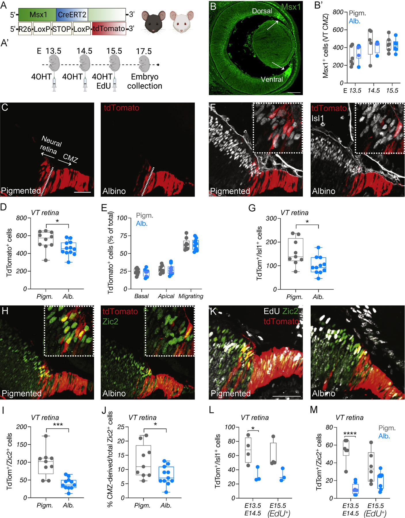
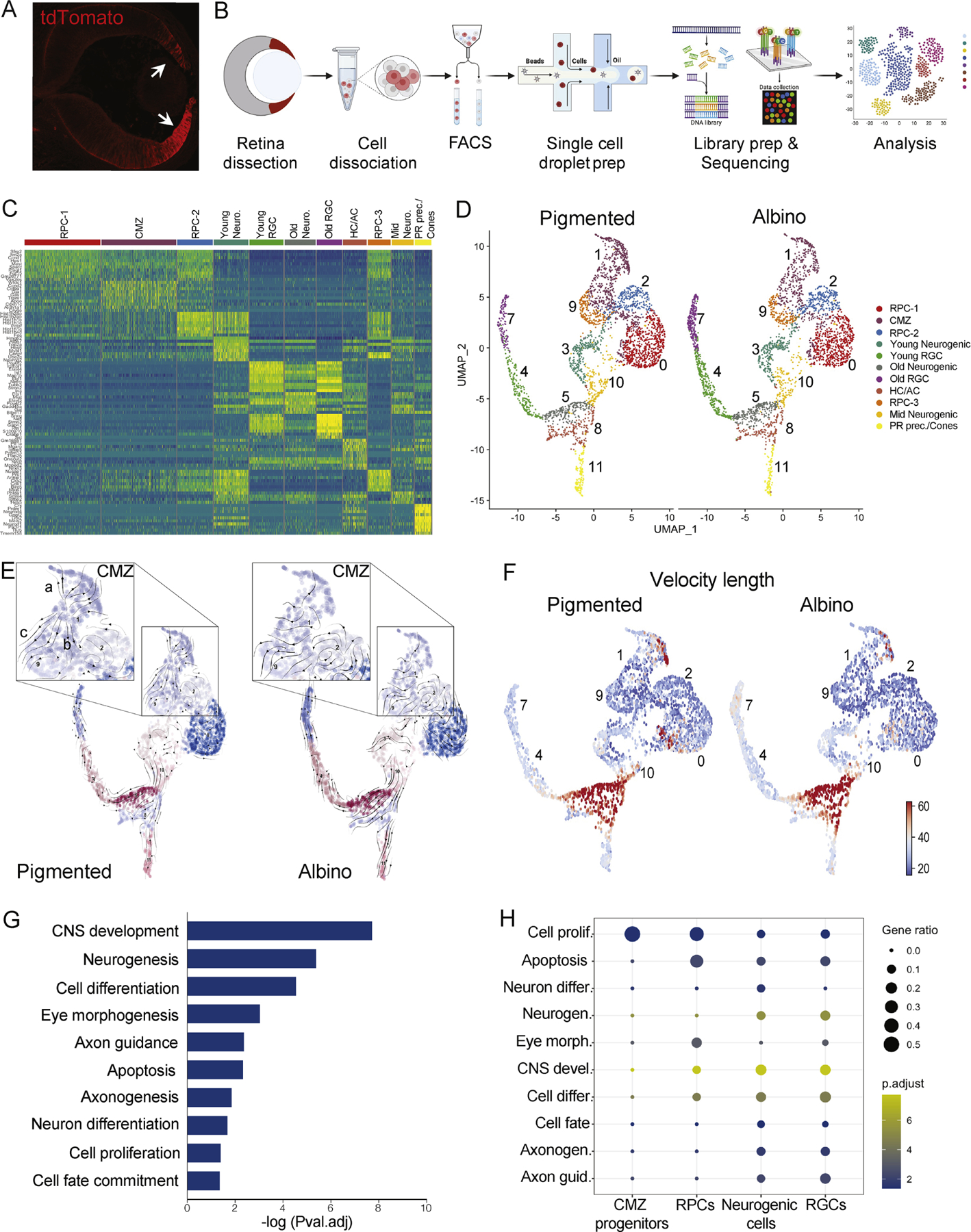
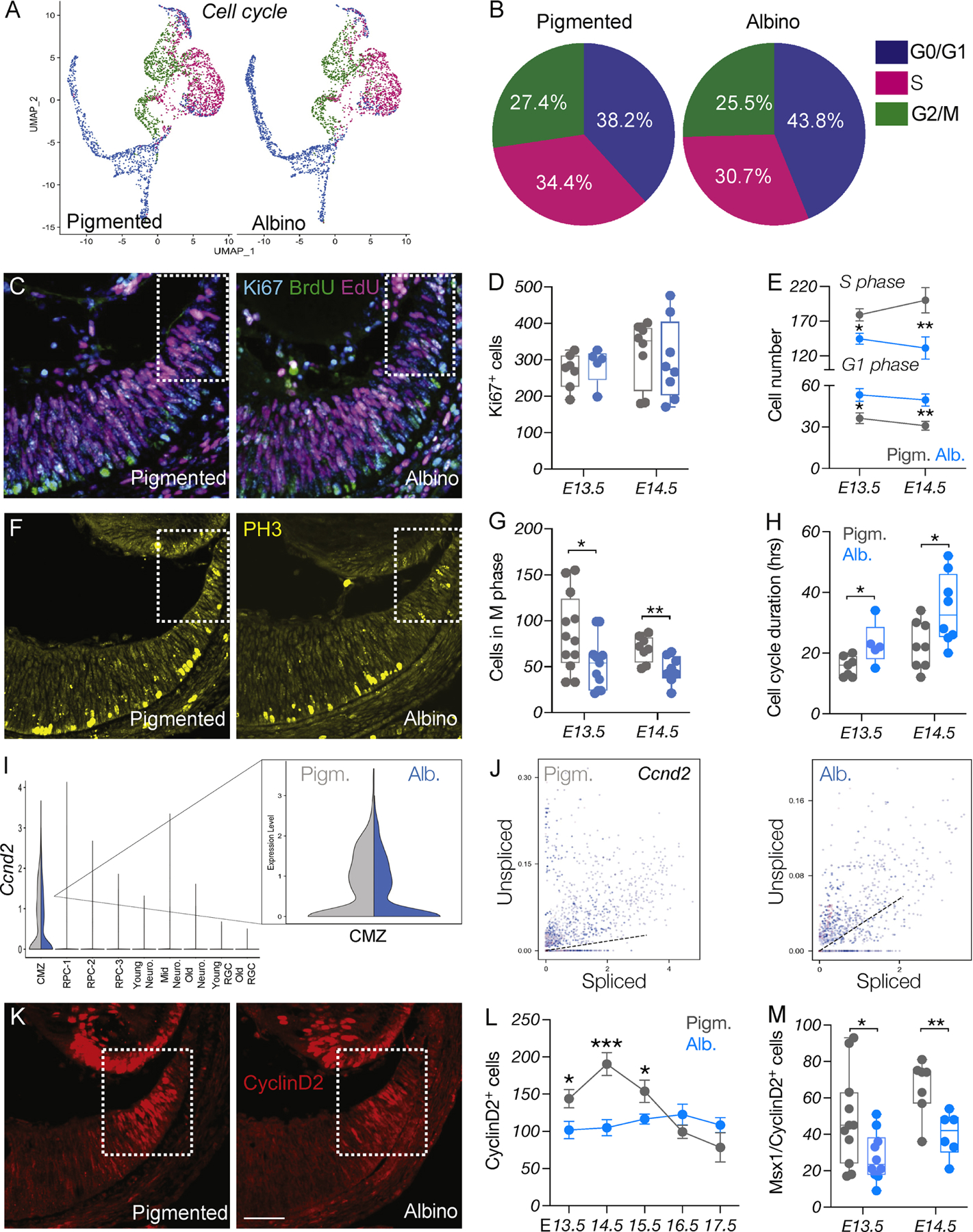
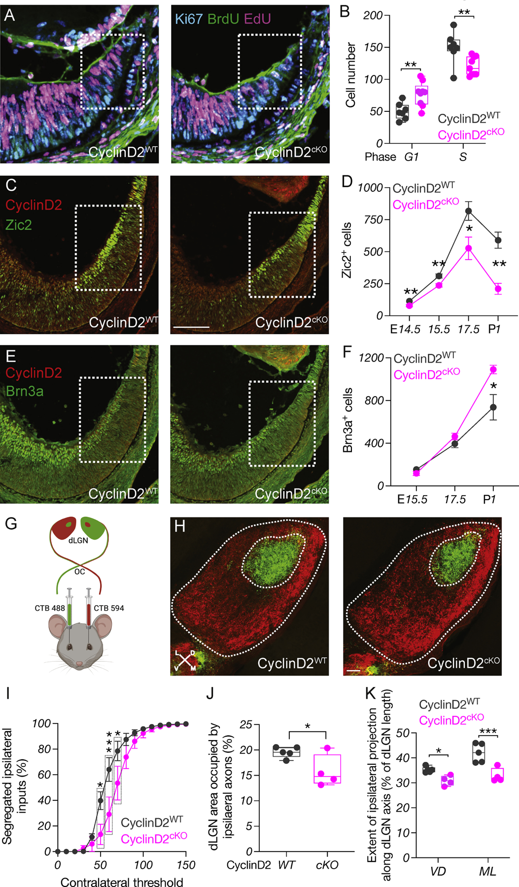
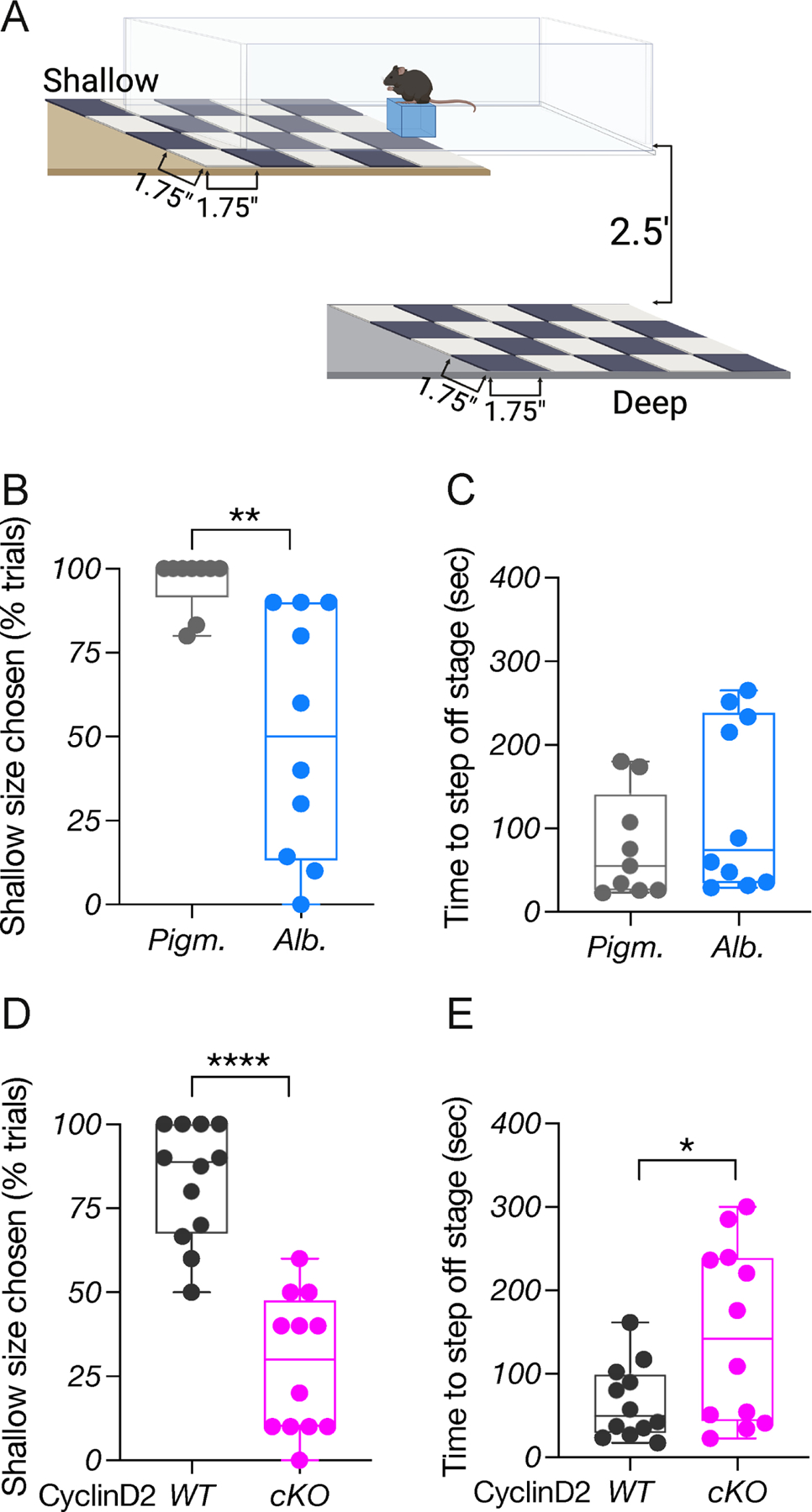
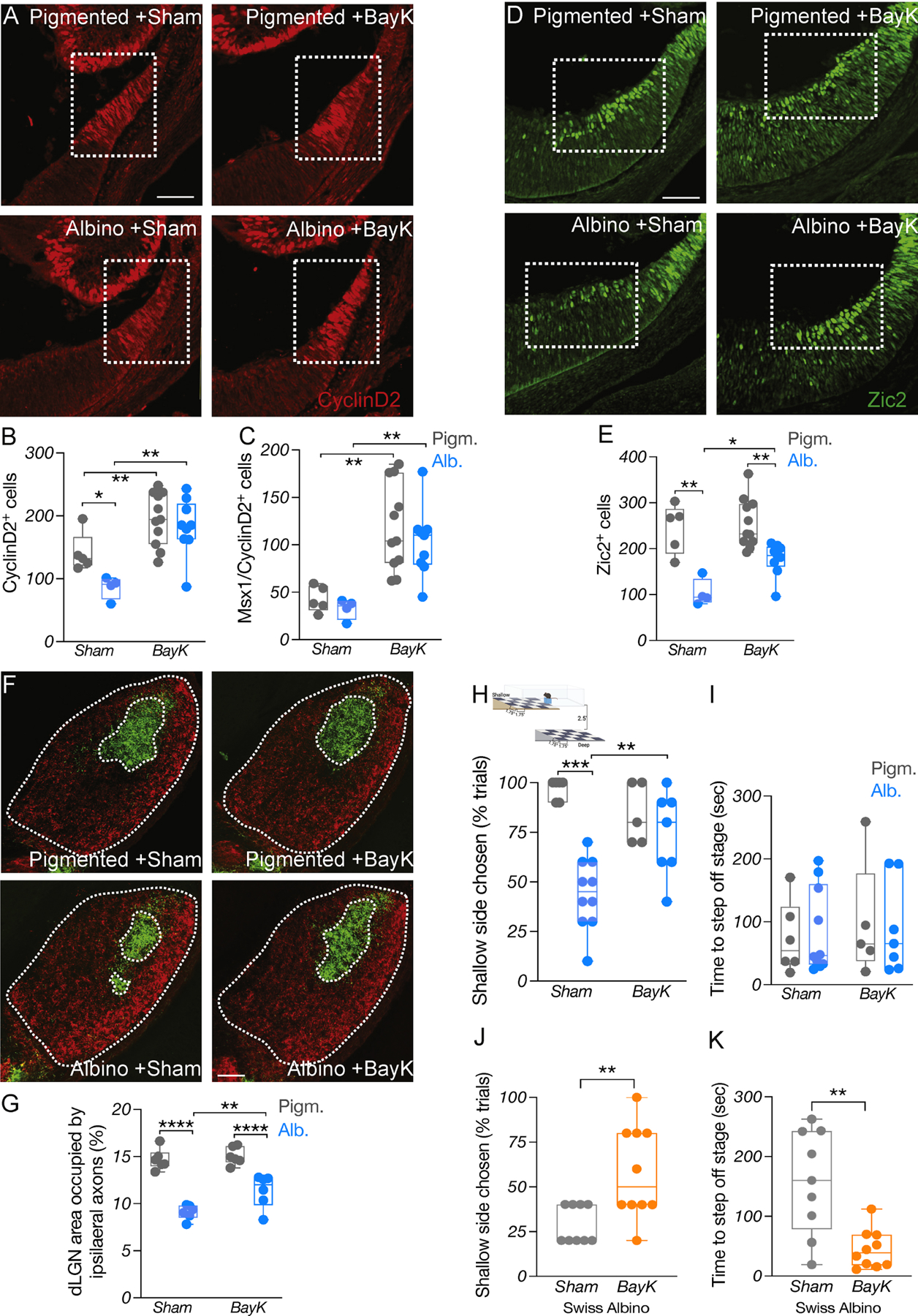
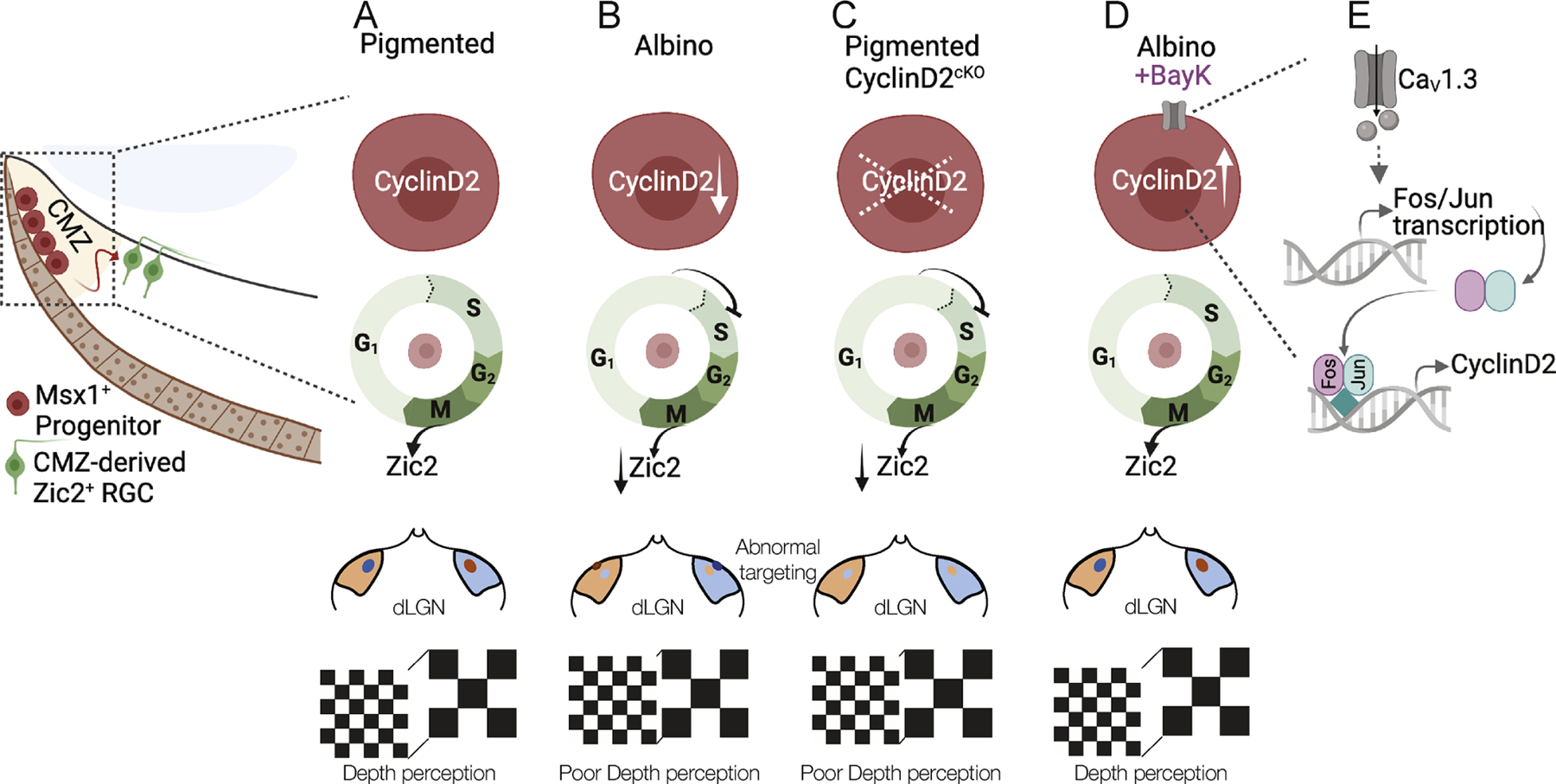
In albinism, aberrations in the ipsi-/contralateral retinal ganglion cell (RGC) ratio compromise the functional integrity of the binocular circuit. Here, we focus on the mouse ciliary margin zone (CMZ), a neurogenic niche at the embryonic peripheral retina, to investigate developmental processes regulating RGC neurogenesis and identity acquisition. We found that the mouse ventral CMZ generates predominantly ipsilaterally projecting RGCs, but this output is altered in the albino visual system because of CyclinD2 downregulation and disturbed timing of the cell cycle. Consequently, albino as well as CyclinD2-deficient pigmented mice exhibit diminished ipsilateral retinogeniculate projection and poor depth perception. In albino mice, pharmacological stimulation of calcium channels, known to upregulate CyclinD2 in other cell types, augmented CyclinD2-dependent neurogenesis of ipsilateral RGCs and improved stereopsis. Together, these results implicate CMZ neurogenesis and its regulators as critical for the formation and function of the mammalian binocular circuit.

Keywords: CyclinD2; RPE; albinism; binocular vision; ciliary margin; ipsilateral projection; neurogenesis; retinal ganglion cells; single-cell RNA seq.


Conflict of interest statement

Declaration of interests The authors declare no competing interests.

Figures

Figure 1:
Figure 1:. Ipsilateral RGC neurogenesis is prominent in the ventral CMZ in pigmented mice but reduced in the albino
A) Schema of elements used to generate Msx1 CreERT2; tdTomato; Tyr mice. A’) Timeline of 4-OHT and EdU injections and sample collection. B) Immunostaining of Msx1 at E14.5. Scale bar: 100μm. B’) Quantification of Msx1+ cells in the pigmented and albino ventral CMZ at E13.5, E14.5 and E15.5. C, F, H) Immunostaining of tdTomato (C), Islet1 (F), and Zic2 (H) in the pigmented and albino ventrotemporal CMZ and retinal crescent at E17.5. Scale bar: 50μm. D, G, I) Quantification of tdTomato+ (D), tdTomato+/Islet1+ (G), tdTomato+/Zic2+ (I) cells in the pigmented and albino ventrotemporal retina at E17.5. E) Quantification of tdTomato+ cells (% of total) residing in the apical, basal or intermediate surface of the pigmented and albino retina at E17.5. J) Quantification of the proportion (% of total) of Zic2+ cells deriving from the CMZ. K) Immunostaining of tdTomato, EdU, and Zic2 in the pigmented and albino ventrotemporal retina and retinal crescent E17.5. Scale bar: 50μm. L-M) Quantification of tdTomato+/Islet1+ (L) and tdTomato+/Zic2+ (M) cells born at E13.5–14.5 (EdU-) or E15.5 (EdU+) in the pigmented and albino ventrotemporal retina at E17.5.
Figure 2:
Figure 2:. scRNA-Seq in the pigmented and albino CMZ
A) tdTomato expression in the CMZ and peripheral retina of αCre; tdTomato mice at E13.5. B) scRNA-Seq work flow. C) Differential gene expression in single-cell clusters. D) UMAP representation of single-cell clusters in the pigmented and albino datasets. E) RNA Velocity vector stream projected on UMAP of single-cell clusters in the pigmented and albino datasets. F) Length of velocity vector quantifying arrow length, embedded on UMAP of single-cell clusters in the pigmented and albino datasets. G) GO terms corresponding to all DEG between pigmented and albino datasets. H) GO term (from G) enrichment across cell states. RPC: retinal progenitor cell, CMZ: ciliary margin zone cell, Neuro: neurogenic cell, RGC: retinal ganglion cell, HC: horizontal cell, AC: amacrine cell, PR: photoreceptor
Figure 3:
Figure 3:. Cell cycle is perturbed in the albino CMZ
A) UMAP representation of single cells based on cell cycle phase. B) Quantification of cells in the G0-G1, S or G2/M phase of the cell cycle, in pigmented and albino datasets. C) Dual-pulse birthdating: Immunostaining of Ki67, BrdU, and EdU in the pigmented and albino ventrotemporal CMZ at E14.5. D) Quantification of Ki67+ (cycling) cells at E13.5 and E14.5. E) Quantification of cells in the G1 and S phases of the cell cycle at E13.5 and E14.5. F) Immunostaining of the mitotic marker PH3 in the pigmented and albino ventrotemporal CMZ at E14.5. G) Quantification of cells in the M phase of the cell cycle (PH3+) at E13.5 and E14.5. H) Quantification of cell cycle duration (hours) at E13.5 and E14.5. I) Violin plot showing downregulation of Ccnd2 in the albino CMZ cell cluster. J) Transcriptional dynamics of Ccnd2 as a ratio of unspliced to spliced transcripts in the pigmented and albino peripheral retina. K) Immunostaining of CyclinD2 in the pigmented and albino ventrotemporal CMZ at E14.5. Scale bar: 100μm. L-M) Quantification of CyclinD2+ (L) from E13.5 to E17.5 and of CyclinD2+/Msx1+ (M) cells at E13.5 and E14.5.
Figure 4:
Figure 4:. CyclinD2-dependent G1/S transition regulates ipsilateral RGC neurogenesis
A) Dual-pulse birthdating: Immunostaining of Ki67, BrdU, and EdU in the CyclinD2WT and CyclinD2cKO ventrotemporal CMZ at E14.5. Scale bar: 100μm. B) Quantification of cells in the G1 and S phases of the cell cycle at E14.5. C) Immunostaining of CyclinD2 and Zic2 in the CyclinD2WT and CyclinD2cKO ventrotemporal retina and CMZ at E15.5. Scale bar: 100μm. D) Quantification of Zic2+ cells at E14.5, E15.5, E17.5 and P1. E) Immunostaining of CyclinD2 and Brn3a in the CyclinD2WT and CyclinD2cKO ventrotemporal retina and CMZ at E15.5. F) Quantification of Brn3a+ cells at E15.5, E17.5 and P1. G) Strategy used for RGC axon tracing to the dLGN. H) Immunostaining of dLGN sections from CyclinD2WT and CyclinD2cKO mice labeled with 488- and 594-CTB at P30. The entire dLGN, as well as the dLGN core receiving ipsilateral input, are outlined. Scale bar: 200μm. D: dorsal, V: ventral, M: medial, L: lateral. Axes indicate the orientation used for quantification along the DV and ML planes. I) Segregation plot: Percent of segregated inputs as a function of contralateral threshold. J) Area of the ipsilateral projection as percentage of total dLGN area. K) Extent of the ipsilateral projection as percentage of length along the DV and ML dLGN axes.
Figure 5:
Figure 5:. Depth perception requires CyclinD2 expression in the CMZ
A) Behavioral platform used for the binocularly-driven visual cliff assay. B) Quantification of depth perception comparing pigmented and albino mice. C) Quantification of time (sec) needed to step off the stage to either side, comparing pigmented and albino mice. D) Quantification of depth perception comparing CyclinD2WT and CyclinD2cKO mice. E) Quantification of time (sec) needed to step off the stage to either side, comparing CyclinD2WT and CyclinD2cKO mice.
Figure 6:
Figure 6:. CyclinD2 upregulation restores binocular circuit formation and function in albino mice
A) Immunostaining of CyclinD2 in the ventrotemporal CMZ of pigmented and albino Sham vs BayK treated mice at E15.5. Scale bar: 100μm. B-C) Quantification of CyclinD2+ (B) and CyclinD2+/Msx1+ (C) cells at E15.5. D) Immunostaining of Zic2 in the ventrotemporal retina of pigmented and albino Sham vs BayK treated mice at E15.5. Scale bar: 100μm. E) Quantification of Zic2+ cells at E15.5. F) Immunostaining of coronal dLGN sections from pigmented and albino Sham vs BayK treated mice labeled with 488- and 647-CTB at P60. The entire dLGN, as well as the dLGN core receiving ipsilateral input, are outlined. Scale bar: 100μm. G) Area of the ipsilateral projection as percentage of total dLGN area. H) Depth perception comparing pigmented and albino Sham vs BayK treated mice. I) Quantification of time (sec) needed to step off the stage to either side during the visual cliff task. J) Quantification of depth perception comparing Swiss albino Sham vs BayK treated mice. K) Quantification of time (sec) needed to step off the stage to either side in the visual cliff task, comparing Swiss albino Sham vs BayK treated mice.
Figure 7:
Figure 7:. Summary
A) The link between CyclinD2 and cell cycle, the ipsilateral RGC hallmark gene (Zic2), retinogeniculate projection, and binocular vision. B) In the albino, CyclinD2 reduction leads to delay in G1/S phase progression from, reduced numbers of Zic2+ RGCs, reduced ipsilateral retinogeniculate projection, and poor depth perception. C) This phenotype is recapitulated when CyclinD2 is deleted from the pigmented peripheral retina. D-E) The binocular circuit-related deficits in the albino retina can be rescued by the delivery of the CaV1 channel agonist BayK-8644, possibly via Fos/Jun transcriptional activation.

Comment in

Similar articles

Cited by

References

    1. AMBROSIO AL, BOYLE JA, ARADI AE, CHRISTIAN KA & DI PIETRO SM 2016. TPC2 controls pigmentation by regulating melanosome pH and size. Proc Natl Acad Sci U S A, 113, 5622–7. - PMC - PubMed
    1. BAKIRI L, LALLEMAND D, BOSSY-WETZEL E & YANIV M 2000. Cell cycle-dependent variations in c-Jun and JunB phosphorylation: a role in the control of cyclin D1 expression. EMBO J, 19, 2056–68. - PMC - PubMed
    1. BALASUBRAMANIAN R, MIN X, QUINN PMJ, LO GIUDICE Q, TAO C, POLANCO K, MAKRIDES N, PEREGRIN J, BOUAZIZ M, MAO Y, WANG Q, DA COSTA BL, BUENAVENTURA D, WANG F, MA L, TSANG SH, FABRE PJ & ZHANG X 2021. Phase transition specified by a binary code patterns the vertebrate eye cup. Science Advances, 7. - PMC - PubMed
    1. BELANGER MC, ROBERT B & CAYOUETTE M 2017. Msx1-Positive Progenitors in the Retinal Ciliary Margin Give Rise to Both Neural and Non-neural Progenies in Mammals. Dev Cell, 40, 137–150. - PubMed
    1. BELLONO NW & OANCEA EV 2014. Ion transport in pigmentation. Arch Biochem Biophys, 563, 35–41. - PMC - PubMed

Publication types

Substances