. 2023 Jan;613(7944):558-564.
doi: 10.1038/s41586-022-05514-2. Epub 2022 Nov 9.

Multiple pathways for SARS-CoV-2 resistance to nirmatrelvir

Affiliations

Multiple pathways for SARS-CoV-2 resistance to nirmatrelvir

Sho Iketani et al. Nature. 2023 Jan.

Abstract

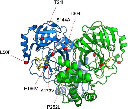
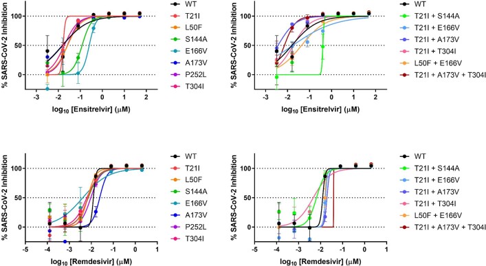
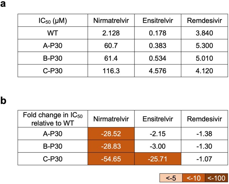
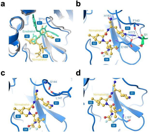
Nirmatrelvir, an oral antiviral targeting the 3CL protease of SARS-CoV-2, has been demonstrated to be clinically useful against COVID-19 (refs. 1,2). However, because SARS-CoV-2 has evolved to become resistant to other therapeutic modalities3-9, there is a concern that the same could occur for nirmatrelvir. Here we examined this possibility by in vitro passaging of SARS-CoV-2 in nirmatrelvir using two independent approaches, including one on a large scale. Indeed, highly resistant viruses emerged from both and their sequences showed a multitude of 3CL protease mutations. In the experiment peformed with many replicates, 53 independent viral lineages were selected with mutations observed at 23 different residues of the enzyme. Nevertheless, several common mutational pathways to nirmatrelvir resistance were preferred, with a majority of the viruses descending from T21I, P252L or T304I as precursor mutations. Construction and analysis of 13 recombinant SARS-CoV-2 clones showed that these mutations mediated only low-level resistance, whereas greater resistance required accumulation of additional mutations. E166V mutation conferred the strongest resistance (around 100-fold), but this mutation resulted in a loss of viral replicative fitness that was restored by compensatory changes such as L50F and T21I. Our findings indicate that SARS-CoV-2 resistance to nirmatrelvir does readily arise via multiple pathways in vitro, and the specific mutations observed herein form a strong foundation from which to study the mechanism of resistance in detail and to inform the design of next-generation protease inhibitors.


Conflict of interest statement

S.I., A.C. and D.D.H. are inventors on patent applications related to the development of inhibitors against the SARS-CoV-2 3CL protease. D.D.H. is a cofounder of TaiMed Biologics and RenBio, consultant to WuXi Biologics and Brii Biosciences and board director for Vicarious Surgical.

Figures

Fig. 1
Fig. 1. Identification of nirmatrelvir resistance in Vero E6 cells.
a, Changes in IC50 during passaging of SARS-CoV-2 with nirmatrelvir. Vero E6 cells were infected in triplicate with SARS-CoV-2 (USA-WA1/2020) and passaged to fresh cells every 3 days for 30 passages (Methods). bd, Validation of nirmatrelvir resistance for the indicated passage from each of the three lineages (A (b), B (c) and C (d)). e, Inhibition of passage 30 viruses from each lineage by remdesivir. f, Mutations in 3CLpro found in the indicated passages from each lineage. Dots indicate WT at that residue. Mutations are shaded according to frequency. g, Residues mutated with passaging in Vero E6 cells overlaid onto the 3CLpro structure with nirmatrelvir bound. The Cα of each mutated residue is denoted by a red sphere. The 3CLpro–nirmatrelvir complex was downloaded from PDB under accession code 7VH8. ae, Error bars denote mean ± s.e.m of four technical replicates.
Fig. 2
Fig. 2. Identification of nirmatrelvir resistance at scale in Huh7-ACE2 cells.
a, Passaging scheme: 480 wells were infected with SARS-CoV-2-mNeonGreen and passaged to fresh Huh7-ACE2 cells every 3–4 days, with the concentration of drug doubled every two passages. b, Validation of nirmatrelvir resistance of three wells from passage 16. These viral populations had the following mutations: 3A8 (T21I, T304I), 1E11 (T21I, N51Y, T304I) and 5A2 (L50F, E166V). See Supplementary Table 1 for exact frequencies. Representative curves from a single experiment from two biologically independent experiments are shown. Error bars denote mean ± s.e.m of three technical replicates. c, Mutations in 3CLpro found in passage 16 from 53 wells. Dots indicate WT at that residue. Mutations are shaded according to frequency. d, Residues mutated in passaging in Huh7-ACE2 cells overlaid onto the 3CLpro structure with nirmatrelvir bound. All 23 mutated residues across all resistant populations are indicated for any individual isolate having between one and six mutations. The Cα of each residue that was mutated is denoted by a red sphere for mutations observed more than ten times, and is denoted by an orange sphere for mutations observed fewer than ten times. The 3CLpro–nirmatrelvir complex was downloaded from PDB under accession code 7VH8.
Fig. 3
Fig. 3. Pathways for SARS-CoV-2 resistance to nirmatrelvir.
a, Phylogenetic tree of sequences from passaging in Huh7-ACE2 cells. Only sequences with mutations are shown. Sequences are denoted as passage number, followed by the well number. Mutations that arose along particular branches are annotated in red; ‘-’ denotes when a mutation appears to have been lost from a particular branch. b, Observed pathways for nirmatrelvir resistance in Huh7-ACE2 cells. The most commonly observed mutations in passage 16 were used to build these pathways (Methods and Supplementary Table 2). Nodes are shaded from dark to light, with founder mutations darker. Percentages indicate the frequency by which child nodes derive from the immediate parental node. Descendent arrows that do not sum to 100% indicate that a proportion did not advance beyond the indicated mutations in the experiment. c, Growth assay with recombinant live SARS-CoV-2 carrying single (top) and combination 3CLpro mutations (bottom). Huh7-ACE2 cells were infected with 0.01 multiplicity of infection (MOI) of virus, and luminescence was quantified at the indicated time points. S144A, E166V and T21I + S144A are statistically significant from WT at 48 h (two-way analysis of variance with Geisser–Greenhouse correction followed by Dunnett’s multiple comparisons test; P = 0.0039, P = 0.0006, P = 0.0006, respectively). Representative curves from a single experiment from two biologically independent experiments are shown. Error bars denote mean ± s.e.m of three technical replicates. RLU, relative luminescence units.
Fig. 4
Fig. 4. Validation of identified mutations in isogenic recombinant SARS-CoV-2.
a, Individual inhibition curves of recombinant live SARS-CoV-2 carrying single (left) and combination 3CLpro mutations (right) by nirmatrelvir. Representative curves from a single experiment from three biologically independent experiments are shown. Error bars denote mean ± s.e.m of three technical replicates. b, Inhibition of recombinant live SARS-CoV-2 carrying single and combination 3CLpro mutations by nirmatrelvir, ensitrelvir and remdesivir. Values shown are fold change of mean values in IC50 relative to inhibition of WT from three biologically independent experiments.
Extended Data Fig. 1
Extended Data Fig. 1. Growth assays with SARS-CoV-2 passaged in Vero E6 cells.
a-d, Growth was quantified for lineage A (a), lineage B (b), lineage C (c), and unpassaged SARS-CoV-2 (d, denoted as WT-P0) in comparison to SARS-CoV-2 passaged without nirmatrelvir for 30 passages (denoted as WT-P30). Vero E6 cells were infected with 200 TCID50 of the indicated viruses and viral RNA was quantified at the indicated time points. e, The slope during the exponential phase (between 11 and 24 h post-infection) of growth for the indicated viruses.
Extended Data Fig. 2
Extended Data Fig. 2. Mutations in the 11 3CLpro cut sites found in passage 16 from the 53 wells passaged in Huh7-ACE2 cells.
Dots indicate wild-type at that cut site. Note that nsp4/5 M(P6’)I = M6I, nsp5/6 S(P6)P = S301P, and nsp5/6 T(P3)I = T304I in 3CLpro.
Extended Data Fig. 3
Extended Data Fig. 3. Mutations studied as isogenic recombinant SARS-CoV-2 overlaid onto the 3CL protease structure.
The Cα of each residue that was mutated is denoted with a red sphere. The 3CLpro-nirmatrelvir complex was downloaded from PDB under accession code 7VH8.
Extended Data Fig. 4
Extended Data Fig. 4. Raw IC50 values for recombinant live SARS-CoV-2 carrying single and combination 3CLpro mutations by nirmatrelvir, ensitrelvir, and remdesivir.
Mean ± SD of three biologically independent experiments are shown.
Extended Data Fig. 5
Extended Data Fig. 5. Individual inhibition curves of recombinant live SARS-CoV-2 carrying single and combination 3CLpro mutations by ensitrelvir and remdesivir.
Representative curves from a single experiment from three biologically independent experiments are shown. Error bars denote mean ± s.e.m of three technical replicates.
Extended Data Fig. 6
Extended Data Fig. 6. Inhibition of passage 30 of SARS-CoV-2 passaged in Vero E6 cells by nirmatrelvir, ensitrelvir, and remdesivir.
a, Raw IC50 values. b, Fold change relative to inhibition of wild-type.
Extended Data Fig. 7
Extended Data Fig. 7. Structural analyses of 3CLpro mutations.
a, Overlay of nirmatrelvir and ensitrelvir binding to 3CLpro. b, Several of the residues involved in direct interaction with nirmatrelvir. c, Several of the residues involved in formation of the S1 subsite. d, Interaction of L167 with nirmatrelvir. In a-d, nirmatrelvir is shown in yellow, enstirelvir is shown in lime green, the 3CLpro-nirmatrelvir complex is shown in marine, and the 3CLpro-ensitrelvir complex is shown in gray. Protomer A is shown in marine and protomer B is shown in green. Hydrogen bonds are indicated as black dashes. The 3CLpro-nirmatrelvir complex and 3CLpro-ensitrelvir complex were downloaded from PDB under accession codes 7VH8 and 7VU6, respectively.
Extended Data Fig. 8
Extended Data Fig. 8. Frequencies of identified 3CLpro mutations in GISAID.
a, All occurrences of the indicated mutations were tabulated from GISAID. b, All occurrences of the indicated mutations were tabulated from GISAID in the three months prior to EUA (9/22/2021 to 12/22/2021) or after EUA (3/26/2022 to 6/26/2022).

Update of

References

    1. Hammond J, et al. Oral nirmatrelvir for high-risk, nonhospitalized adults with Covid-19. N. Engl. J. Med. 2022;386:1397–1408. doi: 10.1056/NEJMoa2118542. - DOI - PMC - PubMed
    1. Owen DR, et al. An oral SARS-CoV-2 M(pro) inhibitor clinical candidate for the treatment of COVID-19. Science. 2021;374:1586–1593. doi: 10.1126/science.abl4784. - DOI - PubMed
    1. Altarawneh HN, et al. Effects of previous infection and vaccination on symptomatic omicron infections. N. Engl. J. Med. 2022;387:21–34. doi: 10.1056/NEJMoa2203965. - DOI - PMC - PubMed
    1. Iketani S, et al. Antibody evasion properties of SARS-CoV-2 Omicron sublineages. Nature. 2022;604:553–556. doi: 10.1038/s41586-022-04594-4. - DOI - PMC - PubMed
    1. Liu L, et al. Striking antibody evasion manifested by the Omicron variant of SARS-CoV-2. Nature. 2022;602:676–681. doi: 10.1038/s41586-021-04388-0. - DOI - PubMed

Publication types