. 2023 Oct 1;22(4):515-526.
doi: 10.2463/mrms.mp.2021-0103. Epub 2022 Nov 8.

Model-based Deep Learning Reconstruction Using a Folded Image Training Strategy for Abdominal 3D T1-weighted Imaging

Affiliations

Model-based Deep Learning Reconstruction Using a Folded Image Training Strategy for Abdominal 3D T1-weighted Imaging

Satoshi Funayama et al. Magn Reson Med Sci. .

Abstract

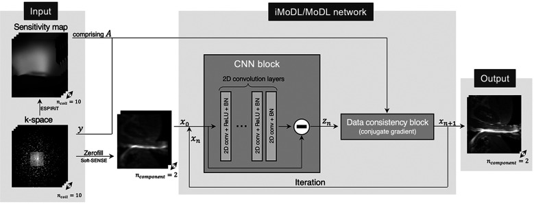
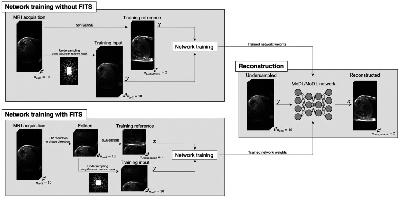
Purpose: To evaluate the feasibility of folded image training strategy (FITS) and the quality of images reconstructed using the improved model-based deep learning (iMoDL) network trained with FITS (FITS-iMoDL) for abdominal MR imaging.

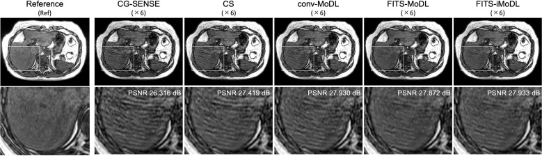
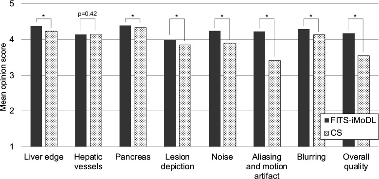
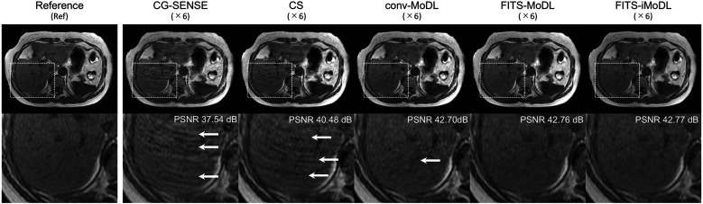
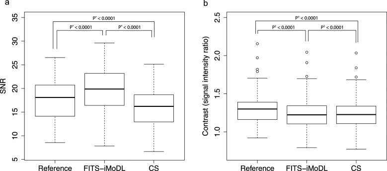
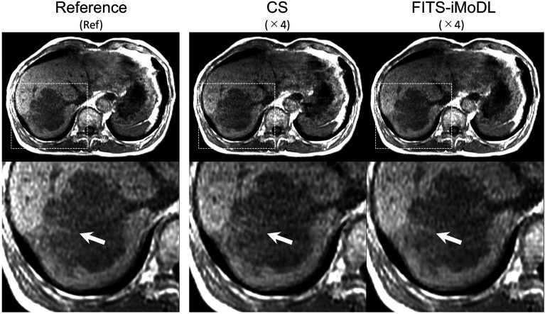
Methods: This retrospective study included abdominal 3D T1-weighted images of 122 patients. In the experimental analyses, peak SNR (PSNR) and structure similarity index (SSIM) of images reconstructed with FITS-iMoDL were compared with those with the following reconstruction methods: conventional model-based deep learning (conv-MoDL), MoDL trained with FITS (FITS-MoDL), total variation regularized compressed sensing (CS), and parallel imaging (CG-SENSE). In the clinical analysis, SNR and image contrast were measured on the reference, FITS-iMoDL, and CS images. Three radiologists evaluated the image quality using a 5-point scale to determine the mean opinion score (MOS).

Results: The PSNR of FITS-iMoDL was significantly higher than that of FITS-MoDL, conv-MoDL, CS, and CG-SENSE (P < 0.001). The SSIM of FITS-iMoDL was significantly higher than those of the others (P < 0.001), except for FITS-MoDL (P = 0.056). In the clinical analysis, the SNR of FITS-iMoDL was significantly higher than that of the reference and CS (P < 0.0001). Image contrast was equivalent within an equivalence margin of 10% among these three image sets (P < 0.0001). MOS was significantly improved in FITS-iMoDL (P < 0.001) compared with CS images in terms of liver edge and vessels conspicuity, lesion depiction, artifacts, blurring, and overall image quality.

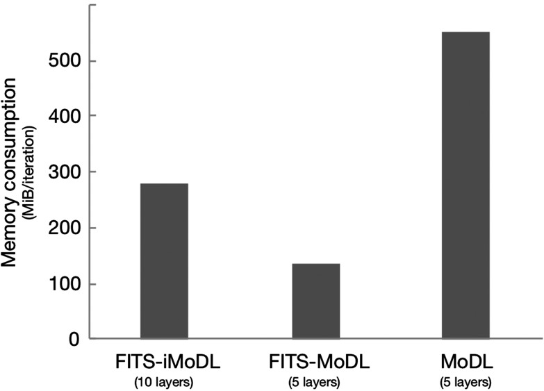
Conclusion: The proposed method, FITS-iMoDL, allowed a deeper MoDL reconstruction network without increasing memory consumption and improved image quality on abdominal 3D T1-weighted imaging compared with CS images.

Keywords: deep learning; image reconstruction; liver imaging; network training.


Conflict of interest statement

Conflicts of Interest

The authors do not have any conflicts of interest.

Figures

Supplementary Figure 1
Memory consumption of each network. FITS-iMoDL showed less memory consumption compared with MoDL although FITS-iMoDL has doubled CNN layers.
Supplementary Figure 2
Visual comparison of out-of-phase images to explore the potentials of the trained networks. These images are the same slice on Fig. 4 and reconstructed using first echo which was not used in the network training and parameter optimization for the CS.
Supplementary Figure 3
MOS of three radiologists. The MOS of the FITS-iMoDL was significantly better than that of CS in the liver edge (P < 0.001), depiction of pancreas (P < 0.001), lesion conspicuity (P < 0.001), noise (P < 0.001), aliasing and motion artifact (P < 0.001), blurring (P < 0.001), and overall quality (P < 0.001). The MOS difference of hepatic vessels is not statistically significant. *: P < 0.001
Fig. 1
Fig. 1
A schema of image cropping (a) and image folding (b) in image domain and frequency domain. Only image folding (b) results in smaller size of image and k-space simultaneously. FT, Fourier transform.
Fig. 2
Fig. 2
The network architecture of the iMoDL/MoDL. This network comprises 2 blocks, CNN block and data consistency block. The number of 2D convolution layers was 10 for iMoDL and 5 for MoDL. CNN, convolutional neural network; iMoDL, the improved model-based deep learning; MoDL, the conventional model-based deep learning.
Fig. 3
Fig. 3
A schema of the network training with and without FITS and image reconstruction. FITS, a folded image training strategy.
Fig. 4
Fig. 4
A representative case for visual comparison of each reconstruction method. An acceleration factor of 6 was applied except to the reference (acceleration factor, 1.5). For CG-SENSE and CS, it is difficult to indicate intrahepatic vessels due to remaining aliasing artifact (arrows). For conv-MoDL, the aliasing is significantly removed but a little is still remaining (arrow). No aliasing artifact is found for FITS-MoDL and FITS-iMoDL. CG-SENSE, conjugate gradient sensitivity encoding; conv-MoDL, conventional model-based deep learning; CS, compressed sensing; FITS-iMoDL, the improved model-based deep learning network trained with a folded image training strategy; FITS-MoDL, the conventional model-based deep learning network trained with a folded image training strategy.
Fig. 5
Fig. 5
Clinical evaluation of SNR and spleen-to-liver signal intensity ratio (SLR) of 75 cases for test set. (a) The difference of measured SNR among reference, FITS-iMoDL, and CS was tested. (b) The equivalence of SLR was also tested. *Paired t-test, †two one-sided t-test with contrast limit of 0.1. CS, compressed sensing; FITS-iMoDL, the improved model-based deep learning network trained with a folded image training strategy.
Fig. 6
Fig. 6
A case of hepatic metastasis from colon cancer. There is a linear hyperintensity on the peripheral area of hepatic metastasis on the reference image (acceleration factor, 1.5). In the FITS-iMoDL image, it has some blurring but can be indicated, whereas it is hard to indicate on CS images. In two readers, the score of lesion conspicuity was improved in the FITS-iMoDL compared to CS. CS, compressed sensing; FITS-iMoDL, the improved model-based deep learning network trained with a folded image training strategy.

Similar articles

Cited by

References

    1. Lee SM, Lee JM, Ahn SJ, Kang HJ, Yang HK, Yoon JH. LI-RADS version 2017 versus version 2018: Diagnosis of hepatocellular carcinoma on gadoxetate disodium-enhanced MRI. Radiology 2019; 292:655–663. - PubMed
    1. van der Pol CB, Lim CS, Sirlin CB, et al. Accuracy of the liver imaging reporting and data system in computed tomography and magnetic resonance image analysis of hepatocellular carcinoma or overall malignancy—a systematic. Gastroenterology 2019; 156:976–986. - PubMed
    1. Lee S, Kim SS, Roh YH, Choi JY, Park MS, Kim MJ. Diagnostic performance of CT/MRI liver imaging reporting and data system v2017 for hepatocellular carcinoma: A systematic review and meta-analysis. Liver Int 2020; 40:1488–1497. - PubMed
    1. Davenport MS, Viglianti BL, Al-Hawary MM, et al. Comparison of acute transient dyspnea after intravenous administration of gadoxetate disodium and gadobenate dimeglumine: Effect on arterial phase image quality. Radiology 2013; 266:452–461. - PubMed
    1. Motosugi U, Bannas P, Bookwalter CA, Sano K, Reeder SB. An investigation of transient severe motion related to gadoxetic acid-enhanced MR imaging. Radiology 2016; 279:93–102. - PMC - PubMed