. 2023 Jan 1;210(1):50-60.
doi: 10.4049/jimmunol.2200349. Epub 2022 Nov 9.

Synergistic Protection against Secondary Pneumococcal Infection by Human Monoclonal Antibodies Targeting Distinct Epitopes

Affiliations

Synergistic Protection against Secondary Pneumococcal Infection by Human Monoclonal Antibodies Targeting Distinct Epitopes

Aaron D Gingerich et al. J Immunol. .

Abstract

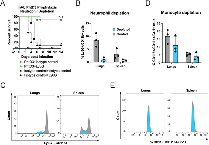
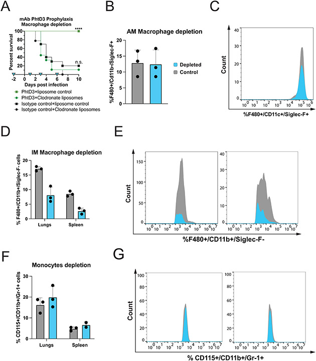
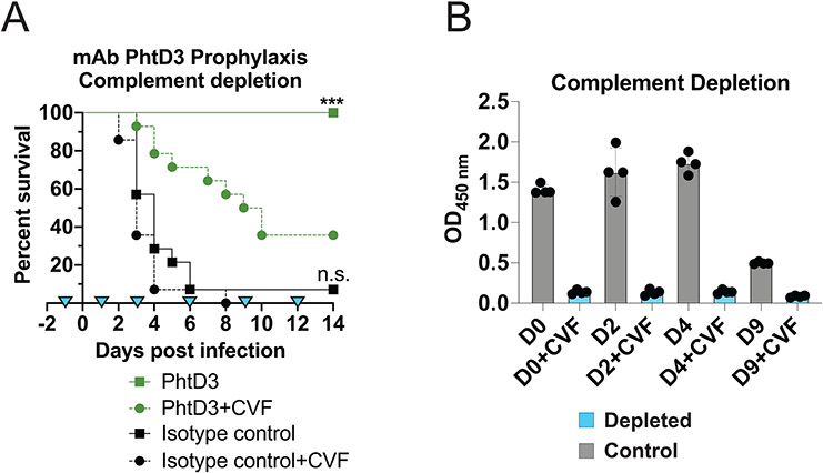
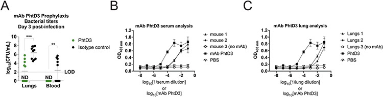
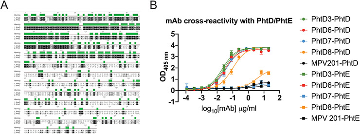
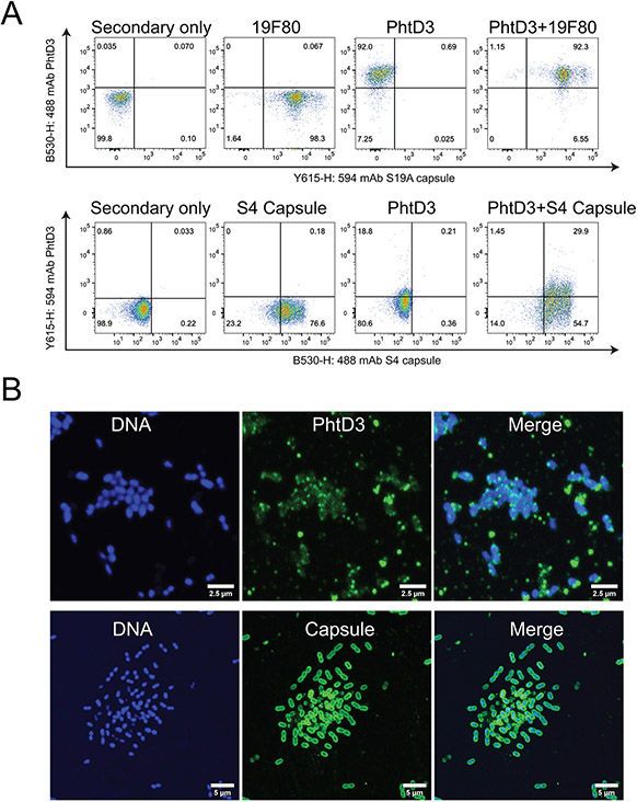
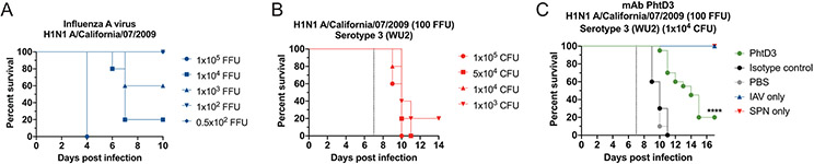
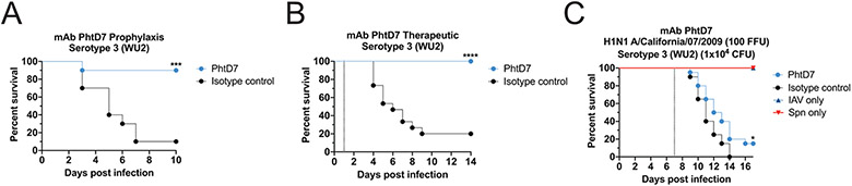
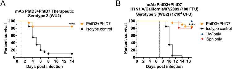
Streptococcus pneumoniae persists as a leading cause of bacterial pneumonia despite the widespread use of polysaccharide-based vaccines. The limited serotype coverage of current vaccines has led to increased incidence of nonvaccine serotypes, as well as an increase in antibiotic resistance among these serotypes. Pneumococcal infection often follows a primary viral infection such as influenza virus, which hinders host defense and results in bacterial spread to the lungs. We previously isolated human monoclonal Abs (mAbs) against the conserved surface Ag pneumococcal histidine triad protein D (PhtD), and we demonstrated that mAbs to this Ag are protective against lethal pneumococcal challenge prophylactically and therapeutically. In this study, we elucidated the mechanism of protection of a protective anti-pneumococcal human mAb, PhtD3, which is mediated by the presence of complement and macrophages in a mouse model of pneumococcal infection. Treatment with mAb PhtD3 reduced blood and lung bacterial burden in mice, and mAb PhtD3 is able to bind to bacteria in the presence of the capsular polysaccharide, indicating exposure of surface PhtD on encapsulated bacteria. In a mouse model of secondary pneumococcal infection, protection mediated by mAb PhtD3 and another mAb targeting a different epitope, PhtD7, was reduced; however, robust protection was restored by combining mAb PhtD3 with mAb PhtD7, indicating a synergistic effect. Overall, these studies provide new insights into anti-pneumococcal mAb protection and demonstrate, to our knowledge, for the first time, that mAbs to pneumococcal surface proteins can protect against secondary pneumococcal infection in the mouse model.


Figures

Figure 1.
Figure 1.. Neutrophil depletion in the pneumococcal infection model with mAb prophylaxis.
(A) Neutrophil depletion does not diminish protection by mAb PhtD3 in an intranasal infection model of pneumococcal serotype 3 (strain WU2) in 6-8 week old C57BL/6 mice. *P=0.0237 via log-rank (Mantel-Cox) test compared to the isotype control+Ly6G group, n.s.=not significant, n=10 mice/group. Blue triangles denote the days of depletion treatment. (B) Comparison of %Ly6G+/CD11b+ cells in the lungs and spleen of neutrophil depleted and control mice. Grey: control animals, Blue: depleted animals. n=3 mice/group. (C) Example histogram of %Ly6G+/CD11b+ cell population counts in the lungs and spleen. Grey: control animals, Blue: depleted animals. Representative of 1 animal each. (D) Comparison of %CD115+/CD11b+/Gr-1+ cells in the lungs and spleen. Grey: control animals, Blue: depleted animals. n=3 mice/group. (E) Example histogram of %CD115+/CD11b+/Gr-1+ cell population counts in the lungs and spleen. Grey: control animals, Blue: depleted animals. Representative of 1 animal each.
Figure 2.
Figure 2.. Macrophage depletion in the pneumococcal infection model with mAb prophylaxis.
(A) Macrophage depletion abolishes protection by mAb PhtD3 in an intranasal infection model of pneumococcal serotype 3 (strain WU2) in 6–8-week-old male C57BL/6 mice, ****P<0.0001 compared to the PhtD3+Clodronate liposome treated group; n.s. compared to the isotype control+clodronate liposome treated group. Analysis conducted via log-rank (Mantel-Cox) test. n=9-10 mice/group. Blue triangles denote the days of depletion treatment. (B) Comparison of %F480+/CD11c+/Siglec-F+ cells in the lungs of mice. Grey: control animals, Blue: depleted animals. n=3 mice/group. (C) Example histogram of %F480+/CD11c+/Siglec-F+ cell population counts of the lungs. Grey: control animals, Blue: depleted animals. Representative of 1 animal each. (D) Comparison of %F480+/CD11b+/Siglec-F− cells in the lungs and spleen. Grey: control animals, Blue: depleted animals. n=3 mice/group. (E) Example histogram of %F480+/CD11b+/Siglec-F− cell population counts of the lungs and spleen. Grey: control animals, Blue: depleted animals. Representative of 1 animal each. (F) Comparison of %CD115+/CD11b+/Gr-1+ cells in the lungs and spleen. Grey: control animals, Blue: depleted animals. n=3 mice/group. (G) Example histogram of the %CD115+/CD11b+/Gr-1+ cell population counts in the lungs and spleen of mice. Grey: control animals, Blue: depleted animals. Representative of 1 animal each.
Figure 3.
Figure 3.. Complement depletion in the pneumococcal infection model with mAb prophylaxis.
(A) Complement depletion partially diminishes protection by mAb PhtD3 in an intranasal infection model of pneumococcal serotype 3 (strain WU2) in 6-8 week old male C57BL/6 mice. ***P=0.0003 compared to the PhtD+CVF group; ns compared to the isotype control mAb+CVF group. Analysis completed via log-rank (Mantel-Cox) test. n=14 mice/group. Blue triangles denote days of depletion treatment. (B) Serum C3 complement levels in CVF and PBS treated mice. Grey: control animals, Blue: depleted animals. n=4 mice/group.
Figure 4.
Figure 4.. Bacterial burden reduction and presence of PhtD mAbs in mice blood and lungs.
(A) mAb PhtD3 reduces lung and blood bacterial titers. Bacterial burden assessed three days after intranasal pneumococcal infection and prophylactic PhtD3 administration, n=10 mice/group, ***P=0.0002; **P=0.009; analysis completed by two-way ANOVA. (B) ELISA binding curves of naïve and PhtD3 treated mouse serum against recombinant PhtD protein. Error bars are the standard deviation of four replicates. (C) ELISA binding curves of naïve and PhtD3 treated mouse lung homogenates against recombinant PhtD protein. Error bars are the standard deviation of four replicates.
Figure 5.
Figure 5.. mAb PhtD3 is cross reactive to both PhtD and PhtE.
(A) Amino acid sequence alignment of protein PhtD and PhtE. (B) ELISA binding curves of PhtD mAbs against recombinant PhtD and PhtE proteins. Error bars are the standard deviation of four replicates.
Figure 6.
Figure 6.. mAb PhtD3 binding to encapsulated bacteria.
(A) Flow cytometry analysis of mAb PhtD3 binding to encapsulated bacteria. Serotype 19A bacteria (strain TCH8431) were labeled with mAb PhtD3-IgG2a (with anti-mouse Fc secondary) and/or the human capsule-specific antibody 19F80 (with anti-human Fc secondary). Serotype 4 bacteria (strain TIGR4) were labeled with mAb PhtD3-IgG1 (with anti-human Fc secondary) and/or the rabbit capsule-specific antibody. Figures are representative of one experiment from triplicate repeats. (B) Binding of the anti-PhtD3 mAb to encapsulated pneumococcal serotype 4 (strain TIGR4). Bacteria were stained with an anti-PhtD3 mAb, followed by a species-specific secondary antibody labeled with Alexa-488 (top panels) or with an anti-serotype 4 capsule labeled with Alexa 488 (bottom panels). The DNA was stained with DAPI. Micrographs were collected with a confocal microscope and projections of ~20 xy optical sections (0.1 μm each) are shown. Data are shown from one of at least two experiments.
Figure 7.
Figure 7.. Co-infection with sublethal influenza A virus followed by Spn causes mortality in mice and is improved by PhtD3 mAb treatment.
(A) Survival of C57BL/6 mice infected with decreasing doses of influenza A virus. n=5 mice/group. (B) Survival of C57BL/6 mice co-infected with influenza A virus at day 0 and pneumococcal serotype 3 (strain WU2) at day 7 (dotted line) in 6-8 week old male mice. n=5 mice/group. (C) Protective efficacy of mAb PhtD3 in a co-infection model. Mice were infected with influenza A virus at day 0 and with WU2 bacteria at day 7 (dotted line). Treatment with mAb PhtD3 occurred 2 hrs prior to pneumococcal infection. ****P<0.0001 via log-rank (Mantel-Cox) test compared to the isotype control mAb group. Co-infected groups had n = 20 mice/group, while IAV and Spn infected had n=5 mice/group.
Figure 8.
Figure 8.. Protective efficacy of mAb PhtD7.
(A) Prophylactic efficacy of mAb PhtD7 in an intranasal infection model of pneumococcal serotype 3 (strain WU2) in 6-8 week old male C57BL/6 mice. ***P=0.0006 via log-rank (Mantel-Cox) test compared to the isotype control group. n=10 mice/group. (B) Treatment efficacy of mAb PhtD7 in an intranasal infection model of pneumococcal serotype 3 (strain WU2) in 6-8 week old male C57BL/6 mice. ****P<0.0001 via log-rank (Mantel-Cox) test compared to the isotype control group. n = 15 mice/group. (C) Protective efficacy of mAb PhtD7 in a co-infection model. Mice were infected with influenza A virus at day 0 and with WU2 bacteria at day 7 (dotted line). *P=0.0264 via log-rank (Mantel-Cox) test compared to the isotype control group. Co-infected groups had n = 20 mice/group, while IAV and Spn infected had n=5 mice/group.
Figure 9.
Figure 9.. Synergistic effects of mAb PhtD3 and mAb PhtD7 co-administration.
(A) Treatment efficacy of mAbs PhtD3+PhtD7 in an intranasal infection model of pneumococcal serotype 3 (strain WU2) in 6-8 week old male C57BL/6 mice. ****P<0.0001 via log-rank (Mantel-Cox) test compared to the isotype control group. n = 20 mice/group. (B) Protective efficacy of mAb PhtD3 and PhtD7 in a co-infection model. Mice were infected with influenza A virus at day 0 and with WU2 bacteria at day 7 (dotted line). ****P<0.0001 via log-rank (Mantel-Cox) test compared to the isotype control group. Co-infected n = 20 mice/group, IAV and Spn infected groups had n=5 mice/group.

Similar articles

Cited by

References

    1. WHO. 2017. Who Publishes List of Bacteria for Which New Antibiotics Are Urgently Needed,. GENEVA.
    1. Bonnave C, Mertens D, Peetermans W, Cobbaert K, Ghesquiere B, Deschodt M, and Flamaing J. 2019. Adult vaccination for pneumococcal disease: a comparison of the national guidelines in Europe. Eur. J. Clin. Microbiol. Infect. Dis. Off. Publ. Eur. Soc. Clin. Microbiol 38: 785–791. - PubMed
    1. Ganaie F, Saad JS, McGee L, van Tonder AJ, Bentley SD, Lo SW, Gladstone RA, Turner P, Keenan JD, Breiman RF, and Nahm MH. 2020. A New Pneumococcal Capsule Type, 10D, is the 100th Serotype and Has a Large cps Fragment from an Oral Streptococcus. MBio 11. - PMC - PubMed
    1. Keller LE, Robinson DA, and McDaniel LS. 2016. Nonencapsulated Streptococcus pneumoniae: Emergence and Pathogenesis. MBio 7: e01792. - PMC - PubMed
    1. Kim L, McGee L, Tomczyk S, and Beall B. 2016. Biological and Epidemiological Features of Antibiotic-Resistant Streptococcus pneumoniae in Pre- and Post-Conjugate Vaccine Eras: a United States Perspective. Clin. Microbiol. Rev 29: 525–552. - PMC - PubMed

Publication types