

Seminal extracellular vesicles subsets modulate gene expression in cumulus cells of porcine in vitro matured oocytes
- PMID: 36351965
- PMCID: PMC9646759
- DOI: 10.1038/s41598-022-22004-7
Seminal extracellular vesicles subsets modulate gene expression in cumulus cells of porcine in vitro matured oocytes
Abstract
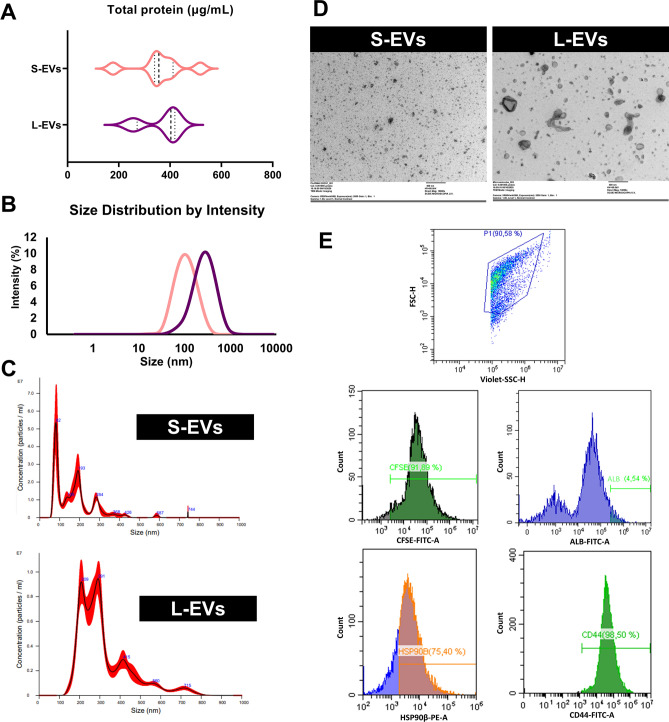
Seminal plasma (SP), a fluid composed mainly by secretions from accessory sex glands, contains a heterogenous population of extracellular vesicles (EVs), involved in several reproductive physiological processes. Seminal plasma has been found to modulate ovary function, in terms of hormone secretion and immune regulation. This study evaluated the potential effect of SP-EV-subsets on the modulation of cumulus-oocyte-complex (COCs) physiology during in vitro maturation (IVM). Two SP-EV-subsets, small-EVs (S-EVs) and large-EVs (L-EVs), were isolated from pig SP by size-exclusion-chromatography. Next, COCs were IVM in the absence (control) or presence of each SP-EV-subset to evaluate their uptake by COCs (PKH67-EVs labelling) and their effect on oocyte and cumulus cells (CCs) (gene expression, and progesterone and estradiol-17β levels). S-EVs and L-EVs were able to bind CCs but not oocytes. Supplementation with L-EVs induced changes (P ≤ 0.05) in the transcript levels of oocyte maturation- (HAS2) and steroidogenesis-related genes (CYP11A1 and HSD3B1) in CCs. No effect on nuclear oocyte maturation and progesterone and estradiol-17β levels was observed when COCs were IVM with any of the two SP-EV-subsets. In conclusion, while SP-EV-subsets can be integrated by CCs during IVM, they do not affect oocyte maturation and only L-EVs are able to modulate CCs function, mainly modifying the expression of steroidogenesis-related genes.
© 2022. The Author(s).
Conflict of interest statement
The authors declare no competing interests.
Figures


Similar articles
-
The effects of plasma-derived extracellular vesicles on cumulus expansion and oocyte maturation in mice.Reprod Biol. 2022 Mar;22(1):100593. doi: 10.1016/j.repbio.2021.100593. Epub 2021 Dec 11. Reprod Biol. 2022. PMID: 34906824
-
Interactions between oocytes and cumulus cells during in vitro maturation of porcine cumulus-oocyte complexes in a chemically defined medium: effect of denuded oocytes on cumulus expansion and oocyte maturation.Theriogenology. 2015 Mar 1;83(4):567-76. doi: 10.1016/j.theriogenology.2014.10.026. Epub 2014 Nov 4. Theriogenology. 2015. PMID: 25467769
-
Estrous cycle impacts microRNA content in extracellular vesicles that modulate bovine cumulus cell transcripts during in vitro maturation†.Biol Reprod. 2020 Feb 14;102(2):362-375. doi: 10.1093/biolre/ioz177. Biol Reprod. 2020. PMID: 31504242
-
Human antral follicles <6 mm: a comparison between in vivo maturation and in vitro maturation in non-hCG primed cycles using cumulus cell gene expression.Mol Hum Reprod. 2013 Jan;19(1):7-16. doi: 10.1093/molehr/gas038. Epub 2012 Sep 6. Mol Hum Reprod. 2013. PMID: 22956770
-
Failure to launch: aberrant cumulus gene expression during oocyte in vitro maturation.Reproduction. 2017 Mar;153(3):R109-R120. doi: 10.1530/REP-16-0426. Epub 2016 Nov 22. Reproduction. 2017. PMID: 27879344 Review.
Cited by
-
Updating Research on Extracellular Vesicles of the Male Reproductive Tract in Farm Animals: A Systematic Review.Animals (Basel). 2024 Oct 31;14(21):3135. doi: 10.3390/ani14213135. Animals (Basel). 2024. PMID: 39518859 Free PMC article. Review.
-
Extracellular vesicles: key mediators in in vitro embryo production.Front Vet Sci. 2025 Aug 20;12:1641966. doi: 10.3389/fvets.2025.1641966. eCollection 2025. Front Vet Sci. 2025. PMID: 40909937 Free PMC article. Review.
-
miR-302d Targeting of CDKN1A Regulates DNA Damage and Steroid Hormone Secretion in Bovine Cumulus Cells.Genes (Basel). 2023 Dec 10;14(12):2195. doi: 10.3390/genes14122195. Genes (Basel). 2023. PMID: 38137018 Free PMC article.
-
Immunophenotype profile by flow cytometry reveals different subtypes of extracellular vesicles in porcine seminal plasma.Cell Commun Signal. 2024 Jan 23;22(1):63. doi: 10.1186/s12964-024-01485-1. Cell Commun Signal. 2024. PMID: 38263049 Free PMC article.
-
Granulosa cell-derived extracellular vesicles mitigate the detrimental impact of thermal stress on bovine oocytes and embryos.Front Cell Dev Biol. 2023 Apr 6;11:1142629. doi: 10.3389/fcell.2023.1142629. eCollection 2023. Front Cell Dev Biol. 2023. PMID: 37091982 Free PMC article.
References
-
- Stahl PD, Raposo G. Extracellular vesicles: Exosomes and microvesicles. Integr. Homeost. Physiol. (Bethesda) 2019;34:169–177. - PubMed
Publication types
MeSH terms
Substances
Grants and funding
- 2017-SGR-1229/Catalan Agency for Management of University and Research Grants, Regional Government of Catalonia, Spain
- 2020-FI-B-00412/Catalan Agency for Management of University and Research Grants, Regional Government of Catalonia, Spain
- MCIN/AEI/10.13039/501100011033, PID2020-113493RB-I00/Ministry of Science and Innovation, Spain
- H2020-MSCA-IF-2019-891382/European Commission (Horizon 2020 Research and Innovation programme)
LinkOut - more resources
Full Text Sources
