. 2022 Nov 9;12(1):19101.
doi: 10.1038/s41598-022-21620-7.

A molecular signature of lung-resident CD8+ T cells elicited by subunit vaccination

Affiliations

A molecular signature of lung-resident CD8+ T cells elicited by subunit vaccination

Naveenchandra Suryadevara et al. Sci Rep. .

Abstract

Natural infection as well as vaccination with live or attenuated viruses elicit tissue resident, CD8+ memory T cell (Trm) response. Trm cells so elicited act quickly upon reencounter with the priming agent to protect the host. These Trm cells express a unique molecular signature driven by the master regulators-Runx3 and Hobit. We previously reported that intranasal instillation of a subunit vaccine in a prime boost vaccination regimen installed quick-acting, CD8+ Trm cells in the lungs that protected against lethal vaccinia virus challenge. It remains unexplored whether CD8+ Trm responses so elicited are driven by a similar molecular signature as those elicited by microbes in a real infection or by live, attenuated pathogens in conventional vaccination. We found that distinct molecular signatures distinguished subunit vaccine-elicited lung interstitial CD8+ Trm cells from subunit vaccine-elicited CD8+ effector memory and splenic memory T cells. Nonetheless, the transcriptome signature of subunit vaccine elicited CD8+ Trm resembled those elicited by virus infection or vaccination. Clues to the basis of tissue residence and function of vaccine specific CD8+ Trm cells were found in transcripts that code for chemokines and chemokine receptors, purinergic receptors, and adhesins when compared to CD8+ effector and splenic memory T cells. Our findings inform the utility of protein-based subunit vaccination for installing CD8+ Trm cells in the lungs to protect against respiratory infectious diseases that plague humankind.


Conflict of interest statement

The authors declare no competing interests.

Figures
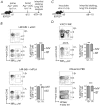
Figure 1
Figure 1
Distribution of immune CD8+ T cells in distinct lung compartments. (A) Experimental design: Mice were prime boost immunized i.p. or i.n. with L4R-b8r and αGC. On the day of harvest, mice were injected i.v. with anti-CD45.2-APC and euthanized after 5 min to allow intravital staining of circulating leukocytes. (B) Lungs were harvested and relative abundance of CD8+ Tem (marginated vasculature, MV) and Trm (interstitial, IST) cells in lungs based on the frequency of staining with anti-CD45.2-APC and B8R70—78/B0702 tetramer-PE. Plots are gated on viable CD8+ T cells. Bar graphs show relative ratio of Tem and Trm. (C) Experimental design: Mice were inoculated i.p. or i.n. route with the virus indicated in panel (D). After 8—10d post-inoculation (p.i.), mice were injected i.v. with anti-CD45.2-APC, and euthanized after 5 min. D. Lungs were harvested and analysed as in panel (B). Cumulative data from 2—3 independent experiments (n = 3–12 mice/group), mean ± SEM.
Figure 2
Figure 2
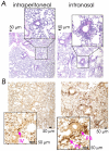
Comparative histologic and immunohistologic localisation of leukocytes responding to subunit vaccination and virus infection. (A) Mice were prime boost immunized i.p. or i.n. with L4R-b8r and αGC and lungs were harvested on d14 post boost. H&E staining of lungs from i.n. immunized mice showed prominent peri-vascular and peri-bronchiolar inflammatory infiltrate (lymphocytes and macrophages) compared to lungs of IP immunized mice. Data represent one of 3 lung sections per mouse from 3 mice per group. (B) Anti-CD3 staining of lung sections performed on d10—14 after booster immunization by i.p. or i.n. route as in Fig. 1. Arrows designate peri-vascular (PV), peri-bronchiolar (PB) and intravascular (IV) T cells. Scale bars, 50 μm. Data are representative of four sections per lung of two mice from two independent experiments per condition.
Figure 3
Figure 3
Prime boost vaccination with rOVA-3 elicits B8R70–78 and D1R808–817-reactive CD8+ T cells. (A) Diagram showing OVA (rOVA-3) construct in which the original cryptic, OT-I and OT-II epitopes were replaced with C4R70–78, B8R70–78 and D1R808—817 epitopes, respectively. A six-histidine tag at the C-terminus of rOVA-3 facilitated purification after expression in E. coli. (B) Mice were primed with rOVA-3 + αGC, boosted twice with the same vaccine, and CD8+ T cells isolated and purified on days shown. (C&D) Intravital staining was performed as in Fig. 1, and B8R70–78/B0702 and D1R808—817/B0702 tetramer-reactive cells were purified by FACS using the gating strategy shown for splenic (C) and pulmonary (D) CD8+ T cells. Purified pB0702 tetramer-reactive cells were used for downstream transcriptomic studies (n = 12 mice; each replicate consisted of cells pooled from 3 mice).
Figure 4
Figure 4
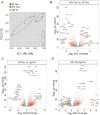
Distinct transcriptome signatures define CD8+ lung IST Trm, MV Tem, and splenic Tm cells. (A) Principal component analysis of CD8+ IST Trm, MV Tem, and splenic Tm transcriptomes. (B–D) Volcano plots showing log2 fold change against -log10 adjusted p value of approximately 17,000 transcripts. Top 20 up and down regulated genes are shown in the volcano plots.
Figure 5
Figure 5
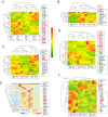
Heat map representation of Trm signature genes. Clusters of differentially expressed transcripts within CD8+ IST Trm, MV Tem, and splenic Tm cell subset were ordered by K-means clustering analysis as described in Materials and Methods. Bulk RNAseq data derived from the three CD8+ memory T cell subsets were used for this analysis. (A) Differential expression of thirty-six previously reported Trm cell signature genes wherein log2 fold change ≥1.5 with adjusted p ≤ 0.05 are shown. Blue side bar, splenic Tm cell-; brown side bar, lung MV Tem cell-; red sidebar, common MV Tem & lung IST Trm-; and maroon sidebar, IST Trm-specific gene expression pattern. (B) Differential expression analysis of transcriptional factors along with Trm signature genes, were performed as in (A) using bulk RNAseq data derived from the three memory T cell subsets. Highlighted are differentially expressed transcripts with log2 fold change ≥1.2 with adjusted p ≤ 0.05 are shown. Sidebars are the same as in (A).
Figure 6
Figure 6
Heat map representation of pathway-specific gene expression changes. Clusters of differentially expressed transcripts specific within the indicated pathways (A–F) were ordered by K-means clustering analysis as described in Materials and Methods. Bulk RNAseq data derived from CD8+ IST Trm, MV Tem, and splenic Tm cell subsets were used for this analysis. (A) Chemokines and chemokine receptors. (B) Integrins. (C) Cell adhesion molecules. (D) Purinergic receptors. (E) inhibitory & activating killer cell lectin-like receptors. (F) T cell effector molecules.
Figure 7
Figure 7
Prime boost vaccination with rOVA-3 without endotoxin depletion elicits a low but substantial level of B8R70–78 and D1R808–817-reactive CD8+ T cell response. (A) B0702tg mice were primed and boosted by the i.n. route, two weeks apart with rOVA-3 mixed with αGC or with αGC alone. Lungs were collected d15 post boost. Cells were stained and gated on live CD8+ T cells and B8R70–79, D1R808-817 tetramer positive cells. Experiments were reproduced at least two times (n = 2 mice) and several times in our published report . (B) B0702tg mice were primed and boosted as in (A). Endotoxins were not depleted from this rOVA-3 preparation used for vaccination. On d15, splenic T cell response to the indicated B0702-restricited epitopes were determined by measuring IFN-γ response in an ELISpot assay. Data represent the mean of triplicate wells; representative of one experiment; (n = 3 mice). (C) The experiment in (B) was repeated with endotoxin-depleted rOVA-3 and the response measured in an IFN-γ specific ELISpot assay using the indicated peptides. Data represent the mean of triplicate wells; representative of two independent experiments; n = 3 mice in each experiment.

Similar articles

Cited by

References

    1. Ariotti S, Beltman JB, Chodaczek G, Hoekstra ME, van Beek AE, Gomez-Eerland R, et al. Tissue-resident memory CD8+ T cells continuously patrol skin epithelia to quickly recognize local antigen. Proc. Natl. Acad. Sci. USA. 2012;109:19739–19744. - PMC - PubMed
    1. Jiang X, Clark RA, Liu L, Wagers AJ, Fuhlbrigge RC, Kupper TS. Skin infection generates non-migratory memory CD8+ T(RM) cells providing global skin immunity. Nature. 2012;483:227–231. - PMC - PubMed
    1. Mackay LK, Rahimpour A, Ma JZ, Collins N, Stock AT, Hafon ML, et al. The developmental pathway for CD103(+)CD8+ tissue-resident memory T cells of skin. Nat. Immunol. 2013;14:1294–1301. - PubMed
    1. Sheridan BS, Pham QM, Lee YT, Cauley LS, Puddington L, Lefrancois L. Oral infection drives a distinct population of intestinal resident memory CD8(+) T cells with enhanced protective function. Immunity. 2014;40:747–757. - PMC - PubMed
    1. Schenkel JM, Fraser KA, Beura LK, Pauken KE, Vezys V, Masopust D. Resident memory CD8 T cells trigger protective innate and adaptive immune responses. Science. 2014 doi: 10.1126/science.1254536. - DOI - PMC - PubMed

Publication types

Substances