. 2023 Feb;22(1):23-41.
doi: 10.1007/s10237-022-01625-x. Epub 2022 Nov 9.

Peristaltic regimes in esophageal transport

Affiliations

Peristaltic regimes in esophageal transport

Guy Elisha et al. Biomech Model Mechanobiol. 2023 Feb.

Abstract

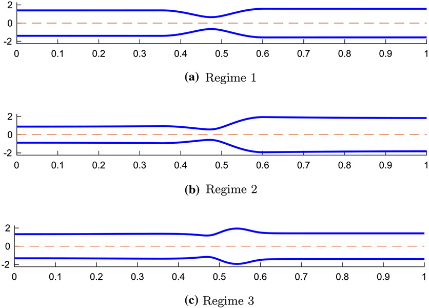
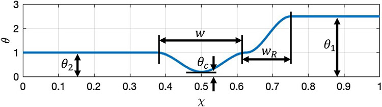
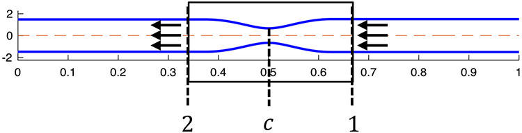
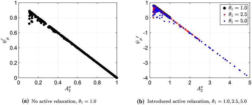
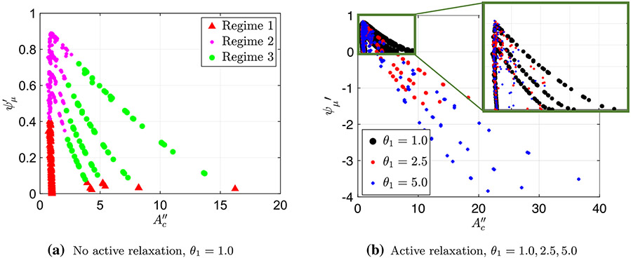
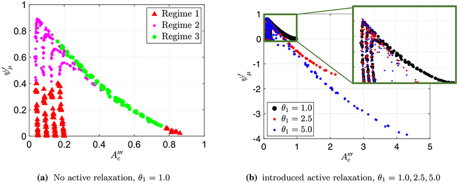
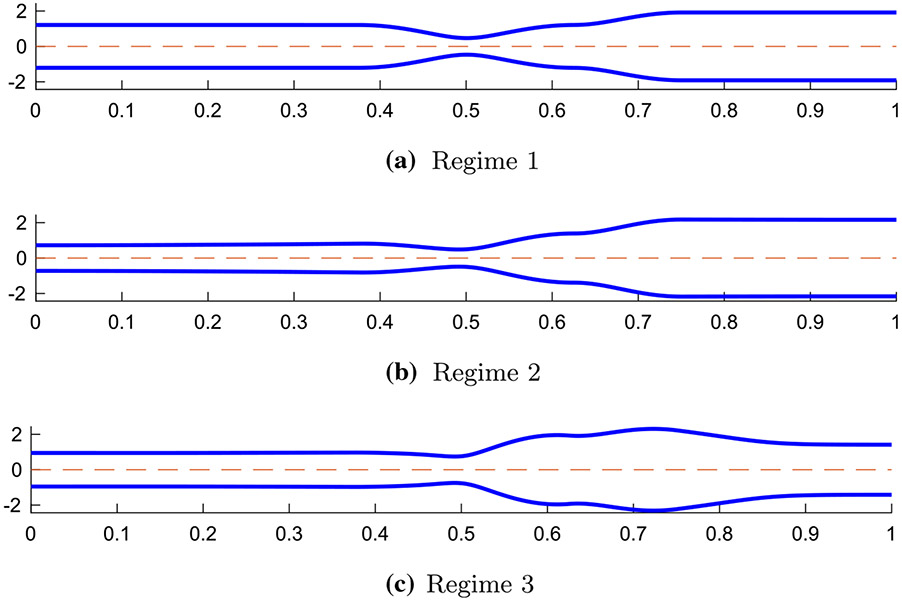
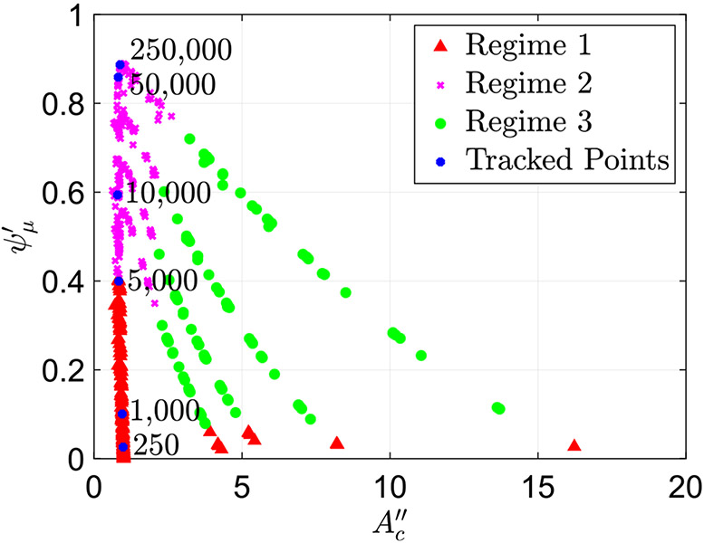
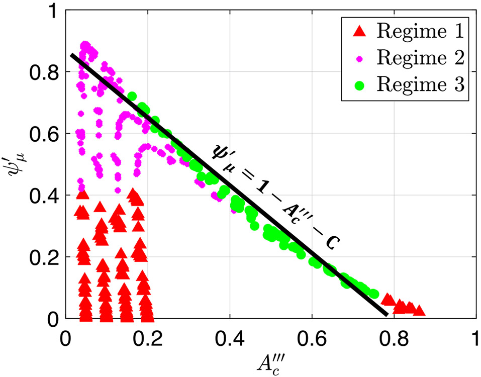
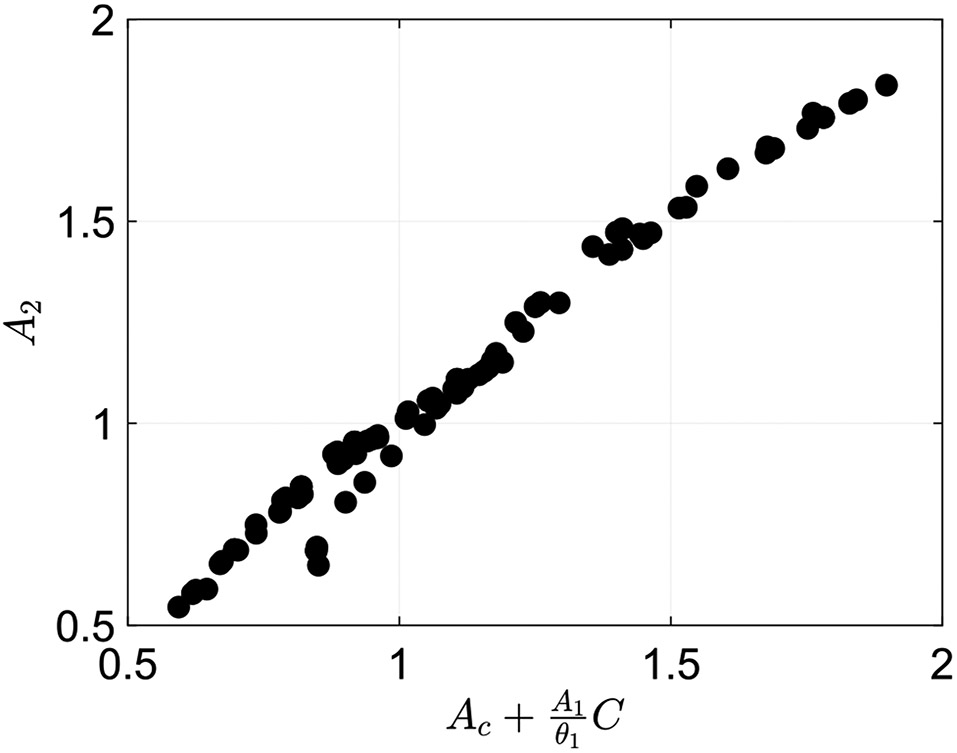
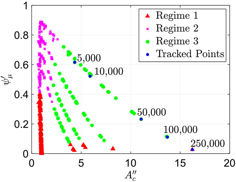
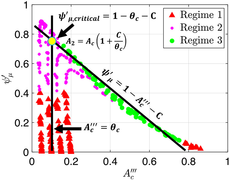
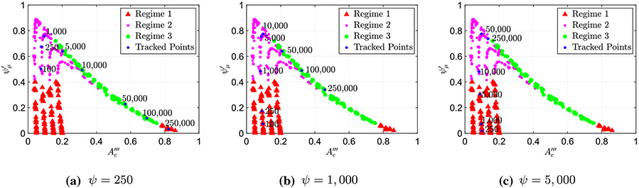
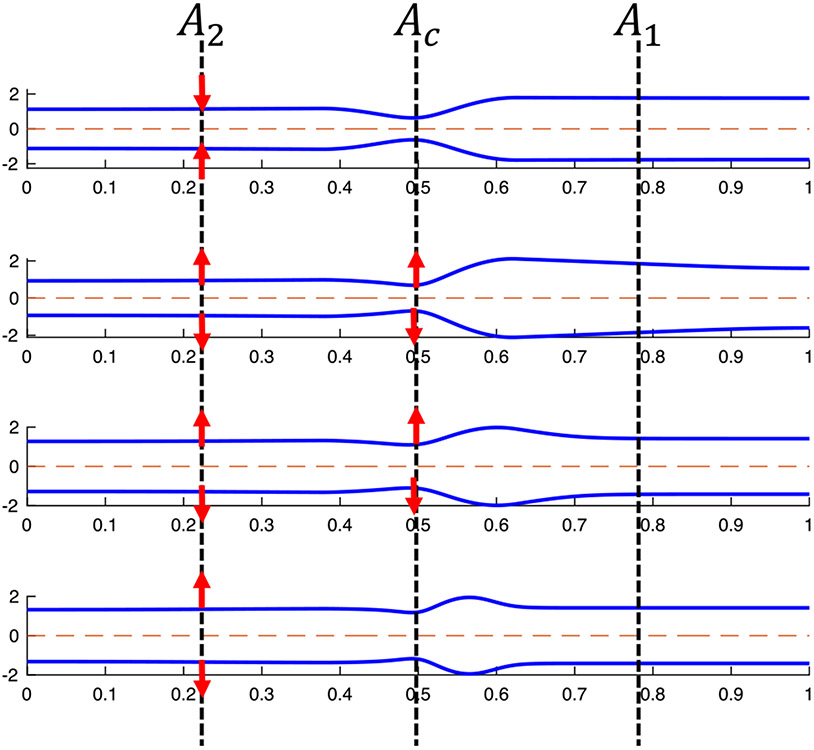
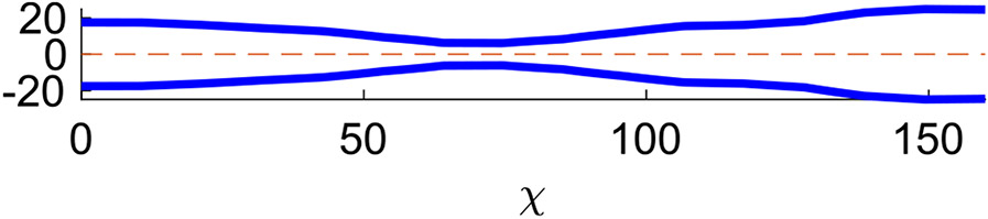
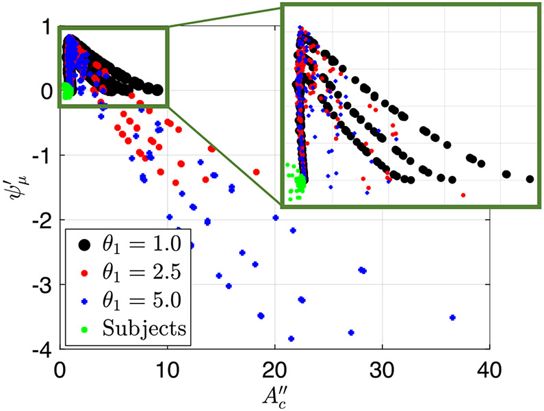
A FLIP device gives cross-sectional area along the length of the esophagus and one pressure measurement, both as a function of time. Deducing mechanical properties of the esophagus including wall material properties, contraction strength, and wall relaxation from these data are a challenging inverse problem. Knowing mechanical properties can change how clinical decisions are made because of its potential for in-vivo mechanistic insights. To obtain such information, we conducted a parametric study to identify peristaltic regimes by using a 1D model of peristaltic flow through an elastic tube closed on both ends and also applied it to interpret clinical data. The results gave insightful information about the effect of tube stiffness, fluid/bolus density and contraction strength on the resulting esophagus shape through quantitive representations of the peristaltic regimes. Our analysis also revealed the mechanics of the opening of the contraction area as a function of bolus flow resistance. Lastly, we concluded that peristaltic driven flow displays three modes of peristaltic geometries, but all physiologically relevant flows fall into two peristaltic regimes characterized by a tight contraction.

Keywords: Elastic tube flow; Esophagus; Fluid–structure interaction; Peristalsis; Reduced-order model.


Conflict of interest statement

Conflict of interest Peter J. Kahrilas, and John E. Pandolfino hold shared intellectual property rights and ownership surrounding FLIP panometry systems, methods, and apparatus with Medtronic Inc. Dustin A. Carlson: Medtronic (Speaking, Consulting). Wenjun Kou: Crospon, Inc. (Consulting). Peter J. Kahrilas: Ironwood (Consulting), Reckitt (Consulting), Johnson & Johnson (Consulting). John E. Pandolfino: Crospon, Inc (stock options), Given Imaging (Consultant, Grant, Speaking), Sandhill Scientific (Consulting, Speaking), Takeda (Speaking), Astra Zeneca (Speaking), Medtronic (Speaking, Consulting), Torax (Speaking, Consulting), Ironwood (Consulting), Impleo (Grant). G. Elisha, S. Halder, S. Acharya, N.A. Patankar: None

Figures

Fig. 1
Fig. 1
Tube geometries of the three physiologically relevant peristaltic regimes identified by Acharya et al. (2021). The shapes were captured from three different simulations at a single time instance in the contractile cycle
Fig. 2
Fig. 2
A plot of the peristaltic activation function θ from Eq. (14) along the tube length at a time instant. Contraction occurs when θ<1 and relaxation occurs when θ>1
Fig. 3
Fig. 3
The geometry corresponding to the reduced-order model
Fig. 4
Fig. 4
Plotting the friction coefficient ψμ and non-dimensional area parameter A2 to show their linear correlation
Fig. 5
Fig. 5
The friction parameter ψμ plotted as a function of the non-dimensional contraction area parameter Ac
Fig. 6
Fig. 6
The friction parameter ψμ was plotted as a function of the non-dimensional contraction area parameter Ac
Fig. 7
Fig. 7
Tube geometry of the three peristaltic regimes in a FLIP device when there is relaxation distal of contraction. The separation into three regimes was done based on the observations similar to those in Fig. 5
Fig. 8
Fig. 8
Plot of the friction parameter ψμ as a function of Ac with six highlighted cases with the same ψ and θc and changing β. The value of β in each case is written on the plot. The plot shows how in peristaltic regimes 1 and 2, friction parameter increases as fluid viscosity parameter increases (β)
Fig. 9
Fig. 9
Plot of the friction parameter ψμ as a function of Ac alongside the line ψμ=1AcC. This plot uses the output data from the simulations where there is no relaxation, θ1=1.0
Fig. 10
Fig. 10
Plot of the actual cross-sectional area at location 2 as a function of the calculated cross-sectional area at location 2 from Eq. (42) for cases classified as peristaltic regime 3. This plot shows an approximate linear line of slope 1, meaning that the model and data match well
Fig. 11
Fig. 11
Plot of the friction parameter ψμ as a function of Ac with five highlighted cases with the same ψ and θc and changing β. The value of β in each case is written on the plot. The plot shows how in peristaltic regimes 3, friction parameter (ψμ) decreases as fluid viscosity parameter (β) increases
Fig. 12
Fig. 12
Plot of the friction parameter ψμ as a function of Ac with the two lines in Eqs. (35) and (39)
Fig. 13
Fig. 13
Three plots of the friction parameter ψμ as a function of Ac, each with a different set of highlighted simulations. Each set shares contractile cycle simulations with the same contraction strength and wall stiffness, with changing fluid resistance parameter
Fig. 14
Fig. 14
Tube geometry at transition
Fig. 15
Fig. 15
Esophagus wall shape of a control at a single time instance (Acharya et al. 2020; Lin et al. 2013; Carlson et al. 2016). The cross sectional data were captures using a FLIP device
Fig. 16
Fig. 16
The friction parameter ψμ plotted as a function of the non-dimensional contraction area parameter Ac for both simulation and clinical FLIP contractile cycles

References

    1. Abo-Elkhair RE, Bhatti MM, Mekheimer KS (2021) Magnetic force effects on peristaltic transport of hybrid bio-nanofluid (aucu nanoparticles) with moderate Reynolds number: an expanding horizon. Int Commun Heat Mass Transf 123:105228. 10.1016/j.icheatmasstransfer.2021.105228 - DOI
    1. Abrahao L Jr, Bhargava V, Babaei A, Ho A, Mittal RK (2011) Swallow induces a peristaltic wave of distension that marches in front of the peristaltic wave of contraction. Neurogastroenterol Motil 23(3):e110–201. 10.1111/j.1365-2982.2010.01624.x - DOI - PubMed
    1. Acharya S, Halder S, Carlson DA, Kou W, Kahrilas PJ, Pandolfino JE, Patankar NA (2020) Assessment of esophageal body peristaltic work using functional lumen imaging probe panometry. Am J Physiol Gastrointest Liver Physiol. 10.1152/ajpgi.00324.2020 - DOI - PMC - PubMed
    1. Acharya S, Kou W, Halder S, Carlson DA, Kahrilas PJ, Pandolfino JE, Patankar NA (2021) Pumping patterns and work done during peristalsis in finite-length elastic tubes. J Biomech Eng. 10.1115/1.4050284 - DOI - PMC - PubMed
    1. Adler DG, Romero Y (2001) Primary esophageal motility disorders. Mayo Clin Proc 76(2):195–200. 10.4065/76.2.195 - DOI - PubMed

LinkOut - more resources