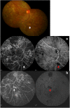
Tubercular serpiginous choroiditis
- PMID: 36352169
- PMCID: PMC9645760
- DOI: 10.1186/s12348-022-00312-3
Tubercular serpiginous choroiditis
Abstract
Tubercular association with serpiginous choroiditis, also called 'serpiginous-like choroiditis' or 'multifocal serpiginoid choroiditis' (MSC) is reported from world over, especially from endemic countries. Though the exact mechanism is not yet clear, a direct or indirect infectious trigger by Mycobacterium tuberculosis (MTB) is believed to cause choroiditis.The link of immune mechanisms with ocular inflammation caused by MTB is emerging, and has been supported by both experimental and human data. The molecular and histopathological findings of tubercular serpiginous-like choroiditis have been demonstrated in clinicopathological reports, as well as in animal models. Young to middle-aged healthy males are more frequently affected. The choroiditis lesions of tubercular serpiginous-like choroiditis evolve as multifocal lesions, affecting the retinal periphery as well as posterior pole. They begin as discrete lesions, and spread in a serpiginoid pattern to become confluent. Fundus imaging including autofluorescence is extremely helpful in monitoring patients for response to therapy. Its diagnosis is essentially clinical. Corroborative evidence is obtained by a positive tuberculin skin test, or a positive QuantiFERON-TB Gold (Cellestis, Carnegie, Victoria, Australia) test, and/or radiological (chest X-ray or chest CT scan) evidence of TB elsewhere in the body. Systemic corticosteroids are the mainstay of therapy to control active inflammation, while ATT helps to reduce recurrence of inflammatory attacks. Immunosuppressive agents are indicated in cases with relentless progression, paradoxical worsening, or recurrent choroiditis.
Keywords: Immune mechanisms; Pathogenesis; Serpiginous choroiditis; Serpiginous-like choroiditis; Tubercular.
© 2022. The Author(s).
Conflict of interest statement
The authors declare that they have no competing interests.
Figures


Similar articles
-
Rare Case of Tubercular Serpiginous-Like Choroiditis.Cureus. 2024 Mar 27;16(3):e57093. doi: 10.7759/cureus.57093. eCollection 2024 Mar. Cureus. 2024. PMID: 38681413 Free PMC article.
-
Tubercular serpiginous-like choroiditis presenting as multifocal serpiginoid choroiditis.Ophthalmology. 2012 Nov;119(11):2334-42. doi: 10.1016/j.ophtha.2012.05.034. Epub 2012 Aug 11. Ophthalmology. 2012. PMID: 22892153
-
Unilateral macular serpiginous-like choroiditis as the initial manifestation of presumed ocular tuberculosis.Int J Retina Vitreous. 2021 Jan 4;7(1):1. doi: 10.1186/s40942-020-00272-7. Int J Retina Vitreous. 2021. PMID: 33397439 Free PMC article.
-
Serpiginous choroiditis and infectious multifocal serpiginoid choroiditis.Surv Ophthalmol. 2013 May-Jun;58(3):203-32. doi: 10.1016/j.survophthal.2012.08.008. Epub 2013 Mar 27. Surv Ophthalmol. 2013. PMID: 23541041 Free PMC article. Review.
-
Tuberculosis-related serpiginous choroiditis: aggressive therapy with dual concomitant combination of multiple anti-tubercular and multiple immunosuppressive agents is needed to halt the progression of the disease.J Ophthalmic Inflamm Infect. 2022 Feb 8;12(1):7. doi: 10.1186/s12348-022-00282-6. J Ophthalmic Inflamm Infect. 2022. PMID: 35132499 Free PMC article. Review.
Cited by
-
Rare Case of Tubercular Serpiginous-Like Choroiditis.Cureus. 2024 Mar 27;16(3):e57093. doi: 10.7759/cureus.57093. eCollection 2024 Mar. Cureus. 2024. PMID: 38681413 Free PMC article.
-
Presumed ocular tuberculosis - need for caution before considering anti-tubercular therapy.Eye (Lond). 2023 Dec;37(18):3716-3717. doi: 10.1038/s41433-023-02628-3. Epub 2023 Jun 14. Eye (Lond). 2023. PMID: 37316712 Free PMC article. No abstract available.
-
Epidemiology of uveitis after tuberculosis in Taiwan - A nationwide population-based cohort study.Taiwan J Ophthalmol. 2024 Jul 22;15(2):252-258. doi: 10.4103/tjo.TJO-D-24-00012. eCollection 2025 Apr-Jun. Taiwan J Ophthalmol. 2024. PMID: 40584193 Free PMC article.
References
-
- Babel J. Geographic and helicoid choroidopathies. Clinical and angiographic study; attempted classification. J Fr Ophtalmol. 1983;6:981–993. - PubMed
-
- Abu el-Asrar AM Serpiginous (geographical) choroiditis. Int Ophthalmol Clin. 1995;35:87–91. - PubMed
-
- Lim WK, Buggage RR, Nussenblatt RB. Serpiginous choroiditis. Surv Ophthalmol. 2005;50:231–244. - PubMed
Publication types
LinkOut - more resources
Full Text Sources

