

[Sacral fragility fractures: risk factors and outcomes after cement sacroplasty]
- PMID: 36352271
- PMCID: PMC9715472
- DOI: 10.1007/s00132-022-04323-9
[Sacral fragility fractures: risk factors and outcomes after cement sacroplasty]
Abstract
Background: The objective of the present study on patients with fragility fractures of the sacrum (FFS) was to assess existing risk factors and clinical outcomes after cement sacroplasty (CSP).
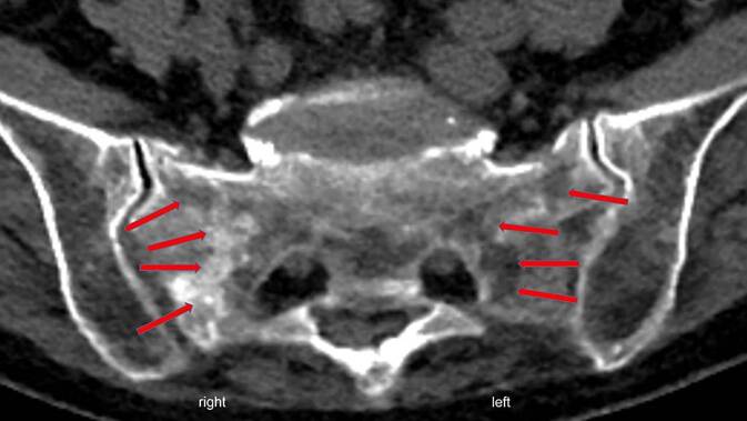
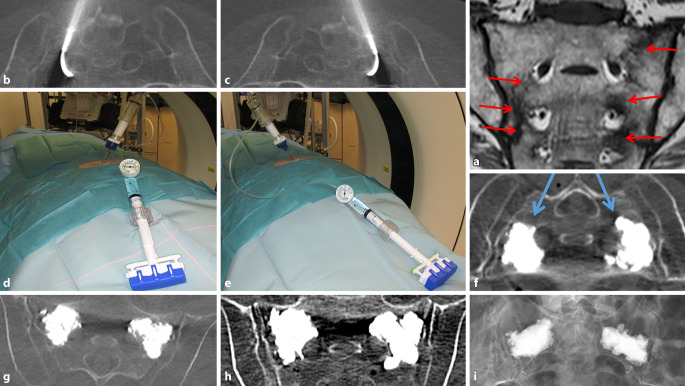
Patients and methods: 68 patients (64 women, 4 men) with previous FFS were followed up retrospectively. CT and MRI images were used to classify fractures according to Denis et al. and Rommens and Hofmann. Bone mineral content was determined by QCT in all patients. Concomitant diseases as well as central and peripheral fractures were recorded, considering the patient's medical history and X‑ray images. Vitamin D levels were also determined. If conservative therapy was unsuccessful, CSP was performed. The results were documented on the basis of pain development, physical independence, patient satisfaction, complication rate and mortality.
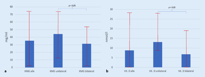
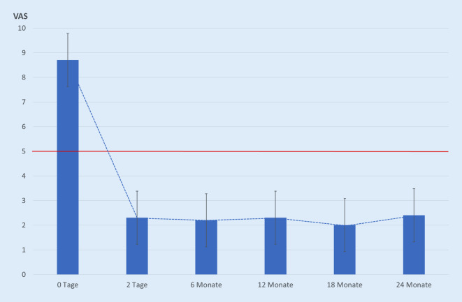
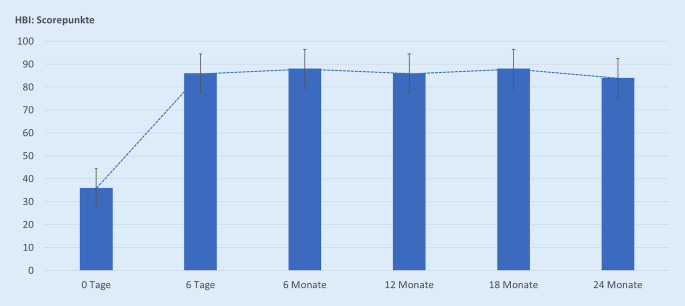
Results: The age of the women in the study was Ø 83.2 (72-99) and that of the men Ø 77.8 (76-85) years. 42.4% had a Denis type 1, 4.2% a Denis type 2, 0% a Denis type 3, 43.3% a Denis type 1-2 and 10.1% a Denis type 1-2-3 fracture zone. FFP type II a to II c fractures were found in 88.2%, FFP type III c in 7.4% and FFP type IV b in 4.4%. Bilateral FFS were found in 68.8%. The average bone mineral content (BMC) was 35.4 (2-74) mg/ml, and the average vitamin D value was 8.8 (0-28) nmol/l. Other osteoporosis-associated fractures were found in around 50% of the patients. After CSP, patients showed a rapid and significant (p < 0.001) reduction in pain and sustained clinical improvement.
Conclusion: FFS fracture risk factors were found to be female gender, advanced age, existing osteoporosis and severe vitamin‑D deficiency. Patients with non-displaced FFS who could not be mobilised due to pain experienced sustained benefit from CSP.
Zusammenfassung: HINTERGRUND: Ziel der Untersuchung bei PatientInnen mit Fragilitätsfrakturen des Os sacrum (FFS) war die Erfassung von vorhandenen Risikofaktoren sowie der klinischen Ergebnisse nach Zementsakroplastie (ZSP).
Patientinnen und methoden: Retrospektiv wurden 68 PatientInnen (64 Frauen, 4 Männer) mit stattgehabten FFS nachuntersucht. Anhand von CT- und MRT-Schnittbildern erfolgte eine Fraktureinteilung nach Denis et al. sowie Rommens und Hofmann. Bei allen PatientInnen wurde eine Knochenmineralgehaltsbestimmung mittels QCT durchgeführt. Unter Berücksichtigung von Anamnese und Röntgenaufnahmen wurden Begleiterkrankungen sowie zentrale und periphere Frakturen miterfasst. Vitamin-D-Werte wurden zusätzlich bestimmt. Nach einem frustranen konservativen Therapieversuch erfolgte eine ZSP. Anhand der Schmerzentwicklung, der körperlichen Selbstständigkeit, der PatientInnen-Zufriedenheit, der Komplikationsrate und der Mortalität wurden die Ergebnisse dokumentiert.
Ergebnisse: Das Alter der Frauen betrugt Ø 83,2 (72–99), dass der Männer Ø 77,8 (76–85) Jahre. Zu 42,4 % fand sich eine Denis-Typ-1-, zu 4,2 % eine Denis-Typ-2-, zu 0 % eine Denis-Typ-3-, zu 43,3 % eine Denis-Typ-1–2- und zu 10,1 % eine Denis-Typ-1–2–3-Frakturzone. Es fand sich ein FFP-Typ-II a-bis -II c-Frakturgeschehen zu 88,2 %, ein FFP-Typ III c zu 7,4 % sowie ein FFP-Typ IV b zu 4,4 %. Bei 68,8 % fanden sich bilaterale FFS. Der Knochenmineralgehalt (KMG) betrug im Ø 35,4 (2–74) mg/ml, der Vitamin-D-Wert im Ø 8,8 (0–28) nmol/l. Weitere osteoporoseassoziierte Frakturen fanden sich in circa 50 %. Nach der ZSP zeigten die PatientInnen eine schnelle und signifikante (p < 0,001) Schmerzreduktion sowie nachhaltige klinische Verbesserung.
Schlussfolgerung: Als Frakturrisikofaktoren von FFS fanden sich das weibliche Geschlecht, das hohe Alter, eine vorhandene Osteoporose und ein schwerer Vitamin-D-Mangel. PatientInnen mit nichtdislozierten FFS, welche schmerzbedingt nicht zu mobilisieren waren, profitierten von einer ZSP nachhaltig.
Keywords: Bone mineral density; Osteoporosis; Os sacrum; Retrospective Study; Vitamin D deficiency.
© 2022. The Author(s).
Similar articles
-
Comparative outcome of different treatment options for fragility fractures of the sacrum.BMC Musculoskelet Disord. 2022 Dec 19;23(1):1106. doi: 10.1186/s12891-022-06039-5. BMC Musculoskelet Disord. 2022. PMID: 36536363 Free PMC article.
-
[Clinical outcome and revenue situation after conservative, interventional and surgical/osteosynthetic treatment of sacral insufficiency fractures].Unfallchirurg. 2021 Jul;124(7):588-597. doi: 10.1007/s00113-020-00932-1. Epub 2020 Dec 10. Unfallchirurg. 2021. PMID: 33301083 German.
-
CT-guided cement sacroplasty (CSP) as pain therapy in non-dislocated insufficiency fractures.Eur J Orthop Surg Traumatol. 2017 Dec;27(8):1045-1050. doi: 10.1007/s00590-017-2001-1. Epub 2017 Jun 26. Eur J Orthop Surg Traumatol. 2017. PMID: 28653101 Free PMC article.
-
Safety and Efficacy of Sacroplasty for Sacral Fractures: A Systematic Review and Meta-Analysis.J Vasc Interv Radiol. 2019 Nov;30(11):1845-1854. doi: 10.1016/j.jvir.2019.06.013. Epub 2019 Oct 3. J Vasc Interv Radiol. 2019. PMID: 31587952
-
Fragility Fractures of the Pelvis and Sacrum: Current Trends in Literature.Hip Pelvis. 2022 Jun;34(2):69-78. doi: 10.5371/hp.2022.34.2.69. Epub 2022 Jun 7. Hip Pelvis. 2022. PMID: 35800130 Free PMC article. Review.
Cited by
-
Retrospective evaluation of percutaneous 3D-navigated screw fixation for fragility fractures of the sacrum: technical notes and four-year experience.Sci Rep. 2023 Jul 28;13(1):12254. doi: 10.1038/s41598-023-39165-8. Sci Rep. 2023. PMID: 37507446 Free PMC article.
References
-
- Andresen R, Radmer S, Kamusella P, Wissgott C, Banzer J, Schober HC. Treatment of Denis 1, 2 and 3 insufficiency fracture zones of the os sacrum. Individual approaches adapted to the course of the fracture in CT-assisted balloon sacroplasty. Osteologie. 2012;21(3):168–173. doi: 10.1055/s-0037-1621680. - DOI
-
- Andresen R, Radmer S, Lüdtke CW, Kamusella P, Görmez M, Wissgott C, Schober HC. Vergleich von konservativer Therapie vs. CT-gesteuerter Ballonsakroplastie bei der Behandlung von Insuffizienzfrakturen des Os sacrum. Osteologie. 2015;24(2):92–98. doi: 10.1055/s-0037-1622046. - DOI
Publication types
MeSH terms
Substances
LinkOut - more resources
Full Text Sources
Medical