. 2023 Jan;20(1):325-337.
doi: 10.1007/s13311-022-01327-3. Epub 2022 Nov 9.

Gpr83 Tunes Nociceptor Function, Controlling Pain

Affiliations

Gpr83 Tunes Nociceptor Function, Controlling Pain

Yerin Kim et al. Neurotherapeutics. 2023 Jan.

Abstract

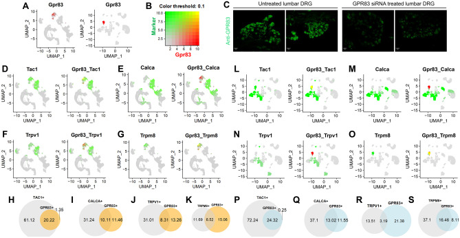
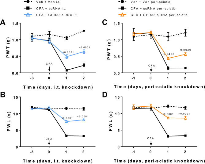
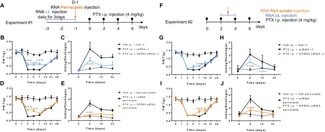
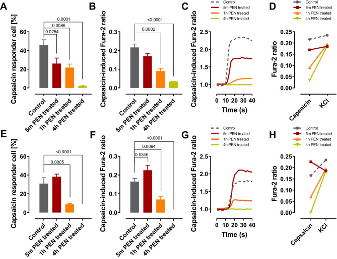
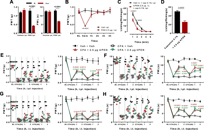
The function of peripheral nociceptors is frequently tuned by the action of G protein-coupled receptors (GPRs) that are expressed in them, which contribute to pain alteration. Expanding new information on such GPRs and predicting their potential outcomes can help to construct new analgesic strategies based on their modulations. In this context, we attempted to present a new GPR not yet acknowledged for its pain association. Gpr83 exhibits relatively high expressions in the peripheral nervous system compared to other tissues when we mined and reconstructed Gene Expression Omnibus (GEO) metadata, which we confirmed using immunohistochemistry on murine dorsal root ganglia (DRG). When Gpr83 expression was silenced in DRG, neuronal and behavioral nociception were all downregulated. Pathologic pain in hind paw inflammation and chemotherapy-induced peripheral neuropathy were also alleviated by this Gpr83 knockdown. Dependent on exposure time, the application of a known endogenous Gpr83 ligand PEN showed differential effects on nociceptor responses in vitro. Localized PEN administration mitigated pain in vivo, probably following Gq/11-involved GPR downregulation caused by the relatively constant exposure. Collectively, this study suggests that Gpr83 action contributes to the tuning of peripheral pain sensitivity and thus indicates that Gpr83 can be among the potential GPR targets for pain modulation.

Keywords: Analgesia; Gpr83; Nociceptor; PEN; Pain.


Conflict of interest statement

The authors declare no competing interests.

Figures

Fig. 1
Fig. 1
Gpr83 expression in a subset of DRG neurons. A Universal manifold approximation and projection (UMAP) representation of graph-based clustered single-cell RNA sequences (GSE139088, left) and single nucleus RNA sequences (GSE154659, right) from mouse dorsal root ganglionic cells revealed Gpr83-expressing subclusters (red). B Two-dimensional color codes used for simultaneously visualizing the co-expressions of Gpr83 (red) and marker (green) RNAs in plots from (D) to (G) and from (L) to (O). C Two representative immunohistochemical images of Gpr83 expression (green) in mouse DRG sections with (right two images) or without Gpr83 small interfering RNA (siRNA) treatment (left two images). DG UMAP representations for visualizing Gpr83 and marker expression in the GSE139088 dataset. Tac1 (D), Calca (E), Trpv1 (F), and Trpm8 (G) were used as markers. HK Venn diagrams of which the relative sizes reflect the numbers of Gpr83 expresser cells and their co-expression profiles with the four markers presented in (D) to (G). LO UMAP representations for visualizing Gpr83 and marker expression in the GSE154659 dataset. Tac1 (L), Calca (M), Trpv1 (N), and Trpm8 (O) were used as markers. PS Venn diagrams of which the relative sizes reflect the numbers of expresser cells of Gpr83 and their co-expression profiles with the four markers presented in (L) to (O).
Fig. 2
Fig. 2
Gpr83 knockdown alters nociceptor functionalities. A Percentage of capsaicin-responsive neurons with or without Gpr83 siRNA treatment or with scrambled RNA (scRNA) treatment in fura-2 intracellular Ca2+ imaging (control group, n = 4 batches; siRNA-treated group, n = 3 batches; scRNA-treated group, n = 3 batches). B Peak increases in fluorescent levels of capsaicin-responsive neurons in (A), with or without Gpr83 siRNA treatment or with scRNA treatment in fura-2 intracellular Ca2+ imaging (control group, n = 317 neurons; siRNA-treated group, n = 158 neurons; scRNA-treated group, n = 109 neurons). Experiments in (A–B) were triplicated. C Percentage of cinnamaldehyde-responsive neurons with or without Gpr83 siRNA treatment or with scRNA treatment in fura-2 intracellular Ca2+ imaging (control group, n = 5 batches; siRNA-treated group, n = 4 batches; scRNA-treated group, n = 5 batches). D Peak increases in fluorescent levels of cinnamaldehyde-responsive neurons in (C) with or without Gpr83 siRNA treatment or with scRNA treatment in fura-2 intracellular Ca2+ imaging (control group, n = 320 neurons; siRNA-treated group, n = 164 neurons; scRNA-treated group, n = 168 neurons). Experiments in (C–D) were triplicated. E A schematic diagram for interfering Gpr83 expression in DRG using a peri-sciatic injection of siRNA. Five animals were used for each experiment. F Time course of the duration of 0.1% capsaicin (CAP)-induced nociceptive behaviors in mice with or without peri-sciatic injection of Gpr83 siRNA and with that of scRNA. CAP was administered intraplantarly in a hind paw. G Total durations of nociceptive responses for 5 min from (F) are quantified in the histogram. H Time course of the duration of 10 mM cinnamaldehyde (CA)-induced nociceptive behaviors in mice with or without peri-sciatic injection of Gpr83 siRNA and with that of scRNA. CA was injected intraplantarly into a hind paw. I Total durations of nociceptive responses for 5 min from (H) are quantified in the histogram. J A schematic diagram for interfering Gpr83 expression in DRG using an intrathecal injection of siRNA. Five animals were used for each experiment. K Time course of the duration of 0.1% CAP-induced nociceptive behaviors in mice with or without peri-sciatic injection of Gpr83 siRNA and with that of scRNA. CAP was intraplantarly injected into a hind paw. L Total durations of nociceptive responses for 5 min from (K) are quantified in the histogram. M Time course of the duration of 10 mM CA-induced nociceptive behaviors in mice with or without peri-sciatic injection of Gpr83 siRNA and with that of scRNA. CA was intraplantarly injected into a hind paw. N Total durations of nociceptive responses for 5 min from (M) are quantified in the histogram. Intracellular fluorescent Ca2+ responses in (A)–(D) were compared with those without RNA injection by one-way ANOVA and Tukey’s test. Effects on the durations of the nociceptive responses in (E)–(N) at every minute were compared with those without RNA injection by two-way ANOVA and Tukey’s test, and the comparisons between total durations were conducted by one-way ANOVA and Tukey’s test
Fig. 3
Fig. 3
Gpr83 knockdown alleviates inflammatory pain. A Time course of von Frey thresholds in complete Freund’s adjuvant (CFA)-inflamed mice. Animals were intrathecally pretreated with siRNA or scRNA, and their thresholds were monitored 24 h and 48 h after CFA injection. The dots in the plot show individual threshold values. B Time course of Hargreaves latencies in CFA-inflamed mice. Animals were treated in the same manner as in (A). The dots in the plot show individual latency values. C Time course of von Frey thresholds in CFA-inflamed mice. Animals were administered peri-sciatically with siRNA or scRNA, and their thresholds were monitored 24 h and 48 h after CFA injection. The dots in the plot show individual threshold values. D Time course of Hargreaves latencies in CFA-inflamed mice. Animals were treated in the same manner as in (C). The dots in the plot show individual latency values
Fig. 4
Fig. 4
Gpr83 knockdown alleviates chemotherapy-induced neuropathic pain. A Summary of the experimental schedule for (B) to (E). Pre-emptive siRNA or scRNA injection was followed by paclitaxel injections (intraperitoneal administration, 4 mg/kg). B Time course of von Frey thresholds in mice treated intrathecally as in (A) (n = 4–5). C Time course of cold sensitivities in mice treated intrathecally as in (A) (n = 4–5). D Time course of von Frey thresholds in mice treated peri-sciatically as in (A) (n = 4–5). E Time course of cold sensitivities in mice treated peri-sciatically as in (A) (n = 4–5). F Summary of the experimental schedule for (G) to (J). siRNA or scRNA was injected in animals during the paclitaxel treatment regimen as shown in (A). G Time course of von Frey thresholds in mice treated intrathecally as in (A) (n = 5). H Time course of cold sensitivities in mice treated intrathecally as in (A) (n = 5). I Time course of von Frey thresholds in mice treated peri-sciatically as in (A) (n = 5). J Time course of cold sensitivities in mice treated peri-sciatically as in (A) (n = 5)
Fig. 5
Fig. 5
Incubation time-dependent differential alteration of nociceptor functions by PEN. A Percentage of capsaicin-responsive neurons under PEN exposures in fura-2 intracellular Ca2+ imaging (untreated group, n = 4 batches; 5-min PEN-treated group, n = 6 batches; 1-h PEN-treated group, n = 8 batches; 4-h PEN-treated group, n = 4 batches). B Peak increases in fluorescent levels of capsaicin-responsive neurons in (A), with or without PEN treatment in fura-2 intracellular Ca2+ imaging (untreated group, n = 453 neurons; 5-min PEN-treated group, n = 455 neurons; 1-h PEN-treated group, n = 508 neurons; 4-h PEN-treated group, n = 172 neurons). Experiments in (A–B) were triplicated. C Representative traces of fura-2 intracellular Ca2+ levels from individual neurons. D Averaged fura-2 intracellular Ca2+ levels upon capsaicin and KCl-induced depolarization (untreated group, n = 453 neurons; 5-min PEN-treated group, n = 455 neurons; 1-h PEN-treated group, n = 508 neurons; 4-h PEN-treated group, n = 172 neurons). E Percentage of capsaicin-responsive neurons under PEN exposures in fura-2 intracellular Ca2+ imaging. When fluorescence increased in response to agonist treatment by 10% or more compared to the baseline before drug treatment using the same raw data with those in (A), we determined that an agonist-induced response had occurred (untreated group, n = 4 batches; 5-min PEN-treated group, n = 6 batches; 1-h PEN-treated group, n = 8 batches; 4-h PEN-treated group, n = 4 batches). B Peak increases in fluorescent levels of capsaicin-responsive neurons in (E), with or without PEN treatment in fura-2 intracellular Ca2+ imaging (untreated group, n = 451 neurons; 5-min PEN-treated group, n = 198 neurons; 1-h PEN-treated group, n = 108 neurons; 4-h PEN-treated group, n = 171 neurons). Experiments in (A–B) were triplicated. C Representative traces of fura-2 intracellular Ca2+ levels from individual neurons. D Averaged fura-2 intracellular Ca2+ levels upon capsaicin and KCl-induced depolarization (untreated group, n = 453 neurons; 5-min PEN-treated group, n = 198 neurons; 1-h PEN-treated group, n = 108 neurons; 4-h PEN-treated group, n = 171 neurons)
Fig. 6
Fig. 6
PEN treatment alleviates pain. A von Frey thresholds (left) and Hargreaves latencies (right) immediately after intraplantarly injection of PEN (2.5 µg/10 µl) in a hind paw. B Time course of von Frey thresholds after intraplantarly injection of PEN (2.5 µg/10 µl) in a hind paw for 150 min. C Time course of the duration of 0.1% capsaicin (CAP)-induced nociceptive behaviors in mice immediately after intraplantarly injection of 2.5 µg/10 µl in a hind paw. CAP was administered intraplantarly in the ipsilateral hind paw. D Total durations of nociceptive responses for 5 min from (C) are quantified in the histogram. E Time course of von Frey thresholds in complete Freund’s adjuvant (CFA)-inflamed mice. Animals were treated intraplantarly with PEN (2.5 µg/10 µl) in the ipsilateral hind paw. The left dot plot shows individual threshold values. F Time course of Hargreaves latencies in CFA-inflamed mice. Animals were treated in the same manner as in (E). The left dot plot shows individual latency values. G Time course of von Frey thresholds in CFA-inflamed mice. Animals were treated intrathecally with PEN (2.5 µg/10 µl). The left dot plot shows individual threshold values. H Time course of Hargreaves latencies in CFA-inflamed mice. Animals were treated in the same manner as in (G). The left dot plot shows individual latency values
Fig. 7
Fig. 7
PEN-induced signal transductions in luciferase assays. A Serum response element (SRE)-mediated luminescence was measured in mGpr83-transfected HEK cells with or without 4-h exposure to mouse PEN (mPEN) or human PEN (hPEN). Experiments were carried out in triplicate (n = 6 batches). B Nuclear factor of activated T cell (NFAT)-mediated luminescence was measured in mGpr83-transfected HEK cells with or without 4-h exposure to mPEN or hPEN. Experiments were carried out in triplicate (n = 19 batches). C cAMP response element (CRE)-mediated luminescence was measured in mGpr83-transfected HEK cells with or without 4-h exposure to mPEN or hPEN. Experiments were carried out in triplicate (n = 17 batches). D Western blots (left) and their quantification (right) of extracellular signal-regulated kinase (ERK) with or without phosphorylation under PEN exposures. Data represent means ± S.E.M. (n = 4, two-way ANOVA)

References

    1. WOOLF, C. J. Capturing novel non-opioid pain targets. Biol Psychiatry. 2020;87:74–81. doi: 10.1016/j.biopsych.2019.06.017. - DOI - PMC - PubMed
    1. Zheng H, Lim JY, Kim Y, Jung ST, Hwang SW. The role of oxytocin, vasopressin, and their receptors at nociceptors in peripheral pain modulation. Front Neuroendocrinol. 2021;63:100942. doi: 10.1016/j.yfrne.2021.100942. - DOI - PubMed
    1. Berta T, Qadri Y, Tan PH, Ji RR. Targeting dorsal root ganglia and primary sensory neurons for the treatment of chronic pain. Expert Opin Ther Targets. 2017;21:695–703. doi: 10.1080/14728222.2017.1328057. - DOI - PMC - PubMed
    1. Cho PS, Lee HK, Choi YI, Choi SI, Lim JY, Kim M, Kim H, Jung SJ, Hwang SW. Gpr171 activation modulates nociceptor functions, alleviating pathologic pain. Biomed. 2021 Mar 5;9(3):256. - PMC - PubMed
    1. Zheng H, Lim JY, Seong JY, Hwang SW. The role of corticotropin-releasing hormone at peripheral nociceptors: implications for pain modulation. Biomed. 2020 Dec 17;8(12):623. - PMC - PubMed

Publication types

Substances