. 2022 Nov 9;17(1):410.
doi: 10.1186/s13023-022-02559-4.

Genetic analysis of 55 cases with fetal skeletal dysplasia

Affiliations

Genetic analysis of 55 cases with fetal skeletal dysplasia

Ying Bai et al. Orphanet J Rare Dis. .

Abstract

Background: Fetal skeletal dysplasia (SD) is a common congenital disability comprising a complex group of skeletal disorders with substantial clinical and genetic heterogeneity. Many of these defects are detected prenatally using ultrasound (US). However, the diagnostic accuracy of the US is limited.

Methods: We recruited 55 unrelated fetuses with US-detected skeletal anomalies and performed sequential tests using copy number variation sequencing, targeted skeletal gene panel sequencing, or whole exome sequencing. The detected variants were validated using Sanger sequencing or multiplex ligation-dependent probe amplification. We conducted breakpoint analysis and structural modeling of variants possibly involved in fetal SD.

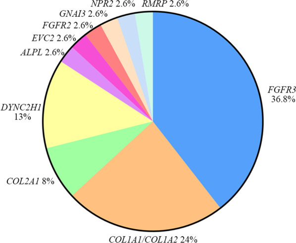
Results: A definitive diagnosis was achieved in 81.82% of affected fetuses (45/55). We identified chromosomal abnormalities in seven cases and 36 variants, of which 18 were novel pathogenic or likely pathogenic in 11 genes in 38 cases. De novo variants were identified in 27 cases (71.05%, 27/38), and one gonosomal mosaicism variant was found in the mother of one fetus. Our case examples demonstrated the high heterogeneity of fetal SDs and the rare fetal SD-associated challenges.

Conclusions: Careful clinical evaluation of fetuses with SD can guide appropriate molecular testing. Our study extends the SD-associated pathogenic variant spectrum and provides useful genetic counselling guidance and an accurate prenatal diagnosis strategy.

Keywords: Breakpoints; Genotype–phenotype analysis; Molecular diagnosis; Novel variants; Skeletal dysplasia.


Conflict of interest statement

The authors have declared no conflicting interests.

Figures

Fig. 1
Fig. 1
Flowchart showing the methods and results of 55 cases enrolled in our study. QF-PCR: quantitative fluorescent PCR; CNV-seq: copy number variation sequencing; Panel: a special NGS gene panel analysis (including 811 congenital skeletal anomalies genes); WES: whole exome sequencing
Fig. 2
Fig. 2
Genes in the present study. Eleven genes were diagnosed in 38 fetuses, including FGFR3(15/38), COL1A1/COL1A2(9/38), COL2A1(3/38), DYNC2H1(5/38), ALPL (1/38), EVC2(1/38), FGFR2(1/38), GNAI3(1/38), NPR2(1/38) and RMRP (1/38)
Fig. 3
Fig. 3
COL2A1 variant caused severe type 2 collagenopathy in case 42 and mother (II2). A The image of US examination of case 42 revealed short limbs. B, C Clinical photograph of the mother with her pelvis inclined on the left side, shortened left lower limb (before extension osteotomy), and shortened fourth toe in the left foot (arrow). D The pedigree map of case 42. E, F Sanger sequencing of case 42 and the mother (II2). G The variant allele fraction and depth of coverage of the COL2A1 pathogenic variant detected by WES in case 42 and the mother. H The variant (COL2A1 c.2303G > T) was highly conserved across different species
Fig. 4
Fig. 4
EVC2 variants caused EVC in case 49. A The image of US examination of case 49 revealed short and curved long bones. B The pedigree map of case 49. C MLPA results of EVC2 in case 49: a heterozygous deletion of EVC2-22 ~ EVC-8. DF Sanger sequencing showed that the proband had a novel variant inherited from the mother. G Schematic presentation of the gross deletion. Arrows indicate genes; Solid horizontal lines indicate retained regions; Broken horizontal lines indicate deleted regions; Multiple primers were designed to be regularly spaced every ~ 25 kb (step 1), ~ 5 kb (step 2), and ~ 1.5 kb (step 3) within breakpoint regions. Long-range PCR with the forward primer of the D131 and the reverse primer of the U4 generates a PCR product of ~ 1.5 kb. H The breakpoint sequences of case 49 and Sanger-sequence of the gross deletion. I UCSC Genome Browser information around the telomeric breakpoint regions (chr4: 5,547,999–5,548,399)
Fig. 5
Fig. 5
GNAI3 variant caused auriculocondylar syndrome (ACS) in case 50. A The pedigree map of case 50. BD Sanger sequencing showed that the proband had a de novo variant. E, F The 3D molecular structure of Gnαi3. The magnified views of the wild-type Gly40 (E) and mutant Val40 (F) are shown respectively. The H-bonds are shown as green dashed lines, and H-bond distances (Å) are shown in red numbers G Gnαi3 domains are depicted in blue: boxes G1–G5. The variant reported here is in red, and previously described variants are in black. H Protein alignment showing conservation of residues GNAI3 p.Gly40Val across multiple species. This mutation occurred at evolutionarily conserved amino acid positions

Similar articles

Cited by

References

    1. Yang K, Shen M, Yan Y, et al. Genetic analysis in fetal skeletal dysplasias by trio whole-exome sequencing. Biomed Res Int. 2019;2019:2492590. - PMC - PubMed
    1. Mortier GR, Cohn DH, Cormier-Daire V, et al. Nosology and classification of genetic skeletal disorders: 2019 revision. Am J Med Genet A. 2019;179(12):2393–2419. doi: 10.1002/ajmg.a.61366. - DOI - PubMed
    1. Lord J, McMullan DJ, Eberhardt RY, et al. Prenatal exome sequencing analysis in fetal structural anomalies detected by ultrasonography (PAGE): a cohort study. Lancet. 2019;393(10173):747–757. doi: 10.1016/S0140-6736(18)31940-8. - DOI - PMC - PubMed
    1. Sun Y, Kong X, Zhao Z, et al. Mutation analysis of 419 family and prenatal diagnosis of 339 cases of spinal muscular atrophy in China. BMC Med Genet. 2020;21(1):133. doi: 10.1186/s12881-020-01069-z. - DOI - PMC - PubMed
    1. Li Q, Zhu X, Wang C, et al. Identification of a rare case with nagashima-type palmoplantar keratoderma and 18q deletion syndrome via exome sequencing and low-coverage whole-genome sequencing. Front Genet. 2021;12:707411. doi: 10.3389/fgene.2021.707411. - DOI - PMC - PubMed

Publication types

LinkOut - more resources