. 2022 Nov 9;26(1):62.
doi: 10.1186/s40824-022-00304-3.

Green tea catechin-grafted silk fibroin hydrogels with reactive oxygen species scavenging activity for wound healing applications

Affiliations

Green tea catechin-grafted silk fibroin hydrogels with reactive oxygen species scavenging activity for wound healing applications

Gyeongwoo Lee et al. Biomater Res. .

Erratum in

Abstract

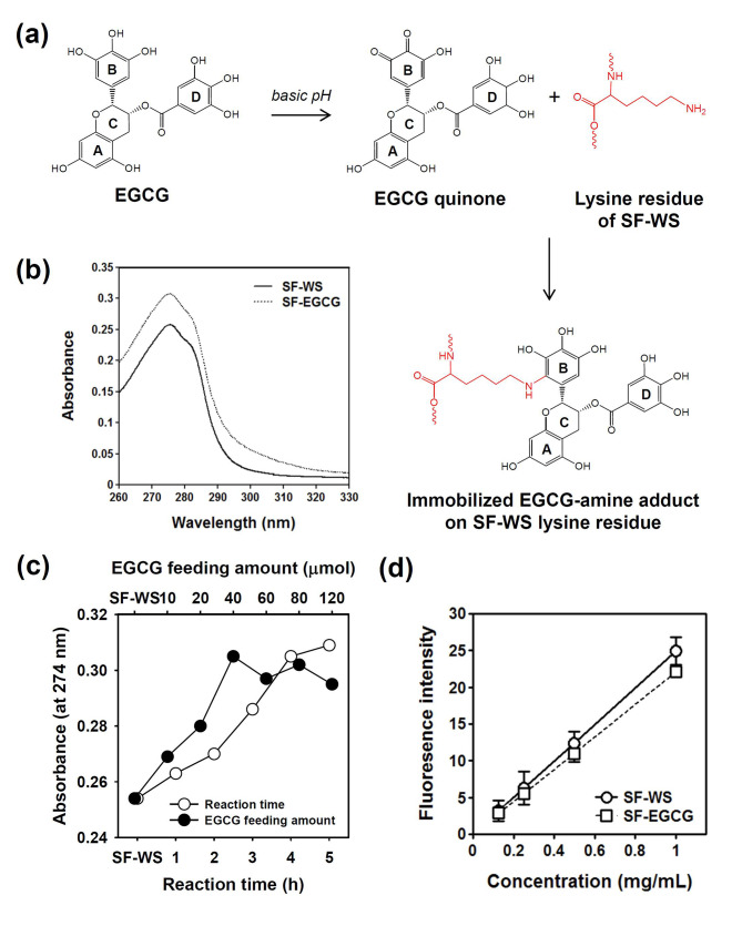
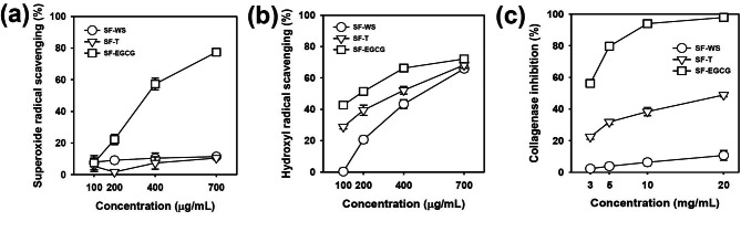
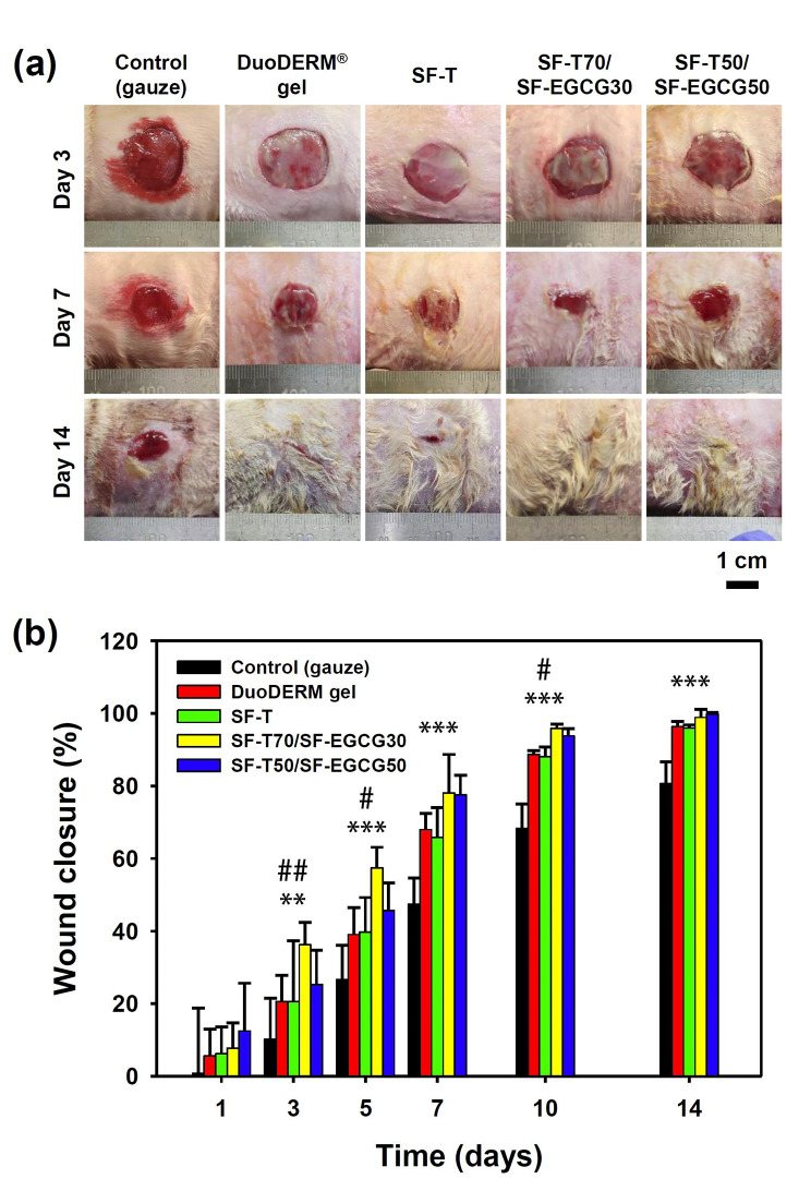
Background: Overproduction of reactive oxygen species (ROS) is known to delay wound healing by causing oxidative tissue damage and inflammation. The green tea catechin, (-)-Epigallocatechin-3-O-gallate (EGCG), has drawn a great deal of interest due to its strong ROS scavenging and anti-inflammatory activities. In this study, we developed EGCG-grafted silk fibroin hydrogels as a potential wound dressing material.

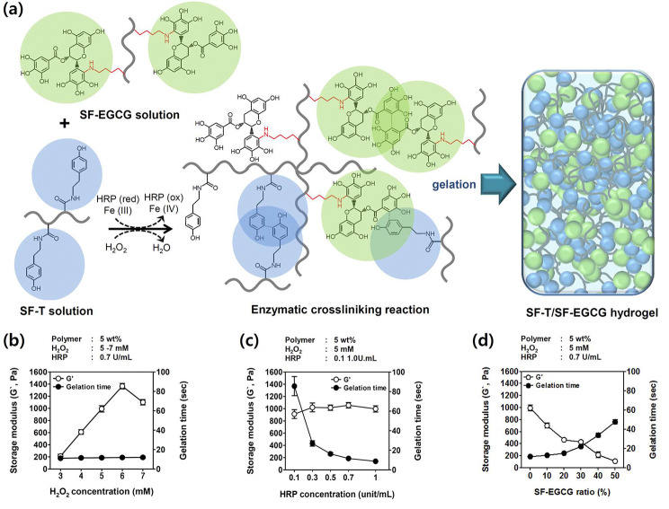
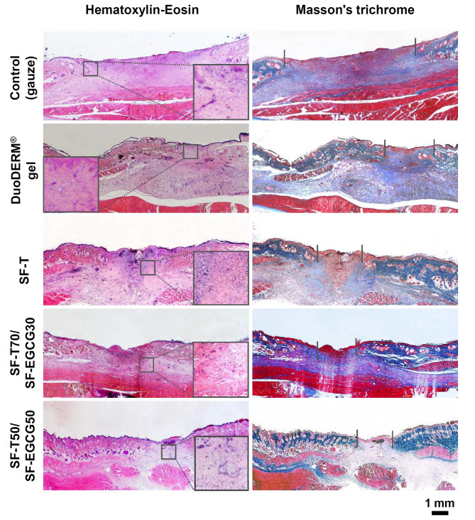
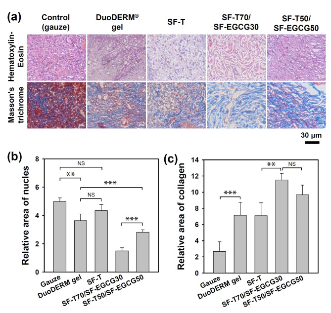
Methods: The introduction of EGCG to water-soluble silk fibroin (SF-WS) was accomplished by the nucleophilic addition reaction between lysine residues in silk proteins and EGCG quinone at mild basic pH. The resulting SF-EGCG conjugate was co-crosslinked with tyramine-substituted SF (SF-T) via horseradish peroxidase (HRP)/H2O2 mediated enzymatic reaction to form SF-T/SF-EGCG hydrogels with series of composition ratios.

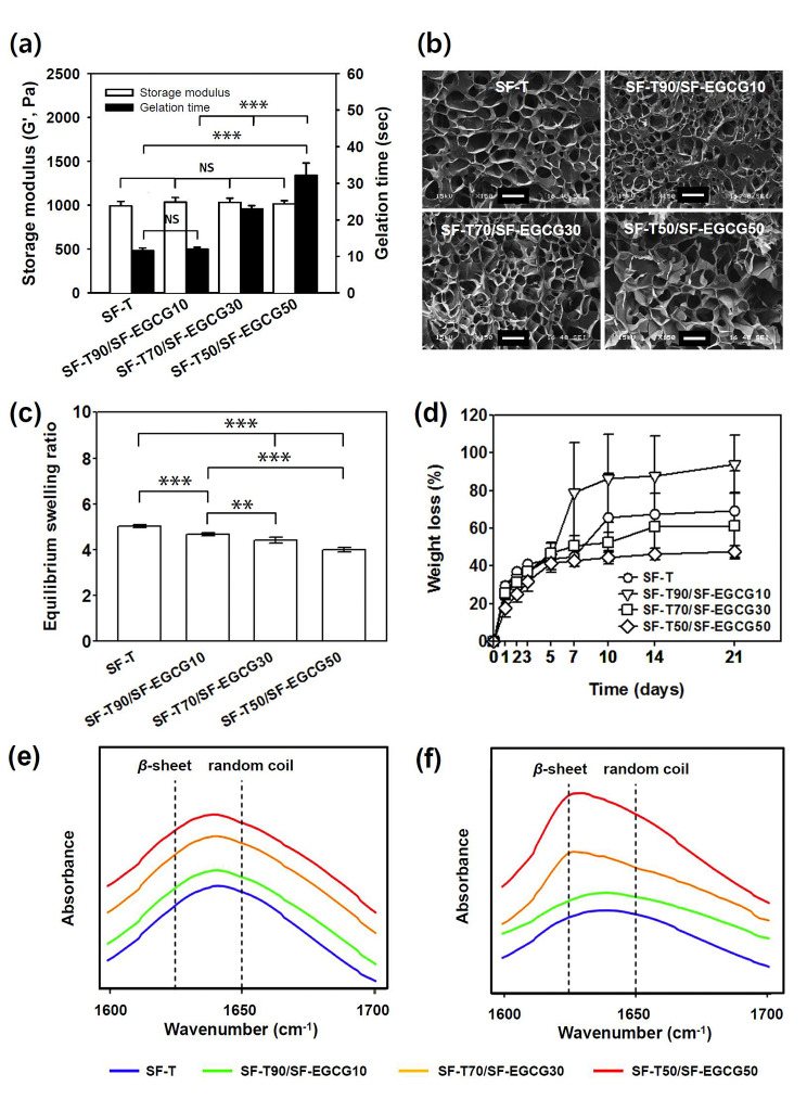
Results: Interestingly, SF-T70/SF-EGCG30 hydrogels exhibited rapid in situ gelation (< 30 s), similar storage modulus to human skin (≈ 1000 Pa) and superior wound healing performance over SF-T hydrogels and a commercial DuoDERM® gel dressings in a rat model of full thickness skin defect.

Conclusion: This study will provide useful insights into a rational design of ROS scavenging biomaterials for wound healing applications.

Keywords: EGCG; Hydrogel; Reactive oxygen species; Silk fibroin; Wound healing.


Conflict of interest statement

The authors declare no competing financial interests.

Figures

Fig. 1
Fig. 1
(a) Scheme of the SF-EGCG conjugate formation through autoxidation of EGCG at basic pH 7.4 and subsequent conjugation reaction of EGCG quinone with a lysine residue of SF-WS. (b) UV-visible spectra of SF-WS and SF-EGCG solutions at an equal concentration (50 µg/mL). (c) Optimization of EGCG feeding amount and reaction time. Absorbance at 274 nm of SF-EGCG conjugates formed at various reaction time (feeding amount of EGCG = 40 µmol) and various feeding amounts of EGCG (reaction time = 4 h). (d) Fluorescence intensity (λex = 390 nm, λem = 515 nm) of SF-WS and SF-EGCG solutions on primary amine content after 1 min of reaction with fluorescamine assay reagent (n = 8)
Fig. 2
Fig. 2
(a) Superoxide anion radical (O2•¯) and (b) hydroxyl radical (•OH) scavenging activity (n = 8) and (c) collagenase inhibitory activity (n = 6) of SF-WS, SF-T and SF-EGCG as a function of concentration
Fig. 3
Fig. 3
(a) Schematic for the chemical formation of SF-T/SF-EGCG composite hydrogels via HRP/H2O2-mediated enzymatic crosslinking reaction. (b) Storage modulus and gelation time of SF-T hydrogels as a function of H2O2 concentration. The concentration of HRP was fixed to 0.7 unit/mL. (c) Storage modulus and gelation time of SF-T hydrogels as a function of HRP concentration. The concentration of H2O2 was fixed to 5 mM. (d) Effect of the addition of SF-EGCG on the storage modulus and gelation time of SF-T/SF-EGCG composite hydrogels. The concentration of H2O2 and HRP was fixed to 5 mM and 0.7 unit/mL, respectively. (Mean ± SD, n = 5)
Fig. 4
Fig. 4
(a) Storage modulus and gelation time of the optimized SF-T hydrogel and SF-T/SF-EGCG composite hydrogels. (Mean ± SD, n = 5, NS: not significant, ***: p < 0.001). (b) Photomicrographs of cross-section of the lyophilized SF-T and SF-T/SF-EGCG composite hydrogels. Scale bar = 100 μm. (c) Equilibrium swelling ratio of the SF-T and SF-T/SF-EGCG composite hydrogels incubated in PBS for 24 h and (d) their weight loss profile in PBS over 21 days. (Mean ± SD, n = 7, **: p < 0.01, ***: p < 0.001). ATR-FTIR spectra of the hydrogels on (e) day 1 and (f) day 9. The dashed lines indicate the amide I band of fibroin β-sheet (1624 cm− 1) and the amide I band of fibroin random coil (1650 cm− 1)
Fig. 5
Fig. 5
(a) Representative photographs of the wound area and (b) wound closure percentage treated with a gauze (control), DuoDERM® gel, SF-T and SF-T/SF-EGCG hydrogels for 14 days after surgery. (Mean ± SD, n = 8, **: p < 0.01 and ***: p < 0.001 for SF-T70/SF-EGCG30 versus the control, #: p < 0.05 and ##: p < 0.01 for SF-T70/SF-EGCG30 versus SF-T).
Fig. 6
Fig. 6
Histological photomicrographs of the wound area harvested after 14 days of treatments with a gauze (control), DuoDERM® gel, SF-T and SF-T/SF-EGCG hydrogels. The left and right panels show hematoxylin-eosin (H&E) staining (pink; cytoplasm, blue purple; nuclei) and Masson’s trichrome staining (pink; cytoplasm, blue; collagen and connective tissues, dark red; keratin and muscle fibers, blue purple; nuclei), respectively. The insets represent inflammatory cells (macrophages, lymphocytes, neutrophils, eosinophils) at the interface between matured and healed wound area
Fig. 7
Fig. 7
(a) High resolution histological microphotographs of regenerated wound area after 14 days of treatments with a gauze (control), DuoDERM® gel, SF-T and, SF-T/SF-EGCG hydrogels. Quantitative image analysis of (b) inflammatory cells and (c) collagen deposition area in the regenerated wound area of each group (Mean ± SD, n = 5, NS: not significant, **: p < 0.01, ***: P < 0.001)

Similar articles

Cited by

References

    1. Darr D, Fridovich I. Free radicals in cutaneous biology. J Invest Dermatol. 1994;102:671–5. doi: 10.1111/1523-1747.ep12374036. - DOI - PubMed
    1. Clark RAF. Oxidative stress and “senescent” fibroblasts in non-healing wounds as potential therapeutic targets. J Invest Dermatol. 2008;128:2361–4. doi: 10.1038/jid.2008.257. - DOI - PubMed
    1. Wlaschek M, Scharffetter-Kochanek K. Oxidative stress in chronic venous leg ulcers. Wound Repair Regen. 2005;13:452–61. doi: 10.1111/j.1067-1927.2005.00065.x. - DOI - PubMed
    1. O’toole EA, Goel M, Woodley DT. Hydrogen peroxide inhibits human keratinocyte migration. Dermatol Surg. 1996;22:525–9. doi: 10.1111/j.1524-4725.1996.tb00368.x. - DOI - PubMed
    1. Weckroth M, Vaheri A, Lauharanta J, Sorsa T, Konttinen YT. Matrix metalloproteinases, gelatinase and collagenase, in chronic leg ulcers. J Invest Dermatol. 1996;106:1119–24. doi: 10.1111/1523-1747.EP12340167. - DOI - PubMed