. 2023 Mar;89(3):937-950.
doi: 10.1002/mrm.29479. Epub 2022 Nov 9.

Simultaneous Optimization of MP2RAGE T1 -weighted (UNI) and FLuid And White matter Suppression (FLAWS) brain images at 7T using Extended Phase Graph (EPG) Simulations

Affiliations

Simultaneous Optimization of MP2RAGE T1 -weighted (UNI) and FLuid And White matter Suppression (FLAWS) brain images at 7T using Extended Phase Graph (EPG) Simulations

Ayşe Sıla Dokumacı et al. Magn Reson Med. 2023 Mar.

Abstract

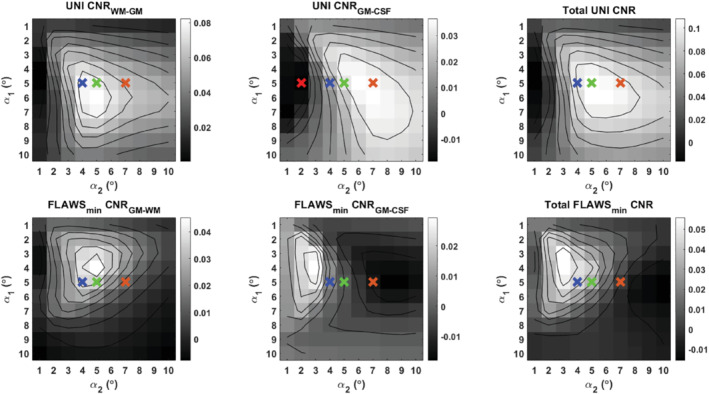
Purpose: The MP2RAGE sequence is typically optimized for either T1 -weighted uniform image (UNI) or gray matter-dominant fluid and white matter suppression (FLAWS) contrast images. Here, the purpose was to optimize an MP2RAGE protocol at 7 Tesla to provide UNI and FLAWS images simultaneously in a clinically applicable acquisition time at <0.7 mm isotropic resolution.

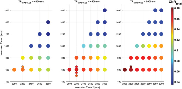
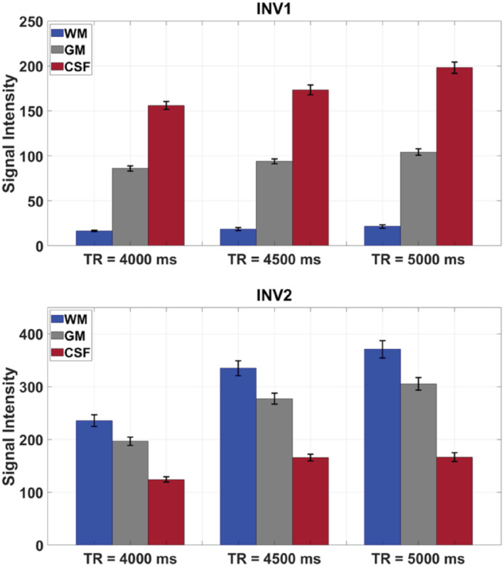
Methods: Using the extended phase graph formalism, the signal evolution of the MP2RAGE sequence was simulated incorporating T2 relaxation, diffusion, RF spoiling, and B1 + variability. Flip angles and TI were optimized at different TRs (TRMP2RAGE ) to produce an optimal contrast-to-noise ratio for UNI and FLAWS images. Simulation results were validated by comparison to MP2RAGE brain scans of 5 healthy subjects, and a final protocol at TRMP2RAGE = 4000 ms was applied in 19 subjects aged 8-62 years with and without epilepsy.

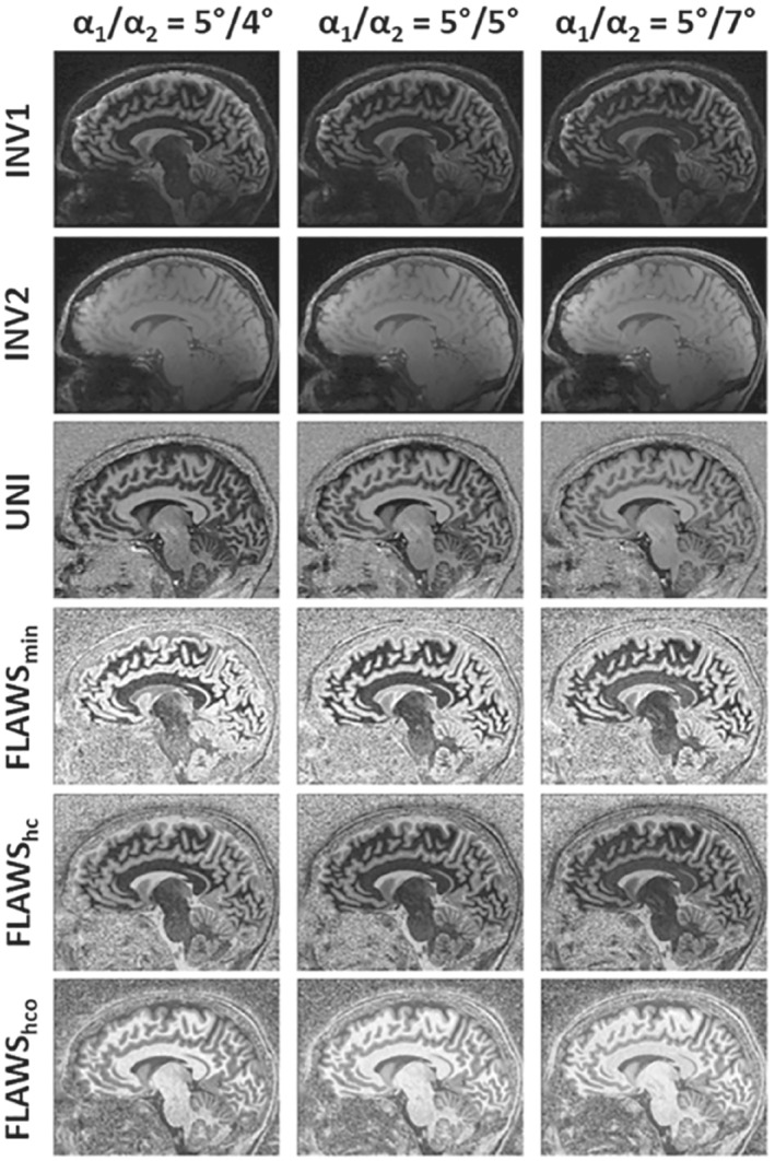
Results: FLAWS contrast images could be obtained while maintaining >85% of the optimal UNI contrast-to-noise ratio. Using TI1 /TI2 /TRMP2RAGE of 650/2280/4000 ms, 6/8 partial Fourier in the inner phase-encoding direction, and GRAPPA factor = 4 in the other, images with 0.65 mm isotropic resolution were produced in <7.5 min. The contrast-to-noise ratio was around 20% smaller at TRMP2RAGE = 4000 ms compared to that at TRMP2RAGE = 5000 ms; however, the 20% shorter duration makes TRMP2RAGE = 4000 ms a good candidate for clinical applications example, pediatrics.

Conclusion: FLAWS and UNI images could be obtained in a single scan with 0.65 mm isotropic resolution, providing a set of high-contrast images and full brain coverage in a clinically applicable scan time. Images with excellent anatomical detail were demonstrated over a wide age range using the optimized parameter set.

Keywords: 7T; FLAWS; MP2RAGE; MRI; ultrahigh field.


Conflict of interest statement

Raphael Tomi‐Tricot is an employee at Siemens Healthineers, and Ronald Mooiweer is seconded to Siemens Healthineers.

Figures

FIGURE 1
FIGURE 1
CNRtotal (taking into account the CNRs of UNI and FLAWSmin) obtained from EPG simulations using different TI1 and TI2 values for 3 choices of TRMP2RAGE. Each CNRtotal value represented by a colored disk is the maximum found over the range of FAs tested, where for each FA the CNR is the average using all B1 + scaling values.  CNR, contrast‐to‐noise ratio; EPG, extended phase graph; FA, flip angle; FLAWS, fluid and white matter suppression; FLAWSm i n, FLAWS minimum image; UNI, uniform image.
FIGURE 2
FIGURE 2
The INV1 and INV2 mean signal intensities at different TRMP2RAGEs for different tissues with the uncertainty of the mean, indicated as the error bars using the 3D segmented data from 4 healthy adults
FIGURE 3
FIGURE 3
The UNI and FLAWSmin CNR plots using different FA combinations at a fixed TI1/TI2 of 650/2220 ms and TRMP2RAGE of 4000 ms. The plots represent the average of simulation results using 10 different B1 + values (50% B1 + to 140% B1 + with steps of 10%). The plots considering individual B1 + values are given in Supporting Information Video S1. The upper row shows the CNRs for the UNI image between WM–GM and GM–CSF, and the total CNR. The lower row shows the CNRs for the FLAWSmin image between the GM–WM and GM–CSF, and the total CNR. The crosses correspond to different FA combinations (red: 5°/2°, blue: 5°/4°, green: 5°/5°, orange: 5°/7°). CNR, contrast‐to‐noise ratio; CS, compressed sensing; GM, gray matter; WM, white matter
FIGURE 4
FIGURE 4
Various contrasts obtained from subject 5 at TRMP2RAGE = 4000 ms using different α 2 values
FIGURE 5
FIGURE 5
The UNI and FLAWS‐related images and the bias fields calculated from the UNI images using (A) the final protocol (B) the MP2RAGE protocol with low sensitivity to B1 + by Marques et al. (C) the protocol optimized for FLAWS contrast by Beaumont et al. The details of the protocols are given in Table 2. It is important to note that the final protocol has a shorter TRMP2RAGE and scan duration and a higher nominal resolution compared to those of the other 2 protocols
FIGURE 6
FIGURE 6
Three areas (each 40 × 40 mm2) having different B1 + values selected from the UNI images that were acquired using (A) the final protocol (B) the MP2RAGE protocol with low sensitivity to B1 + by Marques et al. (C) The protocol optimized for FLAWS contrast by Beaumont et al. (Table 2) to demonstrate the achieved contrasts
FIGURE 7
FIGURE 7
Coronal images with different contrasts acquired at TRMP2RAGE = 4000 ms from (A) 10‐year‐old healthy child, (B) 62‐year‐old healthy adult, and (C) 12‐year‐old pediatric epilepsy patient with focal cortical dysplasia. The lesion is visible in the left hemisphere (e.g., as a hypo‐intense region indicated by the red arrow in the WM of the UNI image). The nominal image resolution was 0.65 mm isotropic. The scan duration (min:s) was 7:18 for pediatric subjects and 8:58 for the adult subject.

Similar articles

Cited by

  • Extent of piriform cortex resection in children with temporal lobe epilepsy.
    Piper RJ, Dasgupta D, Eriksson MH, Ripart M, Moosa A, Chari A, Seunarine KK, Clark CA, Duncan JS, Carmichael DW, Tisdall MM, Baldeweg T. Piper RJ, et al. Ann Clin Transl Neurol. 2023 Sep;10(9):1613-1622. doi: 10.1002/acn3.51852. Epub 2023 Jul 20. Ann Clin Transl Neurol. 2023. PMID: 37475156 Free PMC article.
  • A closer look: pediatric neuroimaging at 7T.
    Barkovich EJ, Cortes-Albornoz MC, Machado-Rivas F, Ferraciolli SF, Afacan O, Jaimes C. Barkovich EJ, et al. Pediatr Radiol. 2025 May;55(6):1071-1087. doi: 10.1007/s00247-025-06231-4. Epub 2025 Apr 21. Pediatr Radiol. 2025. PMID: 40257498 Review.

References

    1. Marques JP, Kober T, Krueger G, van der Zwaag W, Van de Moortele PF, Gruetter R. MP2RAGE, a self bias‐field corrected sequence for improved segmentation and T1‐mapping at high field. Neuroimage. 2010;49:1271‐1281. - PubMed
    1. Mugler JP, Brookeman JR. Three‐dimensional magnetization‐prepared rapid gradient‐echo imaging (3D MPRAGE). Magn Reson Med. 1990;15:152‐157. - PubMed
    1. Van de Moortele PF, Auerbach EJ, Olman C, Yacoub E, Ugurbil K, Moeller S. T1 weighted brain images at 7 Tesla unbiased for Proton Density, T2* contrast and RF coil receive B1 sensitivity with simultaneous vessel visualization. Neuroimage. 2009;46:432‐446. - PMC - PubMed
    1. Tanner M, Gambarota G, Kober T, et al. Fluid and white matter suppression with the MP2RAGE sequence. J Magn Reson Imaging. 2012;35:1063‐1070. - PubMed
    1. Sudhyadhom A, Haq IU, Foote KD, Okun MS, Bova FJ. A high resolution and high contrast MRI for differentiation of subcortical structures for DBS targeting: the fast gray matter acquisition T1 inversion recovery (FGATIR). Neuroimage. 2009;47:T44‐T52. - PubMed

Publication types