

Morphological study of the menisci of the knee joint in human cadaver in Jharkhand population
- PMID: 36353007
- PMCID: PMC9638620
- DOI: 10.4103/jfmpc.jfmpc_2416_21
Morphological study of the menisci of the knee joint in human cadaver in Jharkhand population
Abstract
Introduction: Sports are the leading cause of joint injuries, particularly in the knees. Knee menisci are an important functional unit that aids in load distribution and hence reduces stress on the knee joint. Meniscal morphology provides information on exact size and shape, which is important for meniscal transplantation in cases of meniscal damage. The study's goal is to determine the morphological variation in the shape of menisci, as well as the width and thickness of menisci.
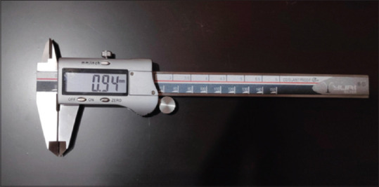
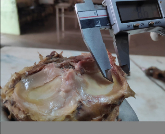
Method: This study was conducted at the Rajendra Institute of Medical Sciences (RIMS) Ranchi, Department of Anatomy. In this study, 100 menisci were taken from 50 adult cadaver knee joints available in the dissection hall.
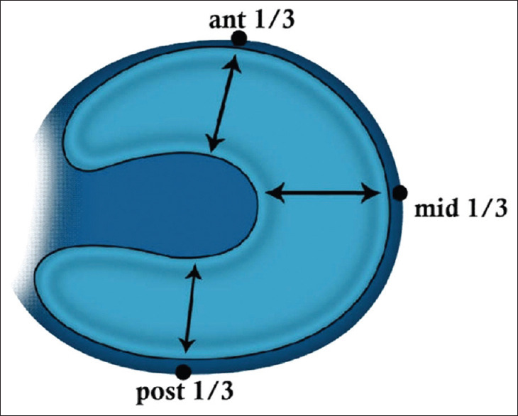
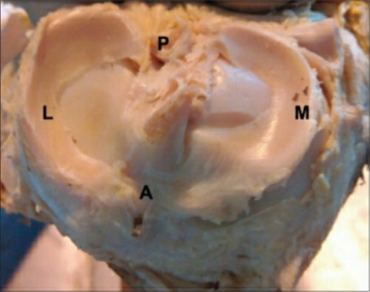
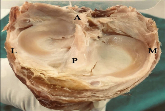
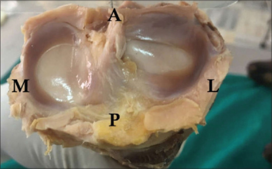
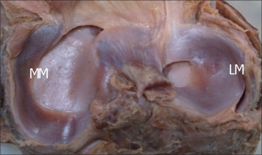
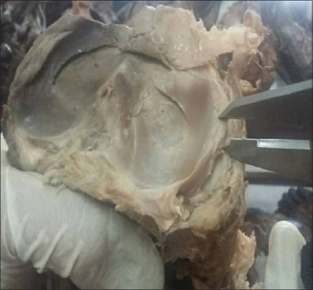
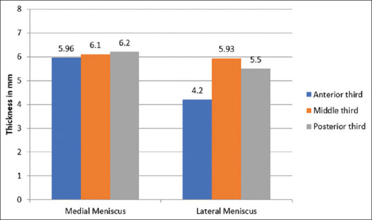
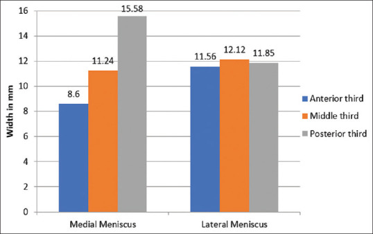
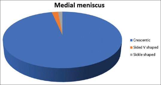
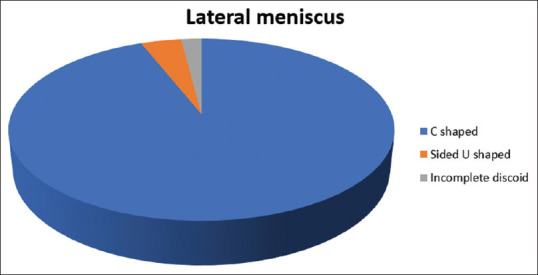
Result: Six morphological kinds of menisci were identified after a morphological and morphometric study of 100 menisci. The most common crescent-shaped menisci (96%) were found in 50 medial menisci (MM), while the most common C-shaped menisci were found in 50 lateral menisci (LM, 94%). There was no statistically significant difference in thickness between the anterior, middle, and posterior thirds of the MM in the morphometric analysis. The thickest section of the lateral meniscus (LM) was in the middle third. There was no significant variation in the width of the LM among the different thirds in the current study. The posterior portion of the medial meniscus (MM), on the other hand, was the widest.
Conclusion: The findings of this study support meniscal anatomy in terms of surgical technique and arthroscopy of the knee joint, as well as contributing to a better understanding of meniscal architecture and meniscal transplantation. As a result, health workers who treat meniscal injuries should be aware of the probable anatomical differences.
Keywords: Knee joint; meniscal anatomy; meniscal injuries.
Copyright: © 2022 Journal of Family Medicine and Primary Care.
Conflict of interest statement
There are no conflicts of interest.
Figures

Similar articles
-
A Cadaveric Study to Define Morphology and Morphometry of Human Knee Menisci in the Region of Central India.Cureus. 2023 Jun 30;15(6):e41174. doi: 10.7759/cureus.41174. eCollection 2023 Jun. Cureus. 2023. PMID: 37525816 Free PMC article.
-
A Study on the Morphometry of a Medial Meniscus in the Knee Joint of Human Cadavers in the South Indian Population.Cureus. 2023 Jul 31;15(7):e42753. doi: 10.7759/cureus.42753. eCollection 2023 Jul. Cureus. 2023. PMID: 37654914 Free PMC article.
-
In Vivo Three-dimensional Morphological Assessment of Adult Knee Menisci: A Computed Tomography-based Approach.In Vivo. 2022 May-Jun;36(3):1416-1423. doi: 10.21873/invivo.12846. In Vivo. 2022. PMID: 35478132 Free PMC article.
-
An anatomical study of the meniscal roots of the knee: landmarks for its surgical reconstruction and implications for knee surgeons.Surg Radiol Anat. 2022 Jul;44(7):971-977. doi: 10.1007/s00276-022-02979-8. Epub 2022 Jul 2. Surg Radiol Anat. 2022. PMID: 35780197
-
Understanding posterior meniscal roots lesions: from basic science to treatment.Rev Bras Ortop. 2017 Jul 26;52(4):463-472. doi: 10.1016/j.rboe.2017.07.005. eCollection 2017 Jun-Jul. Rev Bras Ortop. 2017. PMID: 28884106 Free PMC article. Review.
Cited by
-
A Cadaveric Study to Define Morphology and Morphometry of Human Knee Menisci in the Region of Central India.Cureus. 2023 Jun 30;15(6):e41174. doi: 10.7759/cureus.41174. eCollection 2023 Jun. Cureus. 2023. PMID: 37525816 Free PMC article.
-
Age-Dependent Meniscal and Chondral Damage in Eastern European Women Undergoing First-Time Knee Arthroscopy.Healthcare (Basel). 2025 Jul 26;13(15):1822. doi: 10.3390/healthcare13151822. Healthcare (Basel). 2025. PMID: 40805855 Free PMC article.
-
A Study on the Morphometry of a Medial Meniscus in the Knee Joint of Human Cadavers in the South Indian Population.Cureus. 2023 Jul 31;15(7):e42753. doi: 10.7759/cureus.42753. eCollection 2023 Jul. Cureus. 2023. PMID: 37654914 Free PMC article.
-
Surgical Management of Traumatic Meniscus Injuries.Pathophysiology. 2023 Dec 4;30(4):618-629. doi: 10.3390/pathophysiology30040044. Pathophysiology. 2023. PMID: 38133145 Free PMC article. Review.
References
-
- Standring S. Sacrum. In: Standring S, editor. Grays Anatomy. 40th ed. London: Elsevier Churchill Livingstone; 2008. pp. 1397–9.
-
- Moore KL, Dalley AF. 4th ed. Philadelphia: Lippincott Williams and Wilkins; 1999. Clinically Oriented Anatomy; pp. 690–9.
-
- Fairbank TJ. Knee Joint changes after meniscectomy. J Bone Joint Surg. 1948;30B:664–70. - PubMed
-
- Smillie IS. 4th ed. London: Living Stone; 1975. Injuries of the Knee Joint.
LinkOut - more resources
Full Text Sources
Research Materials