. 2023 Apr 11;51(6):2501-2515.
doi: 10.1093/nar/gkac996.

Internal RNA 2'O-methylation in the HIV-1 genome counteracts ISG20 nuclease-mediated antiviral effect

Affiliations

Internal RNA 2'O-methylation in the HIV-1 genome counteracts ISG20 nuclease-mediated antiviral effect

Priscila El Kazzi et al. Nucleic Acids Res. .

Abstract

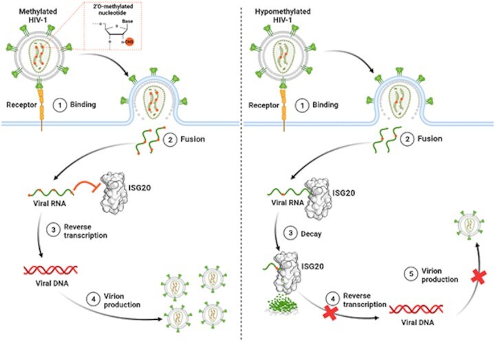
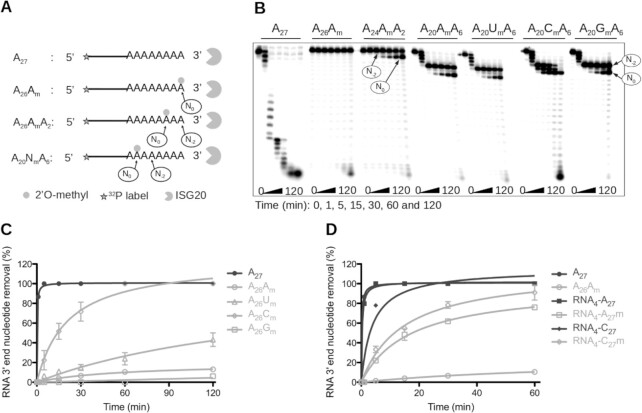
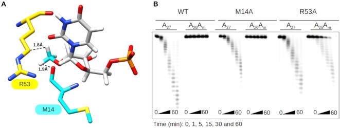
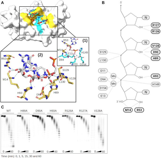
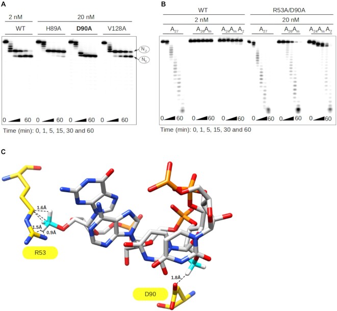
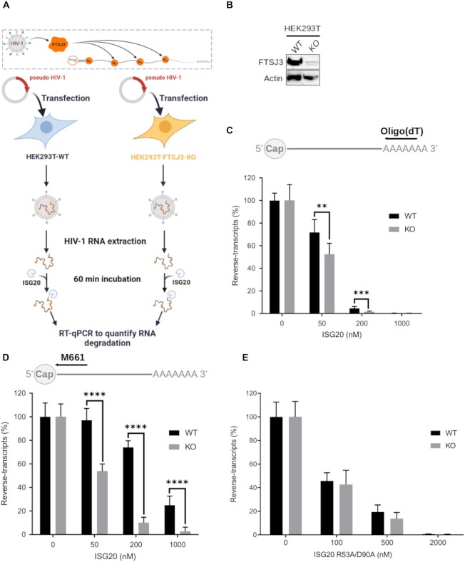
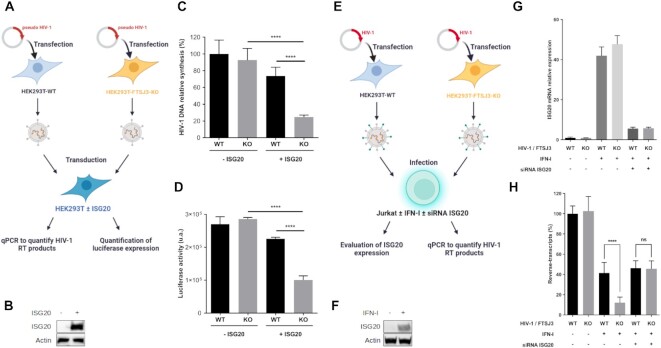
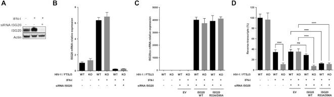
RNA 2'O-methylation is a 'self' epitranscriptomic modification allowing discrimination between host and pathogen. Indeed, human immunodeficiency virus 1 (HIV-1) induces 2'O-methylation of its genome by recruiting the cellular FTSJ3 methyltransferase, thereby impairing detection by RIG-like receptors. Here, we show that RNA 2'O-methylations interfere with the antiviral activity of interferon-stimulated gene 20-kDa protein (ISG20). Biochemical experiments showed that ISG20-mediated degradation of 2'O-methylated RNA pauses two nucleotides upstream of and at the methylated residue. Structure-function analysis indicated that this inhibition is due to steric clash between ISG20 R53 and D90 residues and the 2'O-methylated nucleotide. We confirmed that hypomethylated HIV-1 genomes produced in FTSJ3-KO cells were more prone to in vitro degradation by ISG20 than those produced in cells expressing FTSJ3. Finally, we found that reverse-transcription of hypomethylated HIV-1 was impaired in T cells by interferon-induced ISG20, demonstrating the direct antagonist effect of 2'O-methylation on ISG20-mediated antiviral activity.

Plain language summary

Despite highly effective antiretroviral therapies, the human immunodeficiency virus (HIV-1) remains a major public health threat. Its pathogenesis depends on its ability to establish a persistent infection in cells of the immune system. Our study highlights a new insight into how HIV-1 evades early restriction by the immune system. We showed that 2′O-methylation marks found inside HIV-1 RNA promote viral evasion from the antiviral action of the interferon-stimulated gene 20-kDa protein (ISG20), an innate immune restriction factor with a nuclease activity. By disrupting the level of 2′O-methylation of the HIV-1 genome, we demonstrated that ISG20 impairs the reverse transcription process of hypomethylated viruses, as a result of viral RNA decay.


Figures

Graphical Abstract
Graphical Abstract
Role of HIV-1 RNA 2′O-methylation in limiting ISG20 antiviral action. The presence of methyl on the HIV-1 RNA promotes virus replication by preventing recognition and viral RNA decay by ISG20.
Figure 1.
Figure 1.
Impact of RNA 2′O-methylation on ISG20 nuclease activity. (A–D) Recombinant human ISG20 was incubated with different 5′-radiolabeled RNAs carrying 2′O-methylated residues at various positions in a time-course experiment, and the substrate hydrolysis was followed by PAGE. The degradation kinetics of various substrates in (C and D) were monitored by PAGE separation. Quantification of nucleotide removal, relative to the total RNA length, was done using the FujiImager and Image Gauge analysis software. (A) Schematic representation of the experimental procedure of panel (B). (B) Effect of RNA 2′O-methylation on ISG20 exonuclease activity. The assay was performed using non-methylated A27, and 3′ end 2′O-methylated A26Am, A24AmA2, A20AmA6, A20UmA6, A20CmA6, and A20GmA6. (C) Quantification of non-methylated (A27) and 3′ end 2′O-methylated RNA (A26Am, A26Um, A26Cm, A26Gm) degradation by ISG20. (D) Quantification of non-methylated RNA4-A27 and RNA4-C27 and 3′-end 2′O-methylated RNA4-A27m, RNA4-C27m degradation by ISG20. The results in panel B and C correspond to the mean values and standard deviation of three independent experiments.
Figure 2.
Figure 2.
Molecular basis of ISG20 inhibition by 2′O-methylation at N0. (A) Structural model of ISG20 showing the steric clash between residues R53 (yellow surface) and M14 (cyan surface), and 2′O-methylated UMP (UMP is in grey and 2′O-methyl in cyan). The methylated UMP was built using PubChem Sketcher V2.4 and superimposed on the UMP present in the catalytic pocket of the ISG20 structure (PDB: 1WLJ). (B) The exonuclease activity of wild-type (WT) and M14A and R53A mutant ISG20 on methylated and non-methylated A27 RNAs was monitored by PAGE analysis as in Figure 1.
Figure 3.
Figure 3.
Structural activity relationship of ISG20 RNA binding Domain. (A) Model of ISG20 in interaction with an RNA substrate was built based on superimposition of ISG20 (PDB: 1WLJ) and SDN1 in complex with RNA (PDB: 5Z9X). Surface representation of ISG20 (off-white) containing an RNA from SDN1 structure (sticks). Highlighted in yellow are the residues of the RNA binding domain (RBD) and in cyan the conserved residues of the exonuclease domain. Zoom on the ISG20 catalytic pocket showing the interaction (cyan) between the (1) residues of its catalytic domain (cyan), manganese (purple), and the nucleotide to be excised (grey), (2) residues of its RBD (yellow) and the RNA (grey). (B) Cartoon representing the ISG20 model in interaction with an RNA substrate performed using Biorender in which the residues of ISG20 RBD interacting with the RNA 2′OH are highlighted in bold. (C) Mutagenesis analysis of the residues highlighted in panel (B). The different ISG20 mutants were produced and their exonuclease activity was followed as in Figure 1 using non-methylated A27.
Figure 4.
Figure 4.
The D90 and R53 residues play a key role in ISG20 inhibition by 2′O-methylation. (A) Exonuclease activity of WT ISG20 and the H89A, D90A, and V128A mutants was assessed on A20AmA6, and the RNA degradation products were separated on PAGE and analyzed by autoradiography, as in Figure 1. (B) Exonuclease activity of WT ISG20 and the double mutant R53A/D90A was assessed using the A27, A26Am and A24AmA2 RNA substrates. Degradation was monitored by PAGE and analyzed by autoradiography, as described in Figure 1. (C) Structural model of ISG20 catalytic site (PDB: 1WLJ) with a methylated RNA (the methyl groups were added to the RNA using the chimera build structure option). Residues D90 and R53 are in yellow, the RNA is in grey, the RNA 2′O-methyl moieties in cyan. The model shows the steric hindrance between the distinct methylated nucleotides and the R53 and D90 residues of ISG20.
Figure 5.
Figure 5.
Hypomethylated HIV-1 RNA is sensitive to in vitro ISG20-mediated degradation. (A–D) Parental and FTSJ3-KO HEK293T cells were transfected with HIV-1 molecular clone pNL4-3.Luc.R-E in order to produce HIV-1 virions containing naturally methylated (WT) and hypomethylated (KO) genome, respectively. Upon recovery of viral particles, full-length vRNAs were extracted and incubated with recombinant human ISG20 (WT or double mutant R53A/D90A) for 60 min. Finally, vRNAs degradation by ISG20 was monitored by RT-qPCR. (A) Schematic representation of the experimental procedure. (B) FTSJ3 expression in parental (WT) and FTSJ3-KO (KO) HEK293T cells was assessed by western blotting before transfection of the HIV-1 molecular clone pNL4.3. (C, D) 50 ng of RNA extracted from HIV-1 particles produced in parental (WT) or FTSJ3-KO HEK293T cells was incubated with ISG20 at the indicated concentrations at 37°C for 1h. After ISG20 inactivation (20 min at 70°C), RNA products were reverse-transcribed using a (C) oligo(dT) or (D) the M661 primer, and the reverse-transcripts quantified by qPCR. The relative amount of HIV-1 reverse-transcripts in each condition is shown. (E) The exonuclease activity of ISG20 R53A/D90A was evaluated using HIV-1 RNA extracted from viral particles produced in parental (WT) or FTSJ3-KO HEK293T cells. RNA degradation products were reverse-transcribed using the M661 primer, and the reverse-transcripts quantified by qPCR as in (D). Data are the mean ± standard deviation of three independent experiments performed in triplicate. **P < 0.01, ***P < 0.001 and **** P < 0.0001 (Student's t-test).
Figure 6.
Figure 6.
Restriction of the hypomethylated HIV-1 replication by ISG20. (A–D) ISG20 restricts the reverse-transcription of hypomethylated VSV-G pseudotyped HIV-1. WT and FTSJ3-KO HEK293T cells were used to produce VSV-G-pseudotyped HIV-1 particles containing WT and hypomethylated (KO) genome, respectively. Extracted virions were used to infect 48 h WT HEK293T cells over-expressing or not Flag-tagged human ISG20. At 6 h post-infection, total cell DNA was extracted and HIV-1 reverse-transcripts were quantified by qPCR and HIV-1 infection was estimated by measuring luciferase activity in cell extracts 24h post-infection. (A) Schematic representation of the experimental procedure. (B) HEK293T cells were transfected with an empty plasmid or with a plasmid encoding Flag-tagged human ISG20. The expression of ISG20 was analyzed by western blotting using an anti-Flag antibody. (C) Quantification of HIV-1 reverse-transcripts by qPCR. (D) HIV-1 infection was estimated by measuring luciferase activity in cell extracts 24 h post-infection. (C, D) Data are the mean ± standard deviation of three independent experiments performed in triplicate; **P < 0.01, ***P < 0.001 and **** P < 0.0001 (Student's t-test). (E–H) Jurkat cells were treated or not with 1000 IU/ml human IFN-I for 18 h and transfected with a siRNA control (−) or targeting ISG20 (+), as indicated. At 48 h post-transfection, cells were infected with HIV-1 NL4-3 produced in WT or in FTSJ3-KO HEK293T cells. At 6 h post-infection, cells were harvested and total RNA and DNA were extracted for specific qPCR analysis. (E) Schematic representation of the experimental procedure. (F) Jurkat cells were treated or not with 1000 IU/ml human IFN-I for 18h and ISG20 expression was assessed by western-blot. (G) Evaluation of endogenous ISG20 expression by RT-qPCR. (H) Quantification of HIV-1 RT products by qPCR. (G, H) Data represents the mean of three independent experiments performed in duplicates ± SD. ****P < 0.0001, as determined by one-way ANOVA with Bonferroni post hoc test. ns, non-significant.
Figure 7.
Figure 7.
Restriction profile of ISG20 WT and the R53A/D90A mutant on replication competent HIV-1. (A) HeLa P4C5 cells were treated or not with 1000 IU/ml human IFN-α2a for 18h and transfected with a siRNA control (−) or targeting ISG20 (+), as indicated. At 48h post-transfection, ISG20 expression was assessed by western-blot using anti-ISG20 antibodies. (B–D) HeLa P4C5 cells were treated or not with 1000 IU/ml human IFN-I for 18 h and transfected with a siRNA control (−) or targeting ISG20 (+), as indicated. At 48h post-transfection, cells were infected with HIV-1 NL4-3 produced in WT or in FTSJ3-KO cells. At 6 h post-infection, cells were harvested and total RNA and DNA were extracted for specific qPCR analysis. (B) The expression of endogenous ISG20 upon IFN-I induction was evaluated by RT-qPCR. (C) Quantification of ectopic expression of transfected ISG20 WT and double mutant by RT-qPCR. (D) The amount of HIV-1 RT products was estimated by qPCR. Data represent the mean of three independent experiments performed in duplicate ± SD. ****P < 0.0001, as determined by one-way ANOVA with Bonferroni post hoc test. ns, non-significant.

Similar articles

Cited by

References

    1. Netzband R., Pager C.T.. Epitranscriptomic marks: emerging modulators of RNA virus gene expression. WIREs RNA. 2020; 11:e1576. - PMC - PubMed
    1. McIntyre W., Netzband R., Bonenfant G., Biegel J.M., Miller C., Fuchs G., Henderson E., Arra M., Canki M., Fabris D.et al. .. Positive-sense RNA viruses reveal the complexity and dynamics of the cellular and viral epitranscriptomes during infection. Nucleic Acids Res. 2018; 46:5776–5791. - PMC - PubMed
    1. Kennedy E.M., Courtney D.G., Tsai K., Cullen B.R.. Viral epitranscriptomics. J. Virol. 2017; 91:e02263-16. - PMC - PubMed
    1. Decroly E., Ferron F., Lescar J., Canard B.. Conventional and unconventional mechanisms for capping viral mRNA. Nat. Rev. Microbiol. 2012; 10:51–65. - PMC - PubMed
    1. Decroly E., Canard B.. Biochemical principles and inhibitors to interfere with viral capping pathways. Curr. Opin. Virol. 2017; 24:87–96. - PMC - PubMed

Publication types