. 2022 Dec 20;13(6):e0276022.
doi: 10.1128/mbio.02760-22. Epub 2022 Nov 10.

CotG Mediates Spore Surface Permeability in Bacillus subtilis

Affiliations

CotG Mediates Spore Surface Permeability in Bacillus subtilis

Anella Saggese et al. mBio. .

Abstract

Proteins and glycoproteins that form the surface layers of the Bacillus spore assemble into semipermeable arrays that surround and protect the spore cytoplasm. Such layers, acting like molecular sieves, exclude large molecules but allow small nutrients (germinants) to penetrate. We report that CotG, a modular and abundant component of the Bacillus subtilis spore coat, controls spore permeability through its central region, formed by positively charged tandem repeats. These repeats act as spacers between the N and C termini of the protein, which are responsible for the interaction of CotG with at least one other coat protein. The deletion but not the replacement of the central repeats with differently charged repeats affects the spore resistance to lysozyme and the efficiency of germination-probably by reducing the coat permeability to external molecules. The presence of central repeats is a common feature of the CotG-like proteins present in most Bacillus species, and such a wide distribution of this protein family is suggestive of a relevant role for the structure and function of the Bacillus spore. IMPORTANCE Bacterial spores are quiescent cells extremely resistant to a variety of unphysiological conditions, including the presence of lytic enzymes. Such resistance is also due to the limited permeability of the spore surface, which does not allow lytic enzymes to reach the spore interior. This article proposes that the spore permeability in B. subtilis is mediated by CotG, a modular protein formed by a central region of repeats of positively charged amino acid acting as a "spacer" between the N and C termini. These, in turn, interact with other coat proteins, generating a protein layer whose permeability to external molecules is controlled by the distance between the N and C termini of CotG. This working model is most likely expandable to most sporeformers of the Bacillus genus, since they all have CotG-like proteins, not homologous to CotG of B. subtilis but similarly characterized by central repeats.

Keywords: Bacillus subtilis; endospores; germination; permeability; spore coat.


Conflict of interest statement

The authors declare no conflict of interest.

Figures
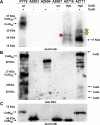
FIG 1
FIG 1
Assembly (A) and phosphorylation (B) of GHyb and (C) its effects on CotB. Western blot analyses of coat proteins extracted from purified spores of the indicated strains were performed on proteins fractionated on 12.5% SDS-PAGE gels. Electrotransferred proteins were then reacted with anti-CotG (A), anti-PKC (B), and anti-CotB (C) antibodies. The type of CotG form and the presence (+) or absence (−) of CotH are indicated. Red and yellow stars represent different GHyb forms.
FIG 2
FIG 2
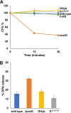
Functional analysis of spores expressing different forms of CotG. (A) Resistance to heat treatment (20 min at 80°C) and (B) DPA release (after 15 min at 100°C) of spores of the wild type (blue symbols) and mutant strains carrying a cotG null mutation (ΔcotG [orange symbols]), a cotG gene with the internal repeats deleted (GΔcentral [gray symbols]), the hybrid cotG gene (GHyb [yellow symbols]), or carrying a cotB null mutation (CotB [green symbols]). One hundred percent is considered the amount of DPA released from spores autoclaved at 120°C for 30 min. Error bars represent standard deviations. Each percentage is the mean of results from three replicate experiments, each performed with a different prepared spore suspension.
FIG 3
FIG 3
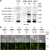
Effect of GHyb on the spore coat assembly of CotC and CotU. (A) Western blot analysis of coat proteins fractionated on 12.5% SDS-PAGE gels, electrotransferred onto a membrane, and incubated with anti-CotC antibody; (B) fluorescence microscopy analysis of spores carrying the fusion protein CotC-GFP. The type of CotG form and the presence (+) or absence (−) of CotH are indicated. Red and white arrows indicate forming and mature spores, respectively. Yellow arrows indicate CotC aggregates in the mother cell cytoplasm. Scale bar, 1 μm.
FIG 4
FIG 4
Immunofluorescence analysis of different strains carrying different forms of CotG with anti-PKC (A) and anti-CotG (B and C) antibodies in the presence (A and B) or absence (C) of CotH. In phase-contrast (PC) panels, the position of the forming spore is indicated by dotted red ovals. The exposure times were 200 ms for panel A and 500 ms for panels B and C. Scale bar, 1 μm.
FIG 5
FIG 5
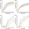
Germination efficiency of spores of the wild type (blue symbols) and mutant strains carrying a cotG null mutation (ΔcotG [orange symbols]), a cotG gene with the internal repeats deleted (GΔcentral [gray symbols]), and the hybrid cotG gene (GHyb [yellow symbols]). The germination was induced using l-Ala-GFK (A and C) or l-Asn-GFK (B and D) and measured as the percentage of loss of the OD600 and by flow cytometry (C and D). Error bars (A and B) are based on the standard deviations of values from four independent experiments. Panels C and D report the percentage of germination obtained from flow cytometry data (Fig. S2).
FIG 6
FIG 6
Lysozyme resistance of spores of the wild type (blue symbols) and mutant strains carrying a cotG null mutation (ΔcotG [orange symbols]), a cotG gene with the internal repeats deleted (GΔcentral [gray symbols]), and the hybrid cotG gene (GHyb [yellow symbols]). Spores were incubated with lysozyme for 6 h at 37°C, and survival was estimated by CFU count. Error bars are based on the standard deviation of values from 4 independent experiments.
FIG 7
FIG 7
(A) Fluorescence microscopy analysis of spores of the wild type and mutant strains carrying a cotG null mutation (ΔcotG), a cotG gene with the internal repeats deleted (GΔcentral), and the hybrid cotG gene (GHyb) after treatment with lysozyme conjugated with rhodamine (Lys-Rd). The exposure time was 100 ms for each panel. Scale bar, 1 μm. PC, phase contrast. (B) Box plots displaying the total corrected cellular fluorescence (TCCF) for 50 different spores of each strain. The limits of each box represent the first and the third quartiles (25 and 75%), and the values outside the boxes represent the maximum and minimum values.
FIG 8
FIG 8
(A) Flow cytometry analysis of 10,000 spores of the wild type and mutant strains carrying a cotG null mutation (ΔcotG), a cotG gene with the internal repeats deleted (GΔcentral), and the hybrid cotG gene (GHyb) after treatment with lysozyme conjugated with rhodamine (Lys-Rd). The black curves represent the nonspecific fluorescence signal of the spores (no Lys-Rd), whereas the red curves represent the fluorescence signal after treatment with Lys-Rd. (B) Quantitative analysis of the fluorescence of over 50 spores by the ImageJ software. The y axis describes the total corrected cellular fluorescence (TCCF) value. The fluorescent intensity threshold is 1 × 103 (red line). a.u., arbitrary units.
FIG 9
FIG 9
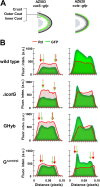
(A) Schematic representation of the localization of GFP in the spore coat of strains carrying GFP fused to inner coat (cotS::gfp), outer coat (cotC::gfp), or crust (cotZ::gfp) proteins; (B) plots of green (GFP) and red (Lys-Rd) fluorescence intensities along the long axis of spores of the strains indicated in panel A. Green and red arrows indicate peaks of GFP and Rd fluorescence intensities and are represented only when the Rd fluorescence signals colocalize or Rd signal is more external than the GFP signal. One pixel corresponds to 1.18 nm. a.u., arbitrary units.
FIG 10
FIG 10
(A) Schematic representation of unphosphorylated and phosphorylated forms of CotG protein; (B) working model of the role of the central repeats of CotG in controlling spore permeability.

References

    1. Riley EP, Schwarz C, Derman AI, Lopez-Garrido J. 2020. Milestones in Bacillus subtilis sporulation research. Microb Cell 8:1–16. doi:10.15698/mic2021.01.739. - DOI - PMC - PubMed
    1. McKenney PT, Driks A, Eichenberger P. 2013. The Bacillus subtilis endospore: assembly and functions of the multilayered coat. Nat Rev Microbiol 11:33–44. doi:10.1038/nrmicro2921. - DOI - PMC - PubMed
    1. Christie G, Setlow P. 2020. Bacillus spore germination: knowns, unknowns and what we need to learn. Cell Signal 74:109729. doi:10.1016/j.cellsig.2020.109729. - DOI - PubMed
    1. Lai E-M, Phadke ND, Kachman MT, Giorno R, Vazquez S, Vazquez JA, Maddock JR, Driks A. 2003. Proteomic analysis of the spore coats of Bacillus subtilis and Bacillus anthracis. J Bacteriol 185:1443–1454. doi:10.1128/JB.185.4.1443-1454.2003. - DOI - PMC - PubMed
    1. Donadio G, Lanzilli M, Sirec T, Ricca E, Isticato R. 2016. Localization of a red fluorescence protein adsorbed on wild type and mutant spores of Bacillus subtilis. Microb Cell Fact 15:153. doi:10.1186/s12934-016-0551-2. - DOI - PMC - PubMed

MeSH terms

Substances

LinkOut - more resources