. 2022 Dec 13;88(23):e0158122.
doi: 10.1128/aem.01581-22. Epub 2022 Nov 10.

The Maize Pathogen Ustilago maydis Secretes Glycoside Hydrolases and Carbohydrate Oxidases Directed toward Components of the Fungal Cell Wall

Affiliations

The Maize Pathogen Ustilago maydis Secretes Glycoside Hydrolases and Carbohydrate Oxidases Directed toward Components of the Fungal Cell Wall

Jean-Lou Reyre et al. Appl Environ Microbiol. .

Abstract

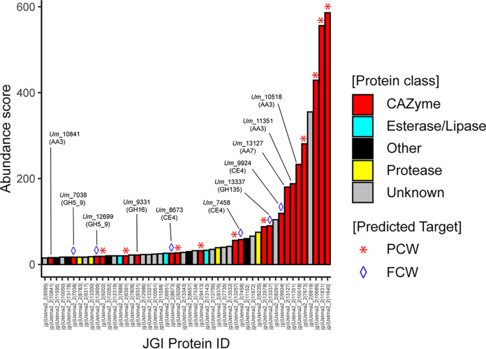
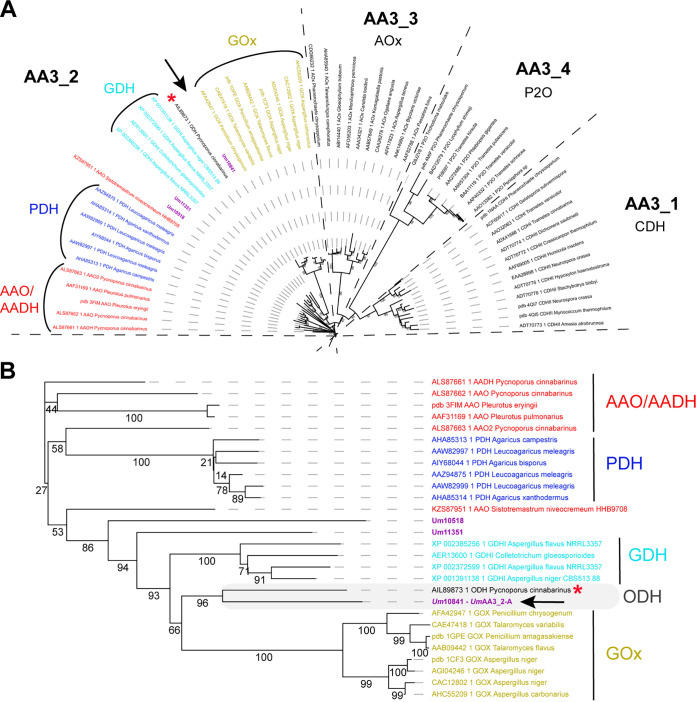
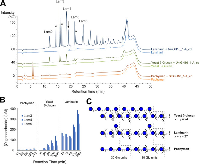
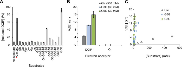
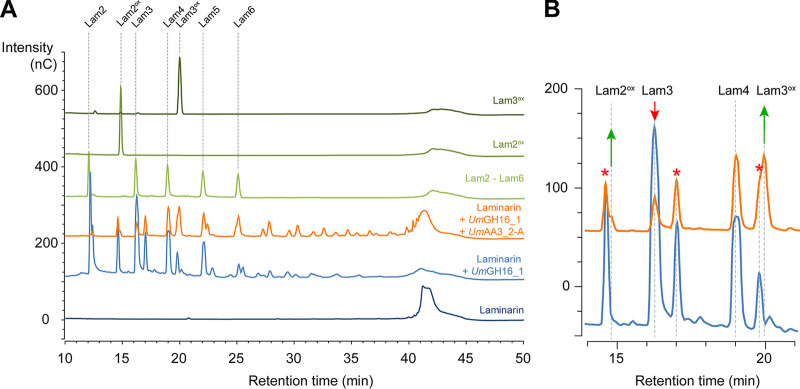
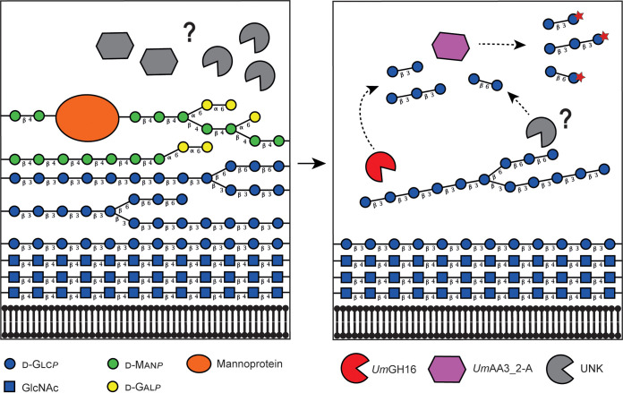
Filamentous fungi are keystone microorganisms in the regulation of many processes occurring on Earth, such as plant biomass decay and pathogenesis as well as symbiotic associations. In many of these processes, fungi secrete carbohydrate-active enzymes (CAZymes) to modify and/or degrade carbohydrates. Ten years ago, while evaluating the potential of a secretome from the maize pathogen Ustilago maydis to supplement lignocellulolytic cocktails, we noticed it contained many unknown or poorly characterized CAZymes. Here, and after reannotation of this data set and detailed phylogenetic analyses, we observed that several CAZymes (including glycoside hydrolases and carbohydrate oxidases) are predicted to act on the fungal cell wall (FCW), notably on β-1,3-glucans. We heterologously produced and biochemically characterized two new CAZymes, called UmGH16_1-A and UmAA3_2-A. We show that UmGH16_1-A displays β-1,3-glucanase activity, with a preference for β-1,3-glucans with short β-1,6 substitutions, and UmAA3_2-A is a dehydrogenase catalyzing the oxidation of β-1,3- and β-1,6-gluco-oligosaccharides into the corresponding aldonic acids. Working on model β-1,3-glucans, we show that the linear oligosaccharide products released by UmGH16_1-A are further oxidized by UmAA3_2-A, bringing to light a putative biocatalytic cascade. Interestingly, analysis of available transcriptomics data indicates that both UmGH16_1-A and UmAA3_2-A are coexpressed, only during early stages of U. maydis infection cycle. Altogether, our results suggest that both enzymes are connected and that additional accessory activities still need to be uncovered to fully understand the biocatalytic cascade at play and its physiological role. IMPORTANCE Filamentous fungi play a central regulatory role on Earth, notably in the global carbon cycle. Regardless of their lifestyle, filamentous fungi need to remodel their own cell wall (mostly composed of polysaccharides) to grow and proliferate. To do so, they must secrete a large arsenal of enzymes, most notably carbohydrate-active enzymes (CAZymes). However, research on fungal CAZymes over past decades has mainly focused on finding efficient plant biomass conversion processes while CAZymes directed at the fungus itself have remained little explored. In the present study, using the maize pathogen Ustilago maydis as model, we set off to evaluate the prevalence of CAZymes directed toward the fungal cell wall during growth of the fungus on plant biomass and characterized two new CAZymes active on fungal cell wall components. Our results suggest the existence of a biocatalytic cascade that remains to be fully understood.

Keywords: CAZymes; Ustilago; beta-glucans; filamentous fungi; fungal cell wall; pathogen; phytopathogens; remodeling.


Conflict of interest statement

The authors declare no conflict of interest.

Figures

FIG 1
FIG 1
Reannotation of the Top50 proteins secreted by U. maydis when cultivated on corn bran. The enzymes are classified according to their abundance in the secretome (after 7 days growth on maize bran; [10]) and a color code identifies the class of protein (see legend in the figure, “Other” refers to all other types of detected proteins). The protein number that is provided corresponds to the JGI protein ID (U. maydis 521 v2.0 strain).
FIG 2
FIG 2
Phylogenetic analysis of GH16 family. Phylogenetic clades, as defined by Viborg et al. (19), are indicated with colored numbers. UmGH16_1-A (indicated by a black arrow) falls within the GH16_1 clade. The tree was inferred using RAxML (100 bootstraps) on the basis of a MSA made with MAFFT.
FIG 3
FIG 3
Phylogenetic analysis of the AA3 family (A) and zoom-in view on the AA3_2 subfamily (B). The AA3s identified in the secretome of U. maydis are shown in purple. The new oligosaccharide dehydrogenase clade, including UmAA3_2-A (indicated by a black arrow), characterized in the present study, and the PcODH (red asterisk), is framed in gray. The tree was inferred using PhyML (bootstrap values, as percentages, are shown on the branches).
FIG 4
FIG 4
Activity of UmGH16_1-A_cd on β-1,3 glucans. (A) The graphs show HPAEC-PAD chromatograms of reaction products released from laminarin, yeast β-glucan and pachyman (10 mg.mL−1 final concentration) by UmGH16_1-A_cd (10 nM). Black arrows indicate reduced β-1,3-gluco-oligosaccharides (see Fig. S7). All reaction mixtures were incubated during 4 h, in citrate phosphate buffer (50 mM, pH 5.5), in a thermomixer (30°C, 1,000 rpm). All experiments were carried out in triplicate but for the sake of clarity, only one replicate is shown. See Fig. S8 for additional control experiments. (B) Time-course release of Lam3-Lam5 oligosaccharides from laminarin, yeast β-glucan and pachyman (same reaction conditions as in panel A; n = 1). (C) Proposed chemical structure of the three tested polymers on the basis of carbohydrate linkage analysis (see Fig. S5 for more details). β3 and β6 represent β-(1,3) and β-(1,6) linkages, respectively.
FIG 5
FIG 5
Activity of UmAA3_2-A. (A) Substrate specificity screening monitored as the reduction of DCIP (400 μM) by UmAA3_2-A (14 nM) in the presence of various substrates (2.5 mM for all, 250 mM when marked with a red star) after 3 h of incubation (see Fig. S11 for substrate nomenclature). (B) Dehydrogenase versus oxidase activity was measured, as, respectively, the reduction of DCIP (400 μM) versus O2 (250 μM) by UmAA3_2-A (110 nM) in the presence of Glucose (500 mM), G3G (30 mM) or G6G (30 mM). (C) [Glucose], [G3G] and [G6G]-dependency of UmAA3_2 initial rate. All reactions were carried out in citrate-phosphate buffer (50 mM, pH 5.5), at 30°C. Data are presented as average values (n = 3, independent biological replicates) and error bars show s.d.
FIG 6
FIG 6
Combined action of UmGH16_1-A and UmAA3_2-A on laminarin. (A) Full HPAEC-PAD chromatograms and (B) zoom-in view on the 11–21 min region comparing products released from laminarin by UmGH16_1 alone (blue line) or in combination with UmAA3_2-A (orange line). The red stars indicate peaks of reduced oligosaccharides already present in the laminarin (see main text).
FIG 7
FIG 7
Proposed reaction scheme illustrating the combined action of UmGH16_1-A and UmAA3_2-A on FCW. Putative enzymatic activities secreted by Ustilago maydis to target its own cell wall. The legend key and the glycosidic linkage between each carbohydrate unit are indicated in the figure (for instance, “β 4” indicates a β-1,4 linkage). In the left-hand side panel, hypothetical enzymatic activities (in gray) degrade the galactomannan and mannoproteins, allowing access to the lower layer of β-1,3/β-1,6-glucans. In the right-hand side panel, the uncovered glucans can act as potential substrate for UmGH16_1-A (shown in red) and other hypothetical hydrolytic activities (in gray), releasing β-1,3 and β-1,6-oligosaccharides oxidizable by UmAA3_2-A (in purple). Another scenario where UmGH16_1-A and UmAA3_2-A would access their substrate by simple diffusion through the FCW, rather than after extracellular secretion, is also possible. In such scenario, extracellular degradation of the first layer of FCW would not be required. For now, it is impossible to settle on the actual trajectory of these enzymes, but, for the sake of clarity, the first layer of mannoprotein and galactomannan is not shown in the right-hand panel.

Similar articles

Cited by

References

    1. Lebreton A, Zeng Q, Miyauchi S, Kohler A, Dai YC, Martin FM. 2021. Evolution of the mode of nutrition in symbiotic and saprotrophic fungi in forest Ecosystems. Annu Rev Ecol Evol Syst 52:385–404. 10.1146/annurev-ecolsys-012021-114902. - DOI
    1. Grigoriev IV, Nikitin R, Haridas S, Kuo A, Ohm R, Otillar R, Riley R, Salamov A, Zhao X, Korzeniewski F, Smirnova T, Nordberg H, Dubchak I, Shabalov I. 2014. MycoCosm portal: gearing up for 1000 fungal genomes. Nucleic Acids Res 42:D699–D704. 10.1093/nar/gkt1183. - DOI - PMC - PubMed
    1. Martinez D, Challacombe J, Morgenstern I, Hibbett D, Schmoll M, Kubicek CP, Ferreira P, Ruiz-Duenas FJ, Martinez AT, Kersten P, Hammel KE, Vanden Wymelenberg A, Gaskell J, Lindquist E, Sabat G, Splinter BonDurant S, Larrondo LF, Canessa P, Vicuna R, Yadav J, Doddapaneni H, Subramanian V, Pisabarro AG, Lavín JL, Oguiza JA, Master E, Henrissat B, Coutinho PM, Harris P, Magnuson JK, Baker SE, Bruno K, Kenealy W, Hoegger PJ, Kües U, Ramaiya P, Lucas S, Salamov A, Shapiro H, Tu H, Chee CL, Misra M, Xie G, Teter S, Yaver D, James T, Mokrejs M, Pospisek M, Grigoriev IV, Brettin T, et al. 2009. Genome, transcriptome, and secretome analysis of wood decay fungus Postia placenta supports unique mechanisms of lignocellulose conversion. Proc Natl Acad Sci USA 106:1954–1959. 10.1073/pnas.0809575106. - DOI - PMC - PubMed
    1. Vanden Wymelenberg A, Gaskell J, Mozuch M, Kersten P, Sabat G, Martinez D, Cullen D. 2009. Transcriptome and secretome analyses of Phanerochaete chrysosporium reveal complex patterns of gene expression. Appl Environ Microbiol 75:4058–4068. 10.1128/AEM.00314-09. - DOI - PMC - PubMed
    1. Eastwood DC, Floudas D, Binder M, Majcherczyk A, Schneider P, Aerts A, Asiegbu FO, Baker SE, Barry K, Bendiksby M, Blumentritt M, Coutinho PM, Cullen D, de Vries RP, Gathman A, Goodell B, Henrissat B, Ihrmark K, Kauserud H, Kohler A, LaButti K, Lapidus A, Lavin JL, Lee Y-H, Lindquist E, Lilly W, Lucas S, Morin E, Murat C, Oguiza JA, Park J, Pisabarro AG, Riley R, Rosling A, Salamov A, Schmidt O, Schmutz J, Skrede I, Stenlid J, Wiebenga A, Xie X, Kües U, Hibbett DS, Hoffmeister D, Högberg N, Martin F, Grigoriev IV, Watkinson SC. 2011. The plant cell wall-decomposing machinery underlies the functional diversity of forest fungi. Science 333:762–765. 10.1126/science.1205411. - DOI - PubMed

Publication types

Supplementary concepts

LinkOut - more resources