Review
. 2022 Nov 22;16(11):17708-17728.
doi: 10.1021/acsnano.2c08164. Epub 2022 Nov 10.

Natural Piezoelectric Biomaterials: A Biocompatible and Sustainable Building Block for Biomedical Devices

Affiliations
Review

Natural Piezoelectric Biomaterials: A Biocompatible and Sustainable Building Block for Biomedical Devices

Ruoxing Wang et al. ACS Nano. .

Abstract

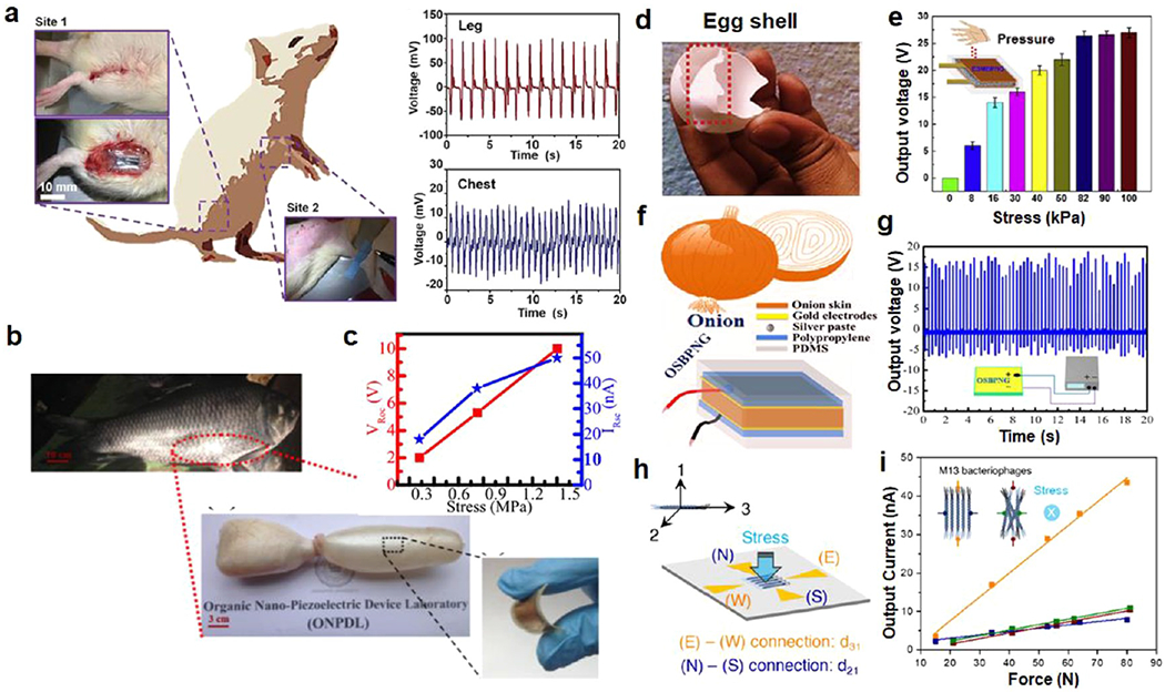
The piezoelectric effect has been widely observed in biological systems, and its applications in biomedical field are emerging. Recent advances of wearable and implantable biomedical devices bring promise as well as requirements for the piezoelectric materials building blocks. Owing to their biocompatibility, biosafety, and environmental sustainability, natural piezoelectric biomaterials are known as a promising candidate in this emerging field, with a potential to replace conventional piezoelectric ceramics and synthetic polymers. Herein, we provide a thorough review of recent progresses of research on five major types of piezoelectric biomaterials including amino acids, peptides, proteins, viruses, and polysaccharides. Our discussion focuses on their structure- and phase-related piezoelectric properties and fabrication strategies to achieve desired piezoelectric phases. We compare and analyze their piezoelectric performance and further introduce and comment on the approaches to improve their piezoelectric property. Representative biomedical applications of this group of functional biomaterials including energy harvesting, sensing, and tissue engineering are also discussed. We envision that molecular-level understanding of the piezoelectric effect, piezoelectric response improvement, and large-scale manufacturing are three main challenges as well as research and development opportunities in this promising interdisciplinary field.

Keywords: amino acids; biomedical devices; flexibility; nanogenerator; natural biomaterials; piezoelectric; polysaccharides; proteins; sustainable materials.


Conflict of interest statement

The authors declare no competing financial interest.

Figures

Figure 1.
Figure 1.
Schematic of the five major types of natural piezoelectric biomaterials and their properties for biomedical applications.
Figure 2.
Figure 2.
Piezoelectric amino acids materials. (a) The general molecular structure of the amino acids. (b) Three atomic structures of glycine. Reprinted with permission under a Creative Commons CC BY License from ref . Copyright 2021 John Wiley and Sons. (c) Calculated piezoelectric coefficients for β-glycine. Experimental d22 and d16 values are marked above relevant columns. (d) Optical image of β-glycine needles. Reprinted with permission from ref . Copyright 2018 Springer Nature. (e) Optical microscopy image of chitosan (CS) film with β-glycine crystals (β-Gly). Reprinted with permission from ref . Copyright 2020 American Chemical Society.
Figure 3.
Figure 3.
Piezoelectric γ-glycine and scaling up. (a) Calculated piezoelectric coefficients for γ-glycine. Experimental d11, d22, and d33 values of d11, d22, and d33 values are marked. Reprinted with permission from ref . Copyright 2018 Springer Nature. (b) The piezoelectric hysteresis loop of γ-glycine microcrystal. Reprinted with permission from ref . Copyright 2012 John Wiley and Sons. (c) The cross-sectional SEM image of a PVA-glycine-PVA film and the corresponding EDS map of nitrogen (N). (d) Schematic illustration of PVA-glycine-PVA sandwich film formation Reprinted with permission from ref . Copyright 2021 American Association for the Advancement of Science. (e) Top: Schematics showing the nucleation site at the convex liquid edge moves to the center flat area when the liquid edge becomes concave. Bottom: Crystal growth kinetics of glycine-PVA films when the liquid film had a convex (green) or concave (red) edge. Reprinted with permission from ref . Copyright 2022 Royal Society of Chemistry.
Figure 4.
Figure 4.
Piezoelectric peptides. (a) PFM image and d33 distribution in two directions indicating opposite piezoelectric polarizations of two FF nanotubes. (b) In-plane piezoresponse angle dependence for PNTs and Y-cut LiNbO3. Reprinted with permission from ref . Copyright 2010 American Chemical Society. (c) Illustration of FF microrods alignment under opposite electric field. (d) Cross-sectional SEM images of FF arrays under positive (up) and negative (bottom) electrical field (scale bars: 100 μm). (e) Statistics of polarization directions of FF microrods under positive, negative, and no electrical field. Reprinted with permission under a Creative Commons CC BY License from ref . Copyright 2016 Springer Nature. (f) Illustration of aligned FF nanotubes growth by Meniscus-driven dip-coating method. Digital photograph and optical microscopy image of FF nanotube array on a flexible substrate. Reprinted with permission from ref . Copyright 2018 American Chemical Society. (g) Field-emission scanning electron microscopy images of 3D-printed FF microstructures. An array of microwalls with an isosceles trapezoid shape (scale bar: 20 μm) and a zigzag wall (scale bar: 5 μm). (h) 3D PFM image of an after-annealed FF spiral pattern (scale bar: 3 μm). Reprinted with permission from ref . Copyright 2021 American Chemical Society. (i) The crystal structure of LDFF. (j) SEM image the LDFF tape. (k) Piezoresponse amplitude as a function of AC voltage of LDFF and lithium niobate (LN). Reprinted with permission from ref . Copyright 2021 John Wiley and Sons.
Figure 5.
Figure 5.
Piezoelectric collagen structure and piezoresponses. (a) Illustration of collagen triple-helix structure formed by three twisted polypeptide chains. Reprinted with permission from ref . Copyright 2018 American Chemical Society. (b) The existence of interstrand hydrogen bonds. Reprinted with permission from ref . Copyright 2011 John Wiley and Sons. (c) Lateral structure of a collagen. Reprinted with permission from ref . Copyright 2022 John Wiley and Sons. (d) PFM topography image (top), piezoresponse amplitude image (mid) and the 2ω signal image (bottom) of a collagen fibril. (e) Shear piezoresponse as a function of applied voltage in the overlap region. Reprinted with permission from ref . Copyright 2009 American Chemical Society. (f) AFM images, corresponding averaged line profiles and amplitude dependence on AC voltage of type I (red) and type II (blue) collagen fibrils. Reprinted with permission from ref . Copyright 2014 AIP Publishing.
Figure 6.
Figure 6.
Piezoelectric silk and elastin. (a) Schematic illustration of components and structures in spider silk. Reprinted with permission from ref . Copyright 2018 Elsevier. (b) The piezoelectric coefficient d14 of silk under different processing parameters (squares: zone drawn; triangles: water-immersion drawn; diamonds: methanol treated). Reprinted with permission from ref . Copyright 2011 John Wiley and Sons. (c) SEM image of electrospun silk fibroin. Reprinted with permission from ref . Copyright 2019 Elsevier. (d) Histological image of fibrous elastin (scale bar: 200 μm). (e) PFM mappings of elastin at high temperature (473 K). Reprinted with permission from ref . Copyright 2014 National Academy of Science.
Figure 7.
Figure 7.
Piezoelectric virus and assembling strategies. (a) Illustration of M13 phage structure with dipole directions indicated by yellow arrows. (b, c) PFM images of monolayer phage film scanning from two directions (indicated by white arrows). (d) Amplitude of piezoresponse–AC voltage curves from periodically poled lithium niobate (PPLN), phage film, and collagen. Reprinted with permission from ref . Copyright 2012 Springer Nature. (e) Illustration of the enforced infiltration strategy for fabrication of vertically aligned M13 phage nanopillars. (f) SEM image of the phage nanopillar-embedded porous template (scale bar = 200 nm) and schematic of phage nanopillars with axial aligned piezoelectricity. Reprinted with permission from ref . Copyright 2015 The Royal Society of Chemistry. (g) Schematic of the template-assisted method for growing vertically polarized phage nanostructure. (h) PFM amplitude image of vertically aligned phages. (i) The out-of-plane PFM amplitude dependence of applied voltage of genetically engineered (6H) and wildtype (WT) phage compared in film and vertical nanopillar forms. Reprinted with permission from ref . Copyright 2019 American Chemical Society.
Figure 8.
Figure 8.
Piezoelectric polysaccharides including cellulose and chitin/chitosan. (a) Schematic of CNC derived from wood pulp. Reprinted with permission from ref . Copyright 2021 Elsevier. (b) The AFM height deflection mapping of CNC film under different voltages. Reprinted with permission from ref . Copyright 2012 American Chemical Society. (c) Vertical displacement as a function of applied voltage for ultrathin CNF films under no alignment, HIMA and EIP alignment, and corona poling conditions. Reprinted with permission from ref . Copyright 2020 American Chemical Society. (d) Schematic of the vertical alignment mechanism. (e) Cross-sectional SEM image and surface PFM image of vertically aligned CNC film. Reprinted with permission from ref . Copyright 2020 American Chemical Society. (f) The molecular structures of chitin and chitosan. Reprinted with permission from ref . Copyright 2022 John Wiley and Sons. (g) Spontaneous polarization of β-chitin nanofibers. The arrow shows the polarization direction. Reprinted with permission from ref . Copyright 2018 Elsevier. (h) Piezoelectric response measured from gelatin, chitin, and collagen nanofibers nanogenerators. Reprinted with permission from ref . Copyright 2018 Elsevier. (i) Average piezoresponse dependence on AC voltage of chitosan film. Reprinted with permission from ref . Copyright 2022 John Wiley and Sons. (j) Cross-sectional SEM image of chitosan film prepared by using formic acid. (k) Average d33 values of chitosan films prepared by dissolving in formic acid dependence of pressure at different sintering temperatures. Reprinted with permission under a Creative Commons CC BY License from ref . Copyright 2017 The Royal Society of Chemistry.
Figure 9.
Figure 9.
Piezoelectric biomaterials for mechanical energy harvesting. (a) Schematic of a Sprague–Dawley rat and photographs of implanted glycine-PVA devices in rats. Corresponding voltage outputs of the glycine-PVA film driven at thigh and chest areas by different muscles. Reprinted with permission from ref . Copyright 2021 American Association for the Advancement of Science. (b) Photographs of the sweet water (Catla Catla) fish and its swim bladder. (c) The output voltages and currents dependence on stress generated by fish swim bladder NG. Reprinted with permission from ref . Copyright 2016 Elsevier. (d) Photograph of eggshell membrane. (e) The output voltages generated from eggshell membrane NG dependence on finger-tapped stress. Reprinted with permission from ref . Copyright 2018 Elsevier. (f) Illustration of the structure of piezoelectric NG made from onionskin (OSBPNG). (g) The generated output voltage from OSBPNG in forward connection. Reprinted with permission from ref . Copyright 2017 Elsevier. (h) Schematic illustration of electrode connections on quadrant electrode piezoelectric NG. (i) The output currents dependence on loading force for aligned phages and random phages. Reprinted with permission from ref . Copyright 2021 Elsevier.
Figure 10.
Figure 10.
Piezoelectric biomaterials for sensing. (a) Illustration of cellulose CNF sensor and the optical photograph of final device. (b) Piezoelectric sensitivity as a function of excitation position. Reprinted with permission from ref . Copyright 2016 American Chemical Society. (c) Schematic structure of carboxymethyl chitosan-based piezoelectric biosensor. (d) The response–time curves of a piezoelectric biosensor with different concentrations of additional IgY in PBS buffer. (e) The 3D sensor responses curve to IgY, lysozyme, and BSA with different concentrations at 60 s. Reprinted with permission from ref . Copyright 2022 Elsevier. (f) Schematic illustration of a self-powered collagen-based humidity sensor and output voltages under varied humidity. Reprinted with permission from ref . Copyright 2018 American Chemical Society.
Figure 11.
Figure 11.
Piezoelectric biomaterials for tissue engineering. (a) Schematic illustration of the kinematic model used to assess the functional recovery of tendon based on gait analyses and the MTR timeline. (b) The statistics of functional recovery in comparison under piezo (EMS) and nonpiezo (MS) conditions for metatarsophalangeal (MTP), knee, and ankle. (c) Histological images of MTR-treated EMS and MS groups after 4 and 8 weeks. Reprinted with permission under a Creative Commons CC BY License from ref . Copyright 2021 John Wiley and Sons. (d) A possible schematic of piezoelectically guided BNCs growth on PEDOT/chitosan nanofibers substrate. (e) SEM image of BNCs cultured on PEDOT/chitosan nanofibers after 1 h of electrical stimulation. (f) Statistics of median neurite length of BNCs with different electrical stimulation time cultured on PEDOT:PSS/chitosan and PEDOT/chitosan substrates. Reprinted with permission from ref . Copyright 2020 Elsevier.

Similar articles

Cited by

References

    1. Katzir S In The Beginnings of Piezoelectricity; Springer: Dordrecht, 2006; pp 15–64.
    1. Shin D-M; Hong SW; Hwang Y-H Recent advances in organic piezoelectric biomaterials for energy and biomedical applications. Nanomaterials 2020, 10, 123. - PMC - PubMed
    1. Jaffe B; Roth R; Marzullo S Piezoelectric properties of lead zirconate-lead titanate solid-solution ceramics. J. Appl. Phys 1954, 25, 809–810.
    1. Berlincourt D; Jaffe H Elastic and piezoelectric coefficients of single-crystal barium titanate. Phys. Rev 1958, 111, 143.
    1. Acosta M; et al. BaTiO3-based piezoelectrics: Fundamentals, current status, and perspectives. Applied Physics Reviews 2017, 4, 041305.

Publication types