Review
. 2022 Oct 30;12(11):941.
doi: 10.3390/bios12110941.

Recent Progress on Solid Substrates for Surface-Enhanced Raman Spectroscopy Analysis

Affiliations
Review

Recent Progress on Solid Substrates for Surface-Enhanced Raman Spectroscopy Analysis

Kun Ge et al. Biosensors (Basel). .

Abstract

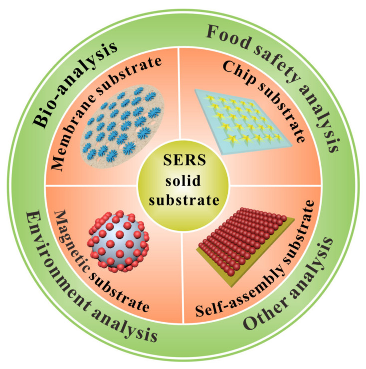
Surface-enhanced Raman spectroscopy (SERS) is a powerful vibrational spectroscopy technique with distinguished features of non-destructivity, ultra-sensitivity, rapidity, and fingerprint characteristics for analysis and sensors. The SERS signals are mainly dependent on the engineering of high-quality substrates. Recently, solid SERS substrates with diverse forms have been attracting increasing attention due to their promising features, including dense hot spot, high stability, controllable morphology, and convenient portability. Here, we comprehensively review the recent advances made in the field of solid SERS substrates, including their common fabrication methods, basic categories, main features, and representative applications, respectively. Firstly, the main categories of solid SERS substrates, mainly including membrane substrate, self-assembled substrate, chip substrate, magnetic solid substrate, and other solid substrate, are introduced in detail, as well as corresponding construction strategies and main features. Secondly, the typical applications of solid SERS substrates in bio-analysis, food safety analysis, environment analysis, and other analyses are briefly reviewed. Finally, the challenges and perspectives of solid SERS substrates, including analytical performance improvement and largescale production level enhancement, are proposed.

Keywords: applications; construction; perspectives; solid substrate; surface-enhanced Raman spectroscopy.


Conflict of interest statement

The authors declare no conflict of interest.

Figures

Figure 1
Figure 1
Various solid SERS substrates employed in analytical applications.
Figure 2
Figure 2
Schematic fabrication of CNF-AuNP membrane substrate (a). © 2022 Elsevier B.V. Adapted with permission from [42]. Schematic illustration of preparation principle of mpg-C3N4/AuCu membrane (b). © 2022 Elsevier B.V. Adapted with permission from [45]. Schematic illustration of fabrication process of Ag NCs@PDMS substrate (c). © 2022 American Chemical Society. Adapted with permission from [79]. Schematic preparation of Ag/Au NWs/PDMS film and its application for on-site detection of pesticides (d). © 2022 Elsevier B.V. Adapted with permission from [80].
Figure 3
Figure 3
Schematic of the fabrication process of the AgNPs/W-Gr membrane (a). © 2022 Elsevier B.V. Adapted with permission from [24]. Schematic preparation of self-assembly gold nanorods (Au NRs) array and detection of pesticides on fruit surface (b). © 2022 Elsevier B.V. Adapted with permission from [87]. Schematic preparation diagram of hierarchical MF/Ag array substrate (c). © 2022 Elsevier B.V. Adapted with permission from [60]. Schematic illustration of preparation of NP-on-WS hierarchical substrate (d). © The Author(s) 2018. Adapted with permission from [61].
Figure 4
Figure 4
Schematic procedure for fabricating of 3D Ag MFs and Ag NMFs on P4VP-g-GO films (a). © 2022 Wiley-VCH Verlag GmbH & Co. KGaA. Adapted with permission from [64]. Schematic illustration of preparation of Au@MIP film (b). © 2022 American Chemical Society. Adapted with permission from [31]. Schematic synthesis of CoFe2O4/Ag substrate (c). © 2022 American Chemical Society. Adapted with permission from [107]. Schematic synthesis of Au-G-AgNs (d). © 2022 American Chemical Society. Adapted with permission from [74].
Figure 5
Figure 5
Schematic fabrication of adhesive tape based solid platform and its applications in rapid sampling, photocontrolled release, and on-site SERS detection (a). © 2022 American Chemical Society. Adapted with permission from [53]. Schematic preparation of Au-coated TiO2 MIO and its detection process (b). © 2022 American Chemical Society. Adapted with permission from [62].
Figure 6
Figure 6
Schematic fabrication of cauliflower-inspired chip-based SERS substrate for simultaneous label-free analysis of multiple mycotoxins (a). © 2022 American Chemical Society. Adapted with permission from [33]. Schematic preparation process of the Ti3C2Tx/DNA/Ag membrane substrate and its application of simultaneous and accurate quantification of multiple antibiotics (b). © 2022 American Chemical Society. Adapted with permission from [49].

Similar articles

Cited by

References

    1. Cai L., Dong J., Wang Y., Chen X. A review of developments and applications of thin-film microextraction coupled to surface-enhanced Raman scattering. Electrophoresis. 2019;40:2041–2049. doi: 10.1002/elps.201800531. - DOI - PubMed
    1. Zhang D., Liang P., Yu Z., Xia J., Ni D., Wang D., Zhou Y., Cao Y., Chen J., Chen J., et al. Self-assembled “bridge” substance for organochlorine pesticides detection in solution based on surface enhanced raman scattering. J. Hazard. Mater. 2020;382:121023. doi: 10.1016/j.jhazmat.2019.121023. - DOI - PubMed
    1. Deng Z., Chen X., Wang Y., Fang E., Zhang Z., Chen X. Headspace thin-film microextraction coupled with surface-enhanced Raman scattering as a facile method for reproducible and specific detection of sulfur dioxide in wine. Anal. Chem. 2015;87:633–640. doi: 10.1021/ac503341g. - DOI - PubMed
    1. Zhang C.Y., Zhao B.C., Hao R., Wang Z., Hao Y.W., Zhao B., Liu Y.Q. Graphene oxide-highly anisotropic noble metal hybrid systems for intensified surface enhanced Raman scattering and direct capture and sensitive discrimination in PCBs monitoring. J. Hazard. Mater. 2020;385:121510. doi: 10.1016/j.jhazmat.2019.121510. - DOI - PubMed
    1. Zhang Z., Wang J., Shanmugasundaram K.B., Yeo B., Moller A., Wuethrich A., Lin L.L., Trau M. Tracking drug-induced epithelial-mesenchymal transition in breast cancer by a microfluidic surface-enhanced raman spectroscopy immunoassay. Small. 2020;16:e1905614. doi: 10.1002/smll.201905614. - DOI - PubMed

MeSH terms

LinkOut - more resources