Review
. 2022 Nov 1;12(11):953.
doi: 10.3390/bios12110953.

Biosensors for the Detection of Enzymes Based on Aggregation-Induced Emission

Affiliations
Review

Biosensors for the Detection of Enzymes Based on Aggregation-Induced Emission

Fengli Gao et al. Biosensors (Basel). .

Abstract

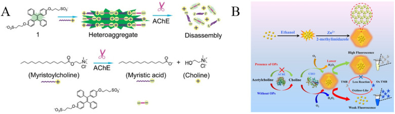
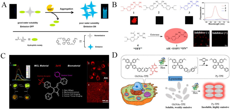
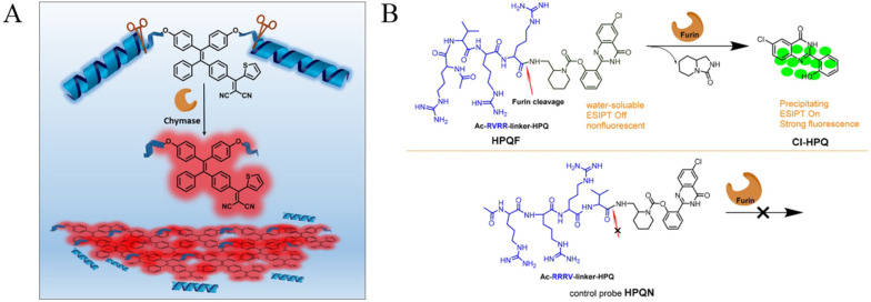
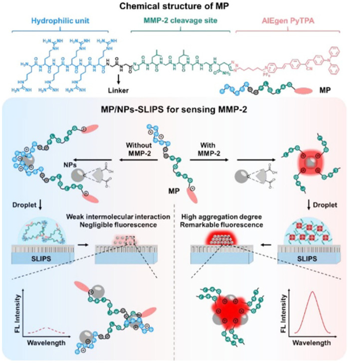
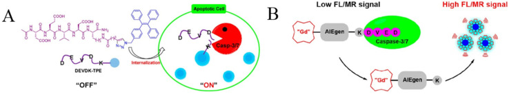
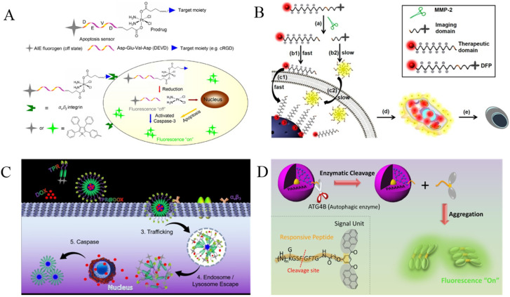
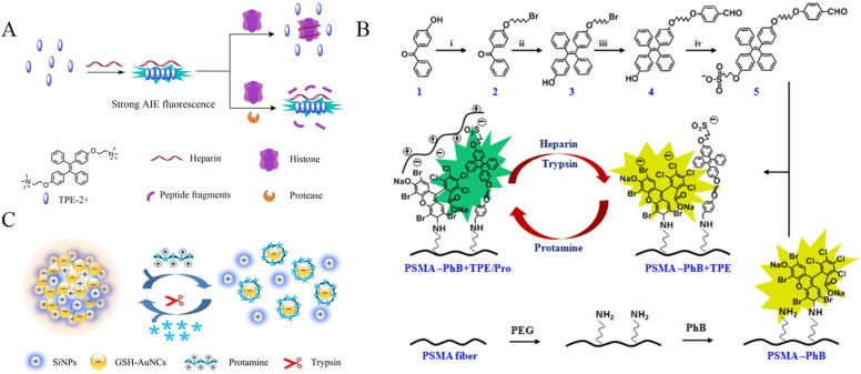
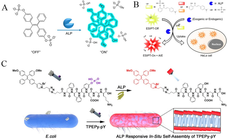
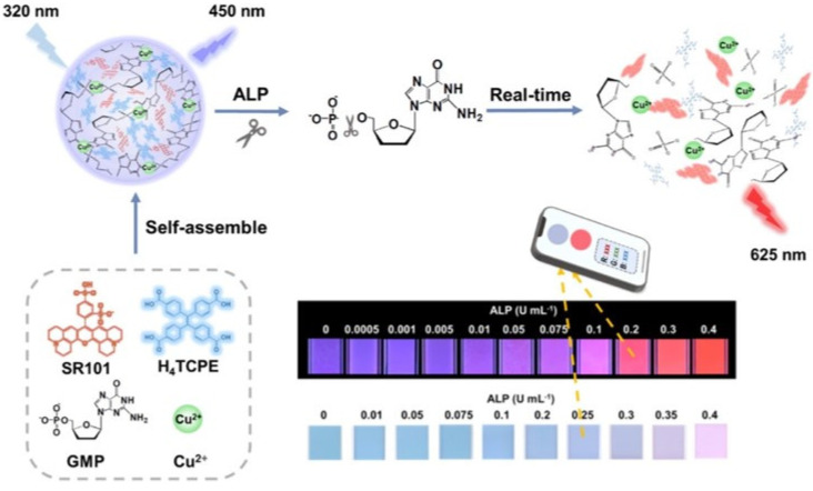
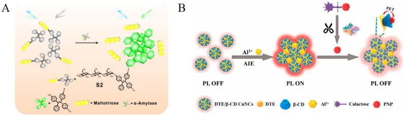
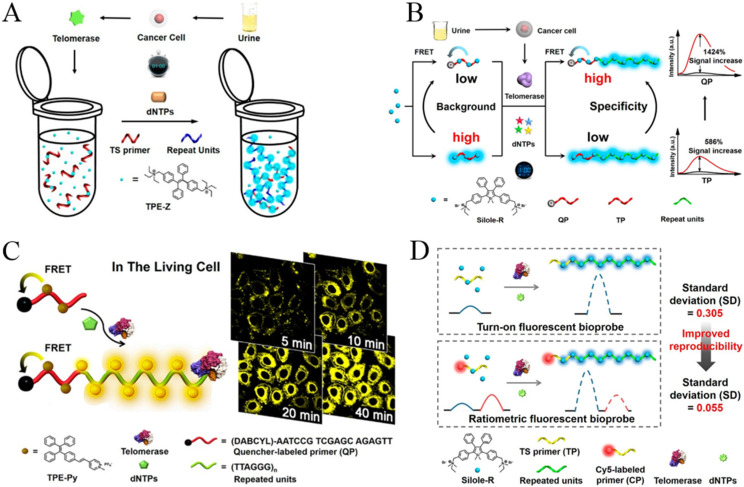
Enzymes play a critical role in most complex biochemical processes. Some of them can be regarded as biomarkers for disease diagnosis. Taking advantage of aggregation-induced emission (AIE)-based biosensors, a series of fluorogens with AIE characteristics (AIEgens) have been designed and synthesized for the detection and imaging of enzymes. In this work, we summarized the advances in AIEgens-based probes and sensing platforms for the fluorescent detection of enzymes, including proteases, phosphatases, glycosidases, cholinesterases, telomerase and others. The AIEgens involve organic dyes and metal nanoclusters. This work provides valuable references for the design of novel AIE-based sensing platforms.

Keywords: aggregation-induced emission; enzymes; fluorescent biosensors; metal nanoclusters; organic dyes.


Conflict of interest statement

The authors declare no conflict of interest.

Figures

Figure 10
Figure 10
(A) Illustration of the formation of heteroaggregate between myristoylcholine and tetraphenylethylene 1 and the disassembly of the aggregate in the presence of AChE. Reprinted with permission from ref. [89]. Copyright 2009, American Chemical Society. (B) Schematic diagram of the mechanism for the detection of OPs. Reprinted with permission from ref. [93]. Copyright 2021, American Chemical Society.
Figure 12
Figure 12
(A) Schematic illustration of the probe’s fluorescent detection for GGT. Reprinted with permission from ref. [102]. Copyright 2016, Elsevier. (B) Schematic illustration of red-emissive fluorophore with AIE and ESIPT characteristics for light-pp sensing of esterase. Reprinted with permission from ref. [105]. Copyright 2014, American Chemical Society. (C) Schematic illustration of rofecoxib-based fluorescent probes for COX-2-targeted bioimaging. Reprinted with permission from ref. [109]. Copyright 2021, American Chemical Society. (D) Detection mechanism of GlcNAc-TPE towards Hex. Reprinted with permission from ref. [110]. Copyright 2019, American Chemical Society.
Figure 1
Figure 1
(A) Schematic illustration of the functional mechanism of the probe TPETH-2(CFTERDn). n = 2 or 3 for chymase sensing. Reprinted with permission from ref. [42]. Copyright 2016, American Chemical Society. (B) Chemical structure of the activatable probe HPQF and its working mechanism with furin (top), and the molecular design strategy of the negative control non-cleavable probe HPQN (bottom). Reprinted with permission from ref. [44]. Copyright 2018, American Chemical Society.
Figure 2
Figure 2
Design of modular peptide-conjugated AIEgen MP and development of the MP/NPs–SLIPS sensing system for sensitively detecting tumor marker MMP-2. Reprinted with permission from ref. [45]. Copyright 2021, American Chemical Society.
Figure 3
Figure 3
(A) Illustration of Ac-DEVDK-TPE for the study of caspase activities. Reprinted with permission from ref. [46]. Copyright 2012, American Chemical Society. (B) Schematic illustration of DPBP as a bioprobe for autophagy detection. Reprinted with permission from ref. [49]. Copyright 2019, American Chemical Society.
Figure 4
Figure 4
(A) Schematic illustration of the targeted theranostic platinum (IV) prodrug with a built-in aggregation-induced emission (AIE) light-up apoptosis sensor for noninvasive, in situ early evaluation of its therapeutic responses. Reprinted with permission from ref. [50]. Copyright 2014, American Chemical Society. (B) Schematic illustration showing the probe DFP for rapid drug delivery and drug release tracking in MMP-2 over-expression living cells. Reprinted with permission from ref. [51]. Copyright 2016, American Chemical Society. (C) Schematic illustration of TPR@DOX for tumor treatment and apoptosis monitoring. Reprinted with permission from Reference [52]. Copyright 2021, American Chemical Society. (D) Caspase-3/7-sensing mechanism of CP1. Reprinted with permission from ref. [53]. Copyright 2017, American Chemical Society.
Figure 5
Figure 5
(A) Schematic illustration of the fluorometric assay for protease activity. Reprinted with permission from ref. [55]. Copyright 2018, Elsevier. (B) Synthetic route of TPE derivatives, schematic drawing of the ratiometric color changes of fibers, and grafting process of PhB on electrospun PSMA fibers. Reprinted with permission from ref. [56]. Copyright 2017, American Chemical Society. (C) Schematic illustration of the mechanism of the ratiometric detection of protamine and trypsin based on a nanohybrid probe. Reprinted with permission from ref. [57]. Copyright 2019, Elsevier.
Figure 6
Figure 6
(A) Illustration of the design principles of ALP assay with a TPE-2PA probe. Reprinted with permission from ref. [59]. Copyright 2013, American Chemical Society. (B) Schematic illustration of HCAP for ALP activity assay in solution and in living cells. Reprinted with permission from ref. [63]. Copyright 2014, American Chemical Society. (C) Schematic illustration of the peptide self-assembly-controlled turn-on probe for sensing ALP activity. Reprinted with permission from ref. [65]. Copyright 2020, American Chemical Society.
Figure 7
Figure 7
Schematic illustration of the real-time monitoring of ALP activity, based on the ratiometric fluorescence response of H4TCPE/SR101/Cu-GMP ICP nanoparticles stemming from the AIE guest and the ACQ guest simultaneously, and its field application for algal bloom warning implanted with a smartphone. Reprinted with permission from ref. [69]. Copyright 2021, American Chemical Society.
Figure 8
Figure 8
(A) Schematic illustration of (top) the proposed mechanism for specific recognition between MBA-stabilized CuNCs and glucose and (bottom) the cleancap-regulated AIE strategy for the imaging of ALP activity. Reprinted with permission from ref. [72]. Copyright 2020, American Chemical Society. (B) Schematic illustration of the detection strategy for ALP activity based on FRET. Reprinted with permission from ref. [73]. Copyright 2019, Elsevier. (C) Schematic illustration of AIEE-based AgNC nanoswitches in response to multiple stimuli and a detection strategy for PPase activity based on ion-triggered switch. Reprinted with permission from ref. [75]. Copyright 2017, American Chemical Society.
Figure 9
Figure 9
(A) Schematic illustration of the sensing mechanisms of the probe S2 in α-amylase activity sensing. Reprinted with permission from ref. [84]. Copyright 2018, American Chemical Society. (B) Schematic representation of the strategy for the determination of β-Gal. Reprinted with permission from ref. [87]. Copyright 2020, American Chemical Society.
Figure 11
Figure 11
(A) Schematic illustration of the AIE-based simple one-pot technique for telomerase activity detection. Reprinted with permission from ref. [97]. Copyright 2015, American Chemical Society. (B) Schematic illustration of the quencher group-induced high specificity fluorescence strategy for the detection of telomerase activity. Reprinted with permission from ref. [98]. Copyright 2015, American Chemical Society. (C) Schematic Illustration of the AIE-based in situ telomerase activity detection and imaging. Reprinted with permission from ref. [99]. Copyright 2016, American Chemical Society. (D) Schematic illustration of the ratiometric fluorescent bioprobe for telomerase activity detection. Reprinted with permission from ref. [100]. Copyright 2016, American Chemical Society.

Similar articles

Cited by

References

    1. Kwek G., Do T.C., Lu X., Lin J., Xing B. Scratching the surface of unventured possibilities with in situ self-assembly: Protease-activated developments for imaging and therapy. ACS Appl. Bio. Mater. 2021;4:2192. doi: 10.1021/acsabm.0c01340. - DOI - PubMed
    1. Yao Y., Zhang Y., Yan C., Zhu W.H., Guo Z. Enzyme-activatable fluorescent probes for beta-galactosidase: From design to biological applications. Chem. Sci. 2021;12:9885. doi: 10.1039/D1SC02069B. - DOI - PMC - PubMed
    1. Luo S., Zhang Y., Situ B., Zheng L. Fluorescence sensing telomerase activity: From extracellular detection to in situ imaging. Sens. Actuat. B Chem. 2018;273:853. doi: 10.1016/j.snb.2018.06.088. - DOI
    1. Feng G., Liao S., Liu Y., Zhang H., Luo X., Zhou X., Fang J. When AIE meets enzymes. Analyst. 2022;147:3958. doi: 10.1039/D2AN00679K. - DOI - PubMed
    1. Hu Z., Li Y., Hussain E., Huang X., Zhang Y., Niu N., Shahzad S.A., Yu C. Black phosphorus nanosheets based sensitive protease detection and inhibitor screening. Talanta. 2019;197:270. doi: 10.1016/j.talanta.2019.01.023. - DOI - PubMed

LinkOut - more resources