

Angiotensin-Converting Enzyme 2-Based Biosensing Modalities and Devices for Coronavirus Detection
- PMID: 36354493
- PMCID: PMC9688389
- DOI: 10.3390/bios12110984
Angiotensin-Converting Enzyme 2-Based Biosensing Modalities and Devices for Coronavirus Detection
Abstract
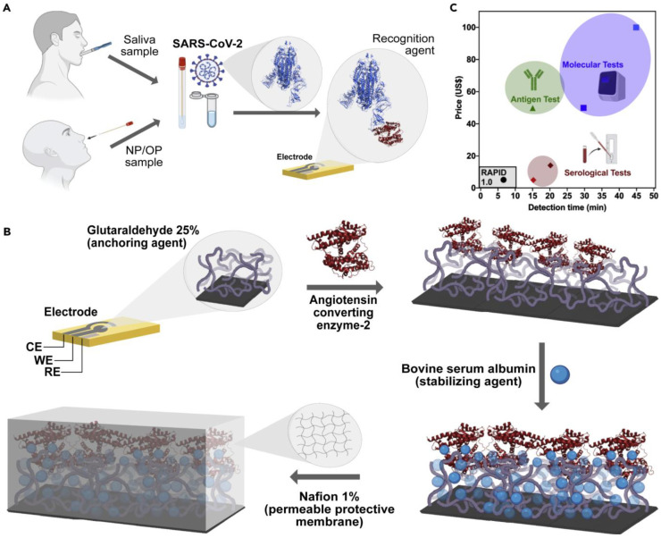
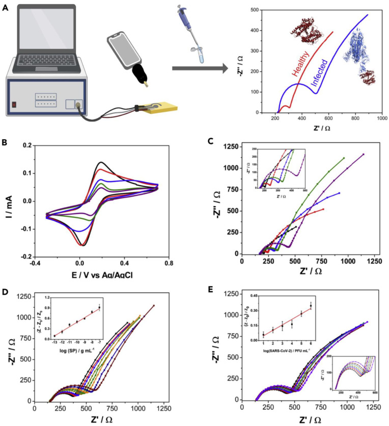
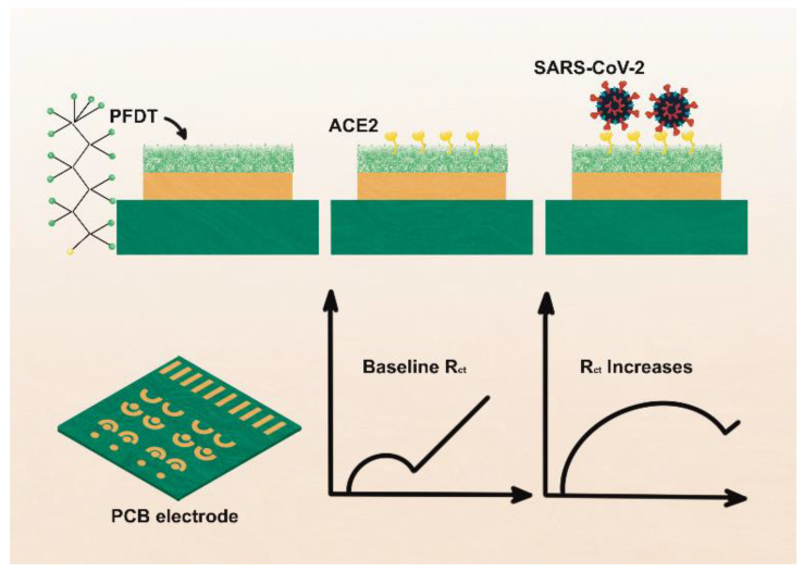
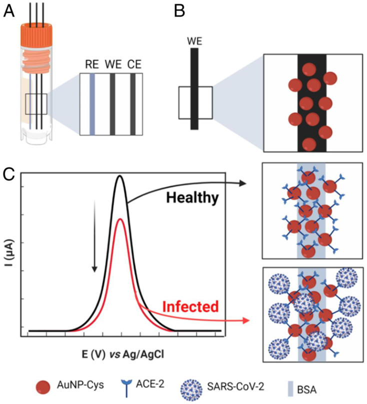
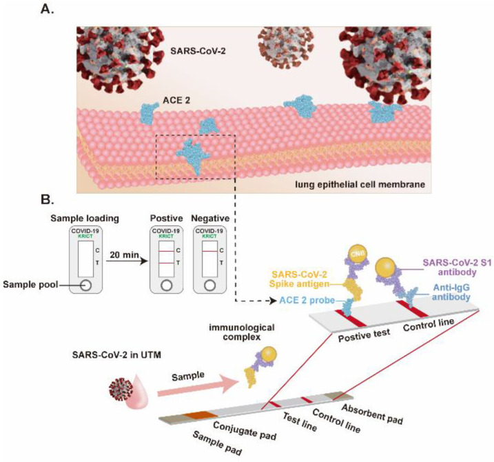
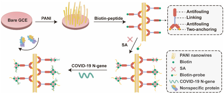
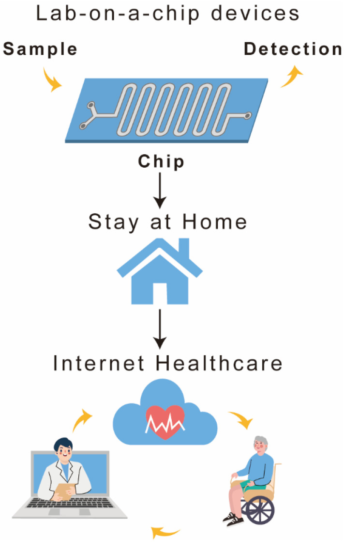
Rapid and cost-effective diagnostic tests for severe acute respiratory syndrome coronavirus 2 (SARS-CoV-2) are a critical and valuable weapon for the coronavirus disease 2019 (COVID-19) pandemic response. SARS-CoV-2 invasion is primarily mediated by human angiotensin-converting enzyme 2 (hACE2). Recent developments in ACE2-based SARS-CoV-2 detection modalities accentuate the potential of this natural host-virus interaction for developing point-of-care (POC) COVID-19 diagnostic systems. Although research on harnessing ACE2 for SARS-CoV-2 detection is in its infancy, some interesting biosensing devices have been developed, showing the commercial viability of this intriguing new approach. The exquisite performance of the reported ACE2-based COVID-19 biosensors provides opportunities for researchers to develop rapid detection tools suitable for virus detection at points of entry, workplaces, or congregate scenarios in order to effectively implement pandemic control and management plans. However, to be considered as an emerging approach, the rationale for ACE2-based biosensing needs to be critically and comprehensively surveyed and discussed. Herein, we review the recent status of ACE2-based detection methods, the signal transduction principles in ACE2 biosensors and the development trend in the future. We discuss the challenges to development of ACE2-biosensors and delineate prospects for their use, along with recommended solutions and suggestions.
Keywords: ACE2 biosensors; COVID-19; SARS-CoV-2; colorimetric sensors; electrochemical detection; low-cost diagnostic.
Conflict of interest statement
The authors declare no conflict of interest.
Figures

Similar articles
-
Research progress of biosensors for detection of SARS-CoV-2 variants based on ACE2.Talanta. 2023 Jan 1;251:123813. doi: 10.1016/j.talanta.2022.123813. Epub 2022 Aug 6. Talanta. 2023. PMID: 35952504 Free PMC article. Review.
-
Angiotensin-converting enzyme 2 (ACE2), SARS-CoV-2 and the pathophysiology of coronavirus disease 2019 (COVID-19).J Pathol. 2020 Jul;251(3):228-248. doi: 10.1002/path.5471. Epub 2020 Jun 10. J Pathol. 2020. PMID: 32418199 Free PMC article. Review.
-
Regulation of Angiotensin-Converting Enzyme 2: A Potential Target to Prevent COVID-19?Front Endocrinol (Lausanne). 2021 Oct 22;12:725967. doi: 10.3389/fendo.2021.725967. eCollection 2021. Front Endocrinol (Lausanne). 2021. PMID: 34745001 Free PMC article. Review.
-
SARS-CoV-2 pandemic and research gaps: Understanding SARS-CoV-2 interaction with the ACE2 receptor and implications for therapy.Theranostics. 2020 Jun 12;10(16):7448-7464. doi: 10.7150/thno.48076. eCollection 2020. Theranostics. 2020. PMID: 32642005 Free PMC article. Review.
-
Angiotensin-Converting Enzyme 2: SARS-CoV-2 Receptor and Regulator of the Renin-Angiotensin System: Celebrating the 20th Anniversary of the Discovery of ACE2.Circ Res. 2020 May 8;126(10):1456-1474. doi: 10.1161/CIRCRESAHA.120.317015. Epub 2020 Apr 8. Circ Res. 2020. PMID: 32264791 Free PMC article. Review.
Cited by
-
Strategies for Increasing the Throughput of Genetic Screening: Lessons Learned from the COVID-19 Pandemic within a University Community.BioTech (Basel). 2024 Jul 11;13(3):26. doi: 10.3390/biotech13030026. BioTech (Basel). 2024. PMID: 39051341 Free PMC article.
-
Unraveling the Dynamics of SARS-CoV-2 Mutations: Insights from Surface Plasmon Resonance Biosensor Kinetics.Biosensors (Basel). 2024 Feb 13;14(2):99. doi: 10.3390/bios14020099. Biosensors (Basel). 2024. PMID: 38392018 Free PMC article. Review.
-
CRISPR-cas technology: A key approach for SARS-CoV-2 detection.Front Bioeng Biotechnol. 2023 Apr 12;11:1158672. doi: 10.3389/fbioe.2023.1158672. eCollection 2023. Front Bioeng Biotechnol. 2023. PMID: 37214290 Free PMC article. Review.
-
Current and Perspective Sensing Methods for Monkeypox Virus.Bioengineering (Basel). 2022 Oct 18;9(10):571. doi: 10.3390/bioengineering9100571. Bioengineering (Basel). 2022. PMID: 36290539 Free PMC article. Review.
-
Unraveling the influence of CRISPR/Cas13a reaction components on enhancing trans-cleavage activity for ultrasensitive on-chip RNA detection.Mikrochim Acta. 2024 Jul 17;191(8):466. doi: 10.1007/s00604-024-06545-4. Mikrochim Acta. 2024. PMID: 39017814
References
-
- Manmana Y., Kubo T., Otsuka K. Recent Developments of Point-of-Care (POC) Testing Platform for Biomolecules. TrAC Trends Anal. Chem. 2021;135:116160. doi: 10.1016/j.trac.2020.116160. - DOI
-
- Deroco P.B., Wachholz Junior D., Kubota L.T. Recent Advances in Point-of-Care Biosensors for the Diagnosis of Neglected Tropical Diseases. Sens. Actuators B Chem. 2021;349:130821. doi: 10.1016/j.snb.2021.130821. - DOI
Publication types
MeSH terms
Substances
Grants and funding
LinkOut - more resources
Full Text Sources
Medical
Miscellaneous
