Case Reports
. 2022 Nov 4;9(11):649.
doi: 10.3390/bioengineering9110649.

Resorbable Membrane Pouch Technique for Single-Implant Placement in the Esthetic Zone: A Preliminary Technical Case Report

Affiliations
Case Reports

Resorbable Membrane Pouch Technique for Single-Implant Placement in the Esthetic Zone: A Preliminary Technical Case Report

Akiyoshi Funato et al. Bioengineering (Basel). .

Abstract

The conventional protocol for lateral guided bone regeneration (GBR) in esthetic areas requires the securing of resorbable collagen membranes using titanium cortical bone pins to immobilize bone grafts. These procedures are highly invasive and can increase patient morbidity and discomfort. Herein, we introduce a minimally invasive novel resorbable membrane pouch technique, wherein collagen membranes can be immobilized by securing them to the periosteum without the need of titanium pins. We describe 11 cases of single-immediate- or delayed-implant placement in the atrophic maxilla esthetic zone. All implants were successful and functional without pain or inflammation and with optimal soft-tissue health and esthetics. Radiographic evaluation with cone-beam computed tomography (CBCT) and esthetic assessment using the pink esthetic score (PES) were performed. At the time of implant placement, the average augmented bone width was 2.8 ± 0.6 mm on CBCT analysis. In all cases, resorption of the augmented bone was confirmed with an average of -1.3 ± 0.8 mm. Soft-tissue outcomes were scored 1 year after permanent restoration. The PES score 1 year after treatment was 11.9 ± 1.4. The resorbable membrane pouch technique with immediate or delayed implant placement for buccal dehiscence in the esthetic area can be predictable and is minimally invasive.

Keywords: case report; connective-tissue graft; dental implant; resorbable membrane pouch techniques.


Conflict of interest statement

The authors declare no conflict of interest. The funders had no role in the design of the study; in the collection, analyses, or interpretation of data; in the writing of the manuscript; or in the decision to publish the results.

Figures

Figure 1
Figure 1
Illustrations of the surgical procedures. (a) ① A vertical releasing incision was made on the distal side of the canine in the same quadrant to create a triangular flap. ② At the implant placement site, a full-thickness flap was elevated and the bone crest was exposed. ③ A full-thickness flap was connected to a partial-thickness flap in the basal part of the maxilla. ④ Periosteum tabs were prepared just apical to the buccal dehiscence in lengths of 2–3 mm. ⑤ Following the pouch preparation, prosthetically driven crestal implant placement was performed. (b) A resorbable membrane was inserted beneath the periosteum tab. The membrane was secured to the periosteum using resorbable sutures. The internal space of the pouch, surrounded by the resorbable membrane and exposed labial implant surface, was treated with a bone graft. The membrane was secured to the palatal wall using resorbable sutures. (c) A connective-tissue graft obtained from the palatal site was placed on the buccal superior aspect and secured to the periosteum or mucosal flap.
Figure 2
Figure 2
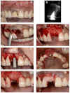
Replacement of a maxillary central incisor with an immediate implant in case #2. (a) Presurgical frontal view. (b) Cross-sectional view of preoperative CBCT. (c) Representation of the intracrevicular partial-thickness incision made around the natural teeth. (d) Preparation of periosteum tabs just apical to the buccal dehiscence in the lengths of 2–3 mm. (e) Insertion of a resorbable membrane beneath the periosteum tab. Execution of implant placement following the preparation of a pouch for lateral GBR. (f) Treatment of the internal space of the pouch surrounded by the resorbable membrane and exposed labial implant surface with a demineralized bovine bone mineral. (g) Covering of the surgical site with the membrane. (h) Securing of the connective-tissue graft to the periosteum. CBCT: cone-beam computed tomography, GBR: guided bone regeneration.
Figure 3
Figure 3
Assessment of the horizontal bone thickness with cross-sectional CBCT images. ① A vertical line was drawn in the center of the implant. ② The horizontal reference line was drawn perpendicular to the vertical line crossing the first thread of implant. ③ The horizontal bone thickness from the implant surface at the first thread to the outermost edge of the buccal bone was measured. CBCT: cone-beam computed tomography.
Figure 4
Figure 4
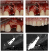
Replacement of a maxillary central incisor with an immediate implant in case #2. (a) CBCT at implant placement. (b) CBCT 1 year later. (c) Frontal view of the final implant restoration. (d) Buccal view of the final implant restoration. CBCT: cone-beam computed tomography.
Figure 5
Figure 5
Replacement of a maxillary central incisor with a delayed implant in case #6. (a) Presurgical frontal view. (b) Placement of the implant 24 weeks after tooth extraction. (c) Securing the resorbable membrane and connective-tissue graft to the periosteum. (d) Frontal view of the final implant restoration. (e) CBCT at implant placement. (f) CBCT 1 year later. CBCT: cone-beam computed tomography.

References

    1. Chen S.T., Wilson T.G., Jr., Hämmerle C.H. Immediate or early placement of implants following tooth extraction: Review of biologic basis, clinical procedures, and outcomes. Int. J. Oral Maxillofac. Implants. 2004;19:12–25. - PubMed
    1. Hürzeler M.B., Zuhr O., Schupbach P., Rebele S.F., Emmanouilidis N., Fickl S. The socket-shield technique: A proof-of-principle report. J. Clin. Periodontol. 2010;37:855–862. doi: 10.1111/j.1600-051X.2010.01595.x. - DOI - PubMed
    1. Zhou X., Yang J., Wu L., Tang X., Mou Y., Sun W., Hu Q., Xie S. Evaluation of the effect of implants placed in preserved sockets versus fresh sockets on tissue preservation and esthetics: A meta-analysis and systematic review. J. Evid. Based. Dent. Pract. 2019;19:101336. doi: 10.1016/j.jebdp.2019.05.015. - DOI - PubMed
    1. Chen S.T., Buser D. Clinical and esthetic outcomes of implants placed in postextraction sites. Int. J. Oral Maxillofac. Implants. 2009;24:186–217. - PubMed
    1. Smeets R., Matthies L., Windisch P., Gosau M., Jung R., Brodala N., Stefanini M., Kleinheinz J., Payer M., Henningsen A., et al. Horizontal augmentation techniques in the mandible: A systematic review. Int. J. Implant Dent. 2022;8:23. doi: 10.1186/s40729-022-00421-7. - DOI - PMC - PubMed

Publication types

LinkOut - more resources