. 2022 Oct 31;44(11):5312-5351.
doi: 10.3390/cimb44110361.

In Silico Identification of Promising New Pyrazole Derivative-Based Small Molecules for Modulating CRMP2, C-RAF, CYP17, VEGFR, C-KIT, and HDAC-Application towards Cancer Therapeutics

Affiliations

In Silico Identification of Promising New Pyrazole Derivative-Based Small Molecules for Modulating CRMP2, C-RAF, CYP17, VEGFR, C-KIT, and HDAC-Application towards Cancer Therapeutics

Fatima Ezzahra Bennani et al. Curr Issues Mol Biol. .

Abstract

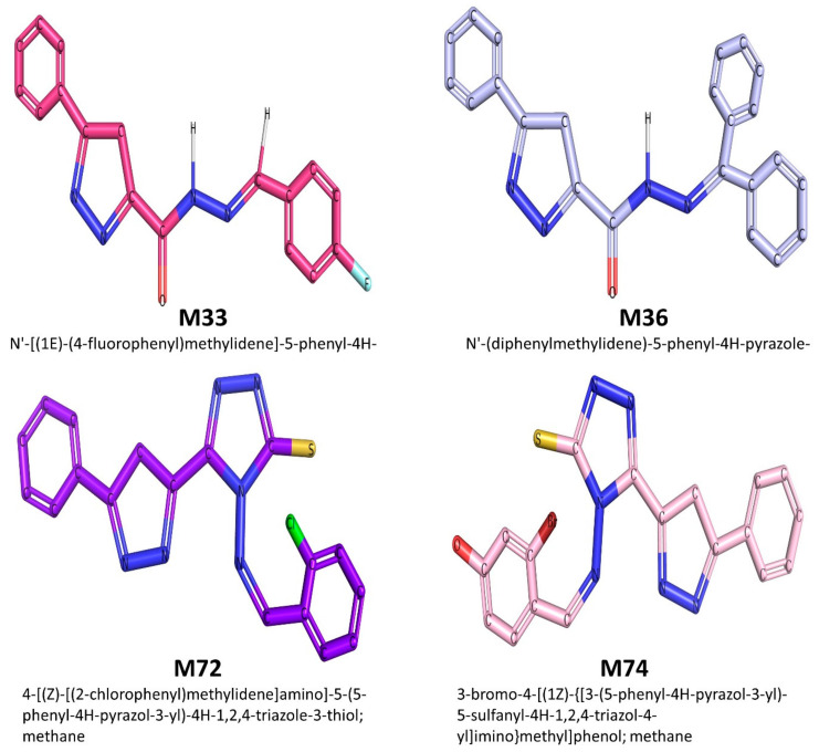
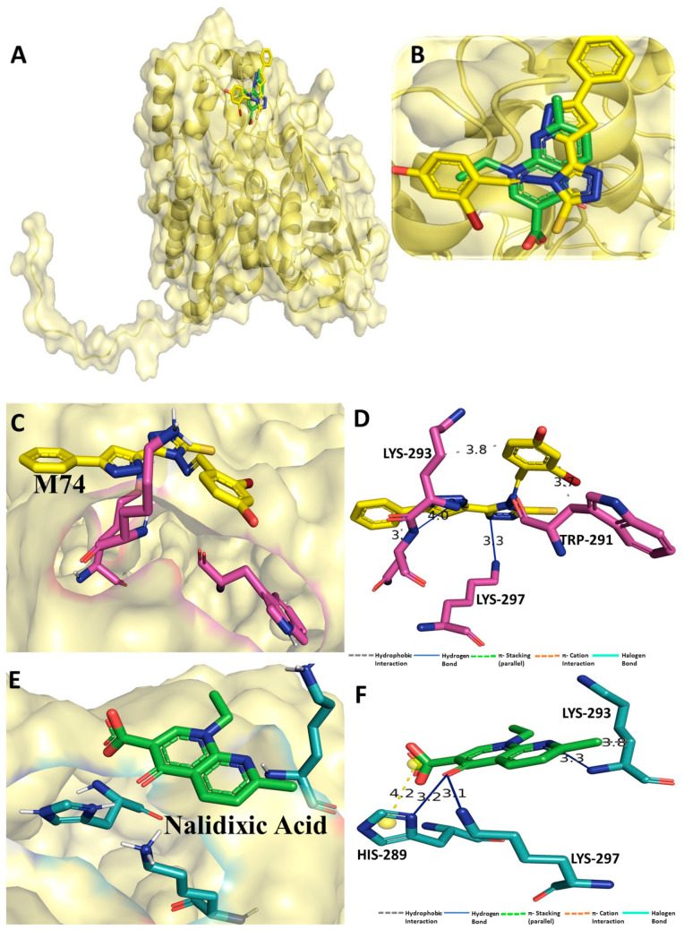
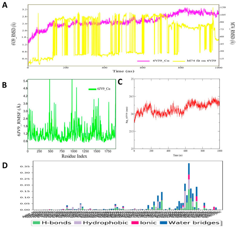
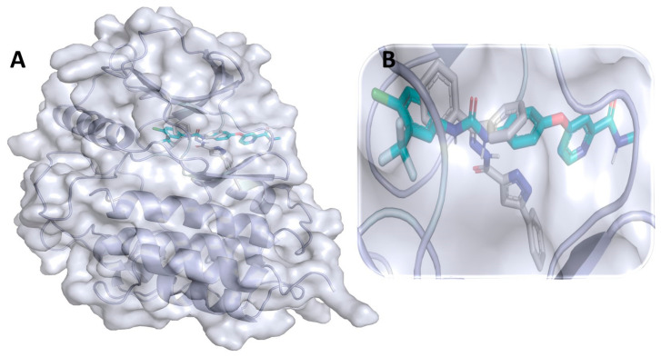
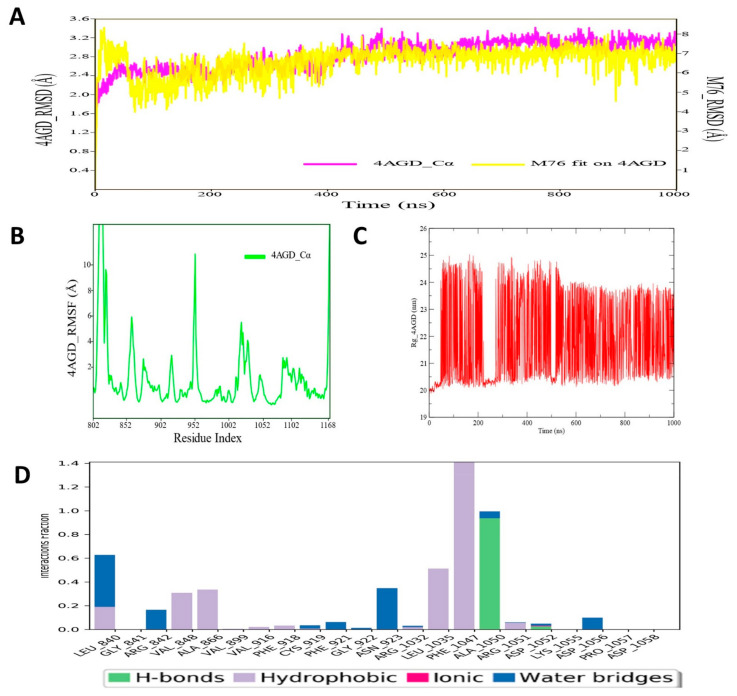
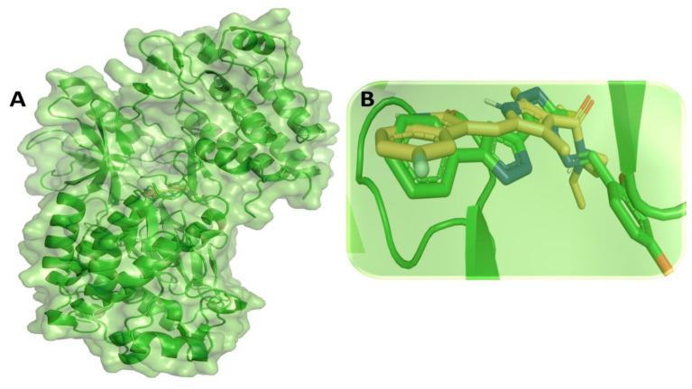
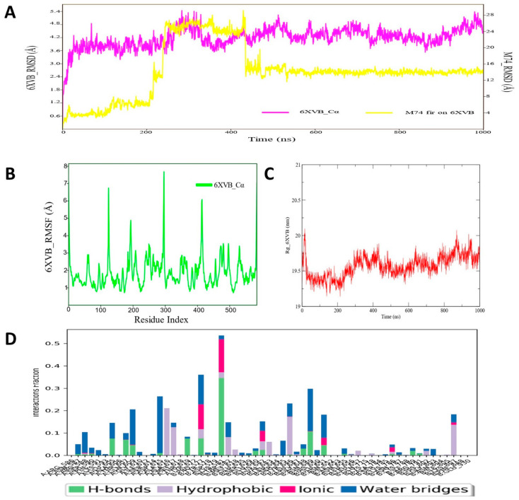
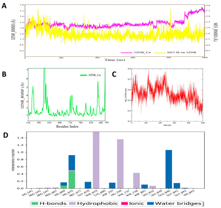
Despite continual efforts being made with multiple clinical studies and deploying cutting-edge diagnostic tools and technologies, the discovery of new cancer therapies remains of severe worldwide concern. Multiple drug resistance has also emerged in several cancer cell types, leaving them unresponsive to the many cancer treatments. Such a condition always prompts the development of next-generation cancer therapies that have a better chance of inhibiting selective target macromolecules with less toxicity. Therefore, in the present study, extensive computational approaches were implemented combining molecular docking and dynamic simulation studies for identifying potent pyrazole-based inhibitors or modulators for CRMP2, C-RAF, CYP17, c-KIT, VEGFR, and HDAC proteins. All of these proteins are in some way linked to the development of numerous forms of cancer, including breast, liver, prostate, kidney, and stomach cancers. In order to identify potential compounds, 63 in-house synthesized pyrazole-derivative compounds were docked with each selected protein. In addition, single or multiple standard drug compounds of each protein were also considered for docking analyses and their results used for comparison purposes. Afterward, based on the binding affinity and interaction profile of pyrazole compounds of each protein, potentially strong compounds were filtered out and further subjected to 1000 ns MD simulation analyses. Analyzing parameters such as RMSD, RMSF, RoG and protein-ligand contact maps were derived from trajectories of simulated protein-ligand complexes. All these parameters turned out to be satisfactory and within the acceptable range to support the structural integrity and interaction stability of the protein-ligand complexes in dynamic state. Comprehensive computational analyses suggested that a few identified pyrazole compounds, such as M33, M36, M72, and M76, could be potential inhibitors or modulators for HDAC, C-RAF, CYP72 and VEGFR proteins, respectively. Another pyrazole compound, M74, turned out to be a very promising dual inhibitor/modulator for CRMP2 and c-KIT proteins. However, more extensive study may be required for further optimization of the selected chemical framework of pyrazole derivatives to yield improved inhibitory activity against each studied protein receptor.

Keywords: cancer targets; molecular docking; molecular dynamic simulation; pyrazole derivatives.


Conflict of interest statement

The authors declare no conflict of interest.

Figures

Figure 1
Figure 1
The top-ranked compounds selected from the 63 synthesized pyrazoles derivatives.
Figure 2
Figure 2
(A) Molecular docking-generated binding orientations of standard compound (nalidixic acid) and the proposed pyrazole-based inhibitor compound M74 in complex with CRMP2 (6JV9) protein; (B) close-up binding interaction view exhibiting similar binding pattern of all the compounds at the active site cavity of CRMP2 (6JV9) protein; (C) binding mode of pyrazole compound M74 in the active site cavity of CRMP2 (6JV9) displayed in surface view representation; (D) molecular binding interaction and orientation of compound M74 with CRMP2 (6JV9) protein; (E) binding mode of standard compound (nalidixic acid) in the active site cavity of CRMP2 (6JV9) displayed in surface view representation; (F) molecular binding interaction and orientation of nalidixic acid with CRMP2 (6JV9) protein.
Figure 3
Figure 3
(A) RMSD profile of CRMP2 (6JV9) protein backbone and compound M74 during 1000 ns simulation span; (B) RMSF profile of CRMP2 (6JV9) protein backbone during 1000 ns simulation span; (C) RoG profile of CRMP2 (6JV9) protein backbone during 1000 ns simulation span; (D) illustration of the CRMP2 (6JV9)–M74 contacts or interactions map monitored during 1000 ns simulation run.
Figure 4
Figure 4
(A) Molecular docking-generated binding orientations of standard compound (sorafenib) and the proposed pyrazole-based inhibitor compound M36 in complex with C-RAF (3OMV) protein; (B) close-up binding interaction view exhibiting similar binding pattern of all the compounds at the active site cavity of C-RAF (3OMV) protein; (C) binding mode of pyrazole compound M36 in the active site cavity of C-RAF (3OMV) in surface view; (D) molecular binding interaction and orientation of compound M36 with C-RAF (3OMV) protein; (E) binding mode of standard compound (sorafenib) in the active site cavity of C-RAF (3OMV) displayed in surface view; (F) molecular binding interaction and orientation of sorafenib with C-RAF (3OMV) protein.
Figure 4
Figure 4
(A) Molecular docking-generated binding orientations of standard compound (sorafenib) and the proposed pyrazole-based inhibitor compound M36 in complex with C-RAF (3OMV) protein; (B) close-up binding interaction view exhibiting similar binding pattern of all the compounds at the active site cavity of C-RAF (3OMV) protein; (C) binding mode of pyrazole compound M36 in the active site cavity of C-RAF (3OMV) in surface view; (D) molecular binding interaction and orientation of compound M36 with C-RAF (3OMV) protein; (E) binding mode of standard compound (sorafenib) in the active site cavity of C-RAF (3OMV) displayed in surface view; (F) molecular binding interaction and orientation of sorafenib with C-RAF (3OMV) protein.
Figure 5
Figure 5
(A) RMSD profile of C-RAF (3OMV) protein backbone and compound M36 during 1000 ns simulation span; (B) RMSF profile of C-RAF (3OMV) protein backbone during 1000 ns simulation span; (C) RoG profile of C-CRAF protein backbone during 1000 ns simulation span; (D) illustration of the C-RAF (3OMV)–M36 contacts or interaction map monitored during 1000 ns simulation run.
Figure 6
Figure 6
(A) Molecular docking-generated binding orientations of all standard compounds (abiraterone acetate, galeterone, orteronel) and the proposed pyrazole-based inhibitor compound M72 in complex with CYP17 (4NKV) protein; (B) close-up binding interaction view exhibiting similar binding pattern of all the standard compounds including M72 at the active site cavity of CYP17 (4NKV) protein; (C) binding mode of pyrazole compound M72 in the active site cavity of CYP17 (4NKV) displayed in surface view; (D) molecular binding interaction and orientation of compound M72 with CYP17 (4NKV) protein; (E) binding mode of standard compound (abiraterone acetate) in the active site cavity of CYP17 (4NKV) displayed in surface view; (F) molecular binding interaction and orientation of abiraterone acetate with CYP17 (4NKV) protein; (G) binding mode of standard compound galeterone in active site cavity of CYP17 (4NKV) displayed in surface view; (H) molecular binding interaction and orientation of galeterone with CYP17 (4NKV) protein; (I) binding mode of standard compound orteronel in active site cavity of CYP17 (4NKV) displayed in surface view; (J) molecular binding interaction and orientation of orteronel with CYP17 (4NKV) protein.
Figure 6
Figure 6
(A) Molecular docking-generated binding orientations of all standard compounds (abiraterone acetate, galeterone, orteronel) and the proposed pyrazole-based inhibitor compound M72 in complex with CYP17 (4NKV) protein; (B) close-up binding interaction view exhibiting similar binding pattern of all the standard compounds including M72 at the active site cavity of CYP17 (4NKV) protein; (C) binding mode of pyrazole compound M72 in the active site cavity of CYP17 (4NKV) displayed in surface view; (D) molecular binding interaction and orientation of compound M72 with CYP17 (4NKV) protein; (E) binding mode of standard compound (abiraterone acetate) in the active site cavity of CYP17 (4NKV) displayed in surface view; (F) molecular binding interaction and orientation of abiraterone acetate with CYP17 (4NKV) protein; (G) binding mode of standard compound galeterone in active site cavity of CYP17 (4NKV) displayed in surface view; (H) molecular binding interaction and orientation of galeterone with CYP17 (4NKV) protein; (I) binding mode of standard compound orteronel in active site cavity of CYP17 (4NKV) displayed in surface view; (J) molecular binding interaction and orientation of orteronel with CYP17 (4NKV) protein.
Figure 7
Figure 7
(A) RMSD profile of CYP17 (4NKV) protein backbone and compound M72 during 1000 ns simulation; (B) RMSF profile of CYP17 (4NKV) protein backbone during 1000 ns simulation; (C) RoG profile of CYP17 (4NKV) protein backbone during 1000 ns simulation; (D) illustration of the CYP17 (4NKV)–M72 contacts or interactions map monitored during 1000 ns simulation.
Figure 8
Figure 8
(A) Molecular docking-generated binding orientations of standard compound (sunitinib) and the proposed pyrazole-based inhibitor compound M76 in complex with VEGFR (4AGD) protein; (B) close-up binding interaction view exhibiting similar binding pattern of all the compounds at the active site cavity of VEGFR (4AGD) protein; (C) binding mode of pyrazole compound M76 in the active site cavity of VEGFR (4AGD) displayed in surface view; (D) molecular binding interaction and orientation of compound M76 with VEGFR (4AGD) protein; (E) binding mode of standard compound (sunitinib) in the active site cavity of VEGFR (4AGD) displayed in surface view; (F) molecular binding interaction and orientation of sunitinib with VEGFR (4AGD) protein.
Figure 8
Figure 8
(A) Molecular docking-generated binding orientations of standard compound (sunitinib) and the proposed pyrazole-based inhibitor compound M76 in complex with VEGFR (4AGD) protein; (B) close-up binding interaction view exhibiting similar binding pattern of all the compounds at the active site cavity of VEGFR (4AGD) protein; (C) binding mode of pyrazole compound M76 in the active site cavity of VEGFR (4AGD) displayed in surface view; (D) molecular binding interaction and orientation of compound M76 with VEGFR (4AGD) protein; (E) binding mode of standard compound (sunitinib) in the active site cavity of VEGFR (4AGD) displayed in surface view; (F) molecular binding interaction and orientation of sunitinib with VEGFR (4AGD) protein.
Figure 9
Figure 9
(A) RMSD profile of VEGFR (4AGD) protein backbone and compound M76 during 1000 ns simulation; (B) RMSF profile of VEGFR (4AGD) protein backbone during 1000 ns simulation; (C) RoG profile of VEGFR (4AGD) protein backbone during 1000 ns simulation; (D) illustration of the VEGFR (4AGD)–M76 contacts or interactions map monitored during 1000 ns simulation.
Figure 10
Figure 10
(A) Molecular docking-generated binding orientations of standard compound (sunitinib) and the proposed pyrazole-based inhibitor compound M74 in complex with c-KIT (6XVB) protein; (B) close-up binding interaction view exhibiting similar binding pattern of all the compounds at the active site cavity of c-KIT (6XVB) protein; (C) binding mode of pyrazole compound M74 in the active site cavity of c-KIT (6XVB) displayed in surface view; (D) molecular binding interaction and orientation of compound M74 with c-KIT (6XVB) protein; (E) binding mode of standard compound (sunitinib) in the active site cavity of c-KIT (6XVB) displayed in surface view; (F) molecular binding interaction and orientation of sunitinib with c-KIT (6XVB) protein.
Figure 10
Figure 10
(A) Molecular docking-generated binding orientations of standard compound (sunitinib) and the proposed pyrazole-based inhibitor compound M74 in complex with c-KIT (6XVB) protein; (B) close-up binding interaction view exhibiting similar binding pattern of all the compounds at the active site cavity of c-KIT (6XVB) protein; (C) binding mode of pyrazole compound M74 in the active site cavity of c-KIT (6XVB) displayed in surface view; (D) molecular binding interaction and orientation of compound M74 with c-KIT (6XVB) protein; (E) binding mode of standard compound (sunitinib) in the active site cavity of c-KIT (6XVB) displayed in surface view; (F) molecular binding interaction and orientation of sunitinib with c-KIT (6XVB) protein.
Figure 11
Figure 11
(A) RMSD profile of c-KIT (6XVB) protein backbone and compound M74 during 1000 ns simulation; (B) RMSF profile of c-KIT (6XVB) protein backbone during 1000 ns simulation; (C) RoG profile of c-KIT (6XVB) protein backbone during 1000 ns simulation; (D) illustration of the c-KIT (6XVB)–M74 contacts or interactions map monitored during 1000 ns simulation.
Figure 12
Figure 12
(A) Molecular docking-generated binding orientations of all standard compounds (SAHA; TMP269) and the proposed pyrazole inhibitor compound M33 in complex with HDAC7 (3ZNR) protein; (B) close-up binding interaction view exhibiting similar binding pattern of all the standard compounds including M33 at the active site cavity of HDAC7 (3ZNR) protein; (C) binding mode of the pyrazole derivative compound M33 in the active site cavity of HDAC7 (3ZNR) displayed in surface view; (D) molecular binding interaction and orientation of compound M33 with HDAC7 (3ZNR) protein; (E) binding mode of standard inhibitor (SAHA) in the active site cavity of HDAC7 (3ZNR) displayed in surface view; (F) molecular binding interaction and orientation of SAHA with HDAC7 (3ZNR) protein; (G) binding mode of standard compound TMP269 in active site cavity of HDAC7 (3ZNR) displayed in surface view; (H) molecular binding interaction and orientation of TMP269 with HDAC7 (3ZNR) protein.
Figure 12
Figure 12
(A) Molecular docking-generated binding orientations of all standard compounds (SAHA; TMP269) and the proposed pyrazole inhibitor compound M33 in complex with HDAC7 (3ZNR) protein; (B) close-up binding interaction view exhibiting similar binding pattern of all the standard compounds including M33 at the active site cavity of HDAC7 (3ZNR) protein; (C) binding mode of the pyrazole derivative compound M33 in the active site cavity of HDAC7 (3ZNR) displayed in surface view; (D) molecular binding interaction and orientation of compound M33 with HDAC7 (3ZNR) protein; (E) binding mode of standard inhibitor (SAHA) in the active site cavity of HDAC7 (3ZNR) displayed in surface view; (F) molecular binding interaction and orientation of SAHA with HDAC7 (3ZNR) protein; (G) binding mode of standard compound TMP269 in active site cavity of HDAC7 (3ZNR) displayed in surface view; (H) molecular binding interaction and orientation of TMP269 with HDAC7 (3ZNR) protein.
Figure 13
Figure 13
(A) RMSD profile of HDAC7 (3ZNR) protein backbone and compound M74 during 1000 ns simulation; (B) RMSF profile of c-KIT (6XVB) protein backbone during 1000 ns simulation; (C) RoG profile of c-KIT (6XVB) protein backbone during 1000 ns simulation; (D) illustration of the c-KIT (6XVB)–M74 contacts or interactions map monitored during 1000 ns simulation run.

Similar articles

Cited by

References

    1. Yacoub E., Abdul-Wahab O.M.S., Al-Shyarba M.H., Mardassi B.B.A. The Relationship between Mycoplasmas and Cancer: Is It Fact or Fiction? Narrative Review and Update on the Situation. J. Oncol. 2021;2021:9986550. doi: 10.1155/2021/9986550. - DOI - PMC - PubMed
    1. Yahya E.B., Alqadhi A.M. Recent trends in cancer therapy: A review on the current state of gene delivery. Life Sci. 2021;269:119087. doi: 10.1016/j.lfs.2021.119087. - DOI - PubMed
    1. Sung H., Ferlay J., Siegel R.L., Laversanne M., Soerjomataram I., Jemal A., Bray F. Global Cancer Statistics 2020: GLOBOCAN Estimates of Incidence and Mortality Worldwide for 36 Cancers in 185 Countries. CA Cancer J. Clin. 2021;71:209–249. doi: 10.3322/caac.21660. - DOI - PubMed
    1. Kocyigit U.M., Budak Y., Gürdere M.B., Tekin Ş., Köprülü T.K., Ertürk F., Ceylan M. Synthesis, characterization, anticancer, antimicrobial and carbonic anhydrase inhibition profiles of novel (3aR,4S,7R,7aS)-2-(4-((E)-3-(3-aryl)acryloyl)phenyl)-3a,4,7,7a-tetrahydro-1H-4,7 methanoisoindole-1,3(2H)-dione derivatives. Bioorg Chem. 2017;70:118–125. doi: 10.1016/j.bioorg.2016.12.001. - DOI - PubMed
    1. Dastan T., Kocyigit U.M., Durna Dastan S., Canturk Kilickaya P., Taslimi P., Cevik O., Cetin A. Investigation of acetylcholinesterase and mammalian DNA topoisomerases, carbonic anhydrase inhibition profiles, and cytotoxic activity of novel bis(α-aminoalkyl)phosphinic acid derivatives against human breast cancer. J. Biochem. Mol. Toxicol. 2017;31:e2171. doi: 10.1002/jbt.21971. - DOI - PubMed