. 2022 Oct 27;13(11):986.
doi: 10.3390/insects13110986.

RNAi-Mediated Manipulation of Cuticle Coloration Genes in Lygus hesperus Knight (Hemiptera: Miridae)

Affiliations

RNAi-Mediated Manipulation of Cuticle Coloration Genes in Lygus hesperus Knight (Hemiptera: Miridae)

Colin S Brent et al. Insects. .

Abstract

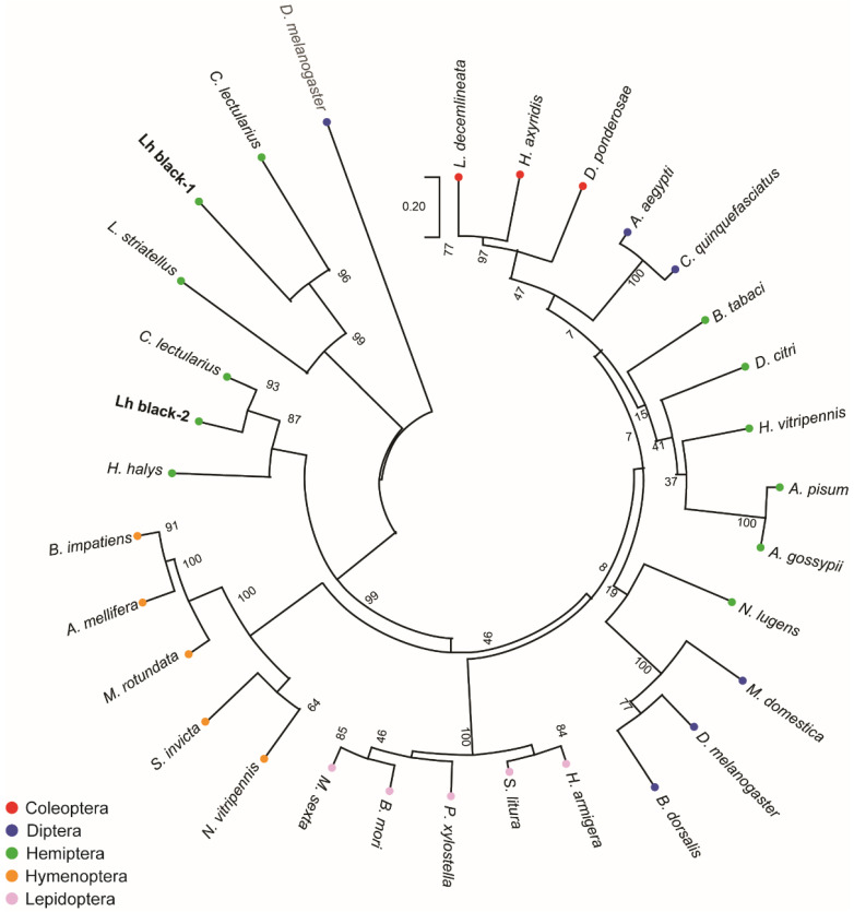
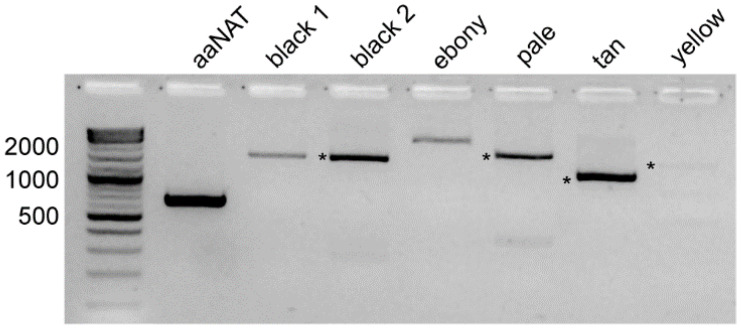
Cuticle coloration in insects is a consequence of the accumulation of pigments in a species-specific pattern. Numerous genes are involved in regulating the underlying processes of melanization and sclerotization, and their manipulation can be used to create externally visible markers of successful gene editing. To clarify the roles for many of these genes and examine their suitability as phenotypic markers in Lygus hesperus Knight (western tarnished plant bug), transcriptomic data were screened for sequences exhibiting homology with the Drosophila melanogaster proteins. Complete open reading frames encoding putative homologs for six genes (aaNAT, black, ebony, pale, tan, and yellow) were identified, with two variants for black. Sequence and phylogenetic analyses supported preliminary annotations as cuticle pigmentation genes. In accord with observable difference in color patterning, expression varied for each gene by developmental stage, adult age, body part, and sex. Knockdown by injection of dsRNA for each gene produced varied effects in adults, ranging from the non-detectable (black 1, yellow), to moderate decreases (pale, tan) and increases (black 2, ebony) in darkness, to extreme melanization (aaNAT). Based solely on its expression profile and highly visible phenotype, aaNAT appears to be the best marker for tracking transgenic L. hesperus.

Keywords: Lygus hesperus; RNAi; cuticular pigmentation; melanin pathway.


Conflict of interest statement

The authors declare no conflict of interest.

Figures

Figure 1
Figure 1
Model of melanin and sclerotin biosynthesis in insect cuticles. Tyrosine is converted by tyrosine hydroxylase (pale) to DOPA, which in turn can be transformed by DDC into dopamine. DOPA and dopamine can be converted into brown and black melanins through several intermediate steps that are promoted by yellow. Dopamine also can be converted by synthases into colorless or yellow sclerotin pigments. Production of clear NADA sclerotins is catalyzed by aaNAT. Production of the yellow to tan NBAD sclerotin is promoted by ebony and black, but its reversion is promoted by tan. Adapted from [17,26].
Figure 2
Figure 2
Phylogenetic analysis of putative black homologs in L. hesperus and other insects. The tree with the highest log likelihood (−14,891.07) is shown. The tree was rooted to the Drosophila melanogaster glutamic acid decarboxylase 1 and is drawn to scale, with branch lengths measured in the number of substitutions per site. Insect orders have been color coded: Coleoptera—red; Diptera—blue; Hemiptera—green; Hymenoptera—orange; and Lepidoptera—pink. Numbers at the nodes indicate bootstrap support across 1000 replicates. Accession numbers for the sequences used are listed in Table S2.
Figure 3
Figure 3
RT-PCR amplification of melanin pathway transcripts. Products were generated from mature adult body cDNAs using primers designed to amplify complete open reading frames. Expected sizes are: aaNAT—669 bp; black 1—1455 bp; black 2—1542 bp; ebony—2343 bp; pale—1680 bp; tan—1131 bp; and yellow—1320 bp. In lanes with multiple bands, products of the expected sizes are marked with an asterisk. All products were subcloned and sequenced.
Figure 4
Figure 4
Expression profile of Lygus hesperus cuticular pigment-associated transcripts. (A) Developmental expression of transcripts from egg (E) through nymphal development (1st–5th), and at varying points in adult maturation including adults at emergence (d0), and at 7 (d7), and 20 days (d20) post-eclosion. (B) Limited sex-specific tissue profile using head (H), thoracic (T), and abdominal (A) segments of reproductively mature adults (i.e., 7 day-old). Amplimers are ~500-bp fragments of the transcripts of interest. Gels shown are representative of results obtained across three replicates.
Figure 5
Figure 5
RT-PCR confirmation of target transcript knockdown by dsRNA-mediated RNAi. Products correspond to ~500-bp fragments of the transcripts of interest amplified from single adults at 7 days post-injection. Gels shown are representative of results obtained across five replicates. Abbreviations are: N (noninjected); V (venus dsRNA injected); T (target dsRNA injected); NT (no template).
Figure 6
Figure 6
Effects of RNA interference-mediated knockdown on Lygus hesperus cuticle coloration. Shown are full-body images (dorsal, lateral and ventral views) of (A) female and (B) male L. hesperus adults at 7 days post-eclosion that were either untreated wildtype (WT) or injected as 5th instars with dsRNA of the control gene venus, or the indicated cuticle pigment-associated gene. Images are representative of three biological replicates consisting of 40 bugs of each sex.
Figure 7
Figure 7
Effects of RNA interference-mediated knockdown on Lygus hesperus cuticle coloration of individual body parts. Shown are the eyes, antennae, pronotum and head, rear right leg, and right forewing of (A) female and (B) male L. hesperus adults at 7 days post-eclosion that were either untreated wildtype (WT) or injected as fifth instars with dsRNA of the control gene venus, or the indicated cuticle pigment-associated gene. Images are representative of three biological replicates consisting of 40 bugs of each sex.

Similar articles

References

    1. Scott D.R. An annotated listing of host plants of Lygus hesperus Knight. Bull. Entomol. Soc. Am. 1977;23:19–22. doi: 10.1093/besa/23.1.19. - DOI
    1. Wheeler A.G. Biology of the Plant Bugs (Hemiptera: Miridae): Pests, Predators, Opportunists. Comstock Publishing Associates; Ithaca, NY, USA: 2001. p. 562.
    1. Naranjo S., Ellsworth P., Dierig D. Impact of Lygus spp. (Hemiptera: Miridae) on damage, yield and quality of lesquerella (Physaria fendleri), a potential new oil-seed crop. J. Econ. Entomol. 2011;104:1575–1583. doi: 10.1603/EC11089. - DOI - PubMed
    1. Ritter R.A., Lenssen A., Blodgett S., Taper M.A. Regional assemblages of Lygus (Heteroptera: Miridae) in Montana canola fields. J. Kans. Entomol. Soc. 2010;83:297–305. doi: 10.2317/JKES0912.23.1. - DOI
    1. Strand L.L. Integrated Pest Management for Strawberries. 2nd ed. University of California Agriculture and Natural Resources Publications; Oakland, CA, USA: 2008. p. 3351.

Grants and funding

LinkOut - more resources