. 2022 Oct 27;8(11):1133.
doi: 10.3390/jof8111133.

Efficacy of Bovine Nail Membranes as In Vitro Model for Onychomycosis Infected by Trichophyton Species

Affiliations

Efficacy of Bovine Nail Membranes as In Vitro Model for Onychomycosis Infected by Trichophyton Species

Marta Elisabetta Eleonora Temporiti et al. J Fungi (Basel). .

Abstract

Onychomycosis is a fungal infection caused by different etiologic agents, including dermatophytes that specifically colonize keratin-rich substrates. The aim of this work was to investigate mechanical modifications of bovine membranes (used as an in vitro nail model) placed in contact with Trichophyton species. Trichophyton strains were isolated from toenails specimens. The procedure was set up by spreading T. rubrum,T. interdigitale, and T. mentagrophytes strains on Petri dishes with minimal and rich media; after that, bovine membranes were placed in the center. After 27 days, T. interdigitale and T. mentagrophytes significantly reduced the thickness of the colonized membranes, whereas two T. rubrum strains showed the highest degradation limited to the small colonized area. These results were confirmed by SEM images of the colonization profile on membranes. Mechanical analyses performed on membranes were used as an innovative method to evaluate the thickness and structural integrity of membranes variation following fungal colonization. In conclusion, mechanical analyses of substrate may be used as a procedure for the development of a new onychomycosis diagnosis test in order to develop personalized and strain-specific treatment.

Keywords: Trichophyton; bovine nail membranes; dermatophytes; onychomycosis.


Conflict of interest statement

The authors declare no conflict of interest.

Figures

Figure 1
Figure 1
Percentage of etiological agents of onychomycosis isolated at the Mycology Laboratory of DSTA (UNIPV, Italy). Filamentous fungi are indicated with blue bars, whereas yeasts with yellow bars.
Figure 2
Figure 2
Growth tests of T. interdigitale 10379 with SDA (first line), ½ SDA (second line), and WA (third line) after 14 days of incubation at 30 °C (frontal view) (A) and after 7 days at 30 °C (back of Petri dishes) (B).
Figure 3
Figure 3
Growth tests of T. rubrum (1000A, 1000B, 10318, 10351, 10356, and 10380), T. mentagrophytes (10326 and 10340), and T. interdigitale (10379) in WA (blue), ½ SDA (orange), and SDA (grey). Error bars indicate the standard deviation of the three replicates used for the tests. ANOVA tests were performed, and statistically significant differences are marked with *.
Figure 4
Figure 4
Time (days) elapsed since first visible contact with membrane of 7 dermatophyte strains in WA (light blue) and ½ SDA (orange). Error bars indicate the standard deviation of the three replicates used for the tests.
Figure 5
Figure 5
T. mentagrophytes 10340 first visible contact with bovine membrane in WA medium at day 6 (a), and T. interdigitale 10379 grown in ½ SDA initial colonization (b).
Figure 6
Figure 6
T. interdigitale 10326 growth on bovine membrane in WA at 10, 12, 15, and 27 days (from left to right).
Figure 7
Figure 7
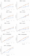
Membrane coverage percentage by T. rubrum 1000A (a) and 10318 (b), T. mentagrophytes 10326 (c), T. mentagrophytes 10340 (d), T. interdigitale 10379 (e) in WA (blue line) and ½ SDA (orange line).
Figure 8
Figure 8
Percentage decrease in the CA value of membranes before and after fungal colonization.
Figure 9
Figure 9
Membrane thickness at time zero (t0) and after 27 days of fungal colonization (t27) in ½ SDA (a) and in WA (b). Error bars indicate the standard deviation of the three replicates used for the tests. Two-sample paired t-test were performed, and statistically significant differences are marked with *.
Figure 9
Figure 9
Membrane thickness at time zero (t0) and after 27 days of fungal colonization (t27) in ½ SDA (a) and in WA (b). Error bars indicate the standard deviation of the three replicates used for the tests. Two-sample paired t-test were performed, and statistically significant differences are marked with *.
Figure 10
Figure 10
SEM image of control membrane without fungal growth (a); SEM image of T. mentagrophytes 10326 mycelium on bovine membrane after 27 days (b).
Figure 11
Figure 11
SEM images of colonization and growth on membrane by T. mentagrophytes 10326, in different magnifications. In each image, a membrane portion not yet colonized is visible on the left (black arrows), and the already developed mycelium on the right (white arrows). In the center, there is the point of contact between the fungus and the membrane, which at higher magnifications can be seen to be degraded (yellow arrows).

Similar articles

Cited by

References

    1. Gupta A., Mays R., Versteeg S., Piraccini B., Takwale A., Shemer A., Babaev M., Grover C., Di Chiacchio N., Taborda V., et al. Global perspectives for the management of onychomycosis. Int. J. Dermatol. 2019;58:1118–1129. doi: 10.1111/ijd.14346. - DOI - PubMed
    1. Piraccini B.M., Alessandrini A. Onychomycosis: A review. J. Fungi. 2015;1:30–43. doi: 10.3390/jof1010030. - DOI - PMC - PubMed
    1. Kaur R., Kashyap B., Bhalla P. Onychomycosis–Epidemiology, diagnosis and management. Indian J. Med. Microbiol. 2008;26:108–116. doi: 10.1016/S0255-0857(21)01924-1. - DOI - PubMed
    1. Leung A.K.C., Lam J.M., Leong K.F., Hon K.L., Barankin B., Leung A.A.M., Wong A.H.C. Onychomycosis: An Updated Review. Recent Pat. Inflamm. Allergy Drug Discov. 2020;14:32–45. - PMC - PubMed
    1. Gupta A., Versteeg S., Shear N., Piguet V., Tosti A., Piraccini B. A pratical guide to curing onychomycosis: How to maximize cure at the patient, organism, treatment and environmental level. Am. J. Clin. Dermatol. 2019;20:123–133. doi: 10.1007/s40257-018-0403-4. - DOI - PubMed

LinkOut - more resources