

Oncolytic Vaccinia Virus Carrying Aphrocallistes vastus Lectin (oncoVV-AVL) Enhances Inflammatory Response in Hepatocellular Carcinoma Cells
- PMID: 36354990
- PMCID: PMC9696330
- DOI: 10.3390/md20110667
Oncolytic Vaccinia Virus Carrying Aphrocallistes vastus Lectin (oncoVV-AVL) Enhances Inflammatory Response in Hepatocellular Carcinoma Cells
Abstract
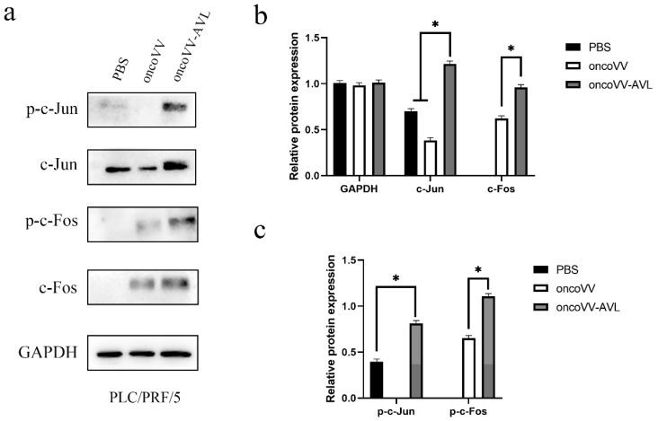
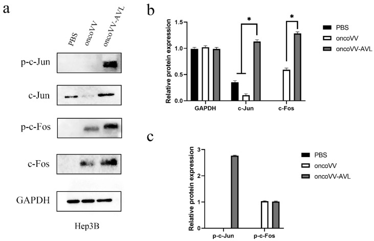
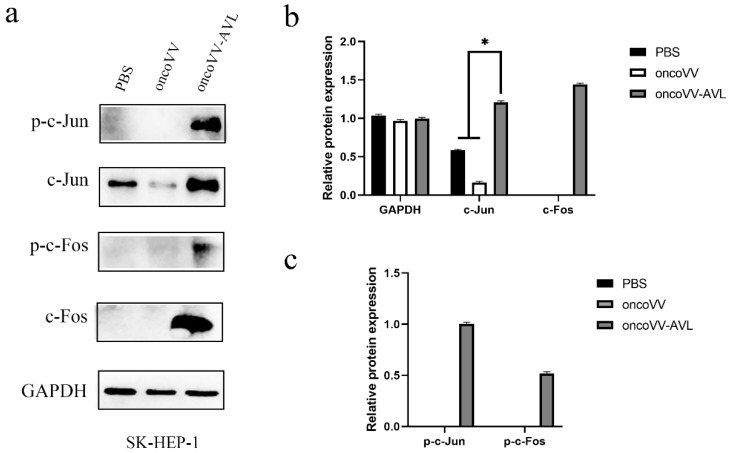
Aphrocallistes vastus lectin (AVL) is a C-type marine lectin derived from sponges. Our previous study demonstrated that oncolytic vaccinia virus carrying AVL (oncoVV-AVL) significantly enhanced the cytotoxicity of oncoVV in cervical cancer, colorectal cancer and hepatocellular carcinoma through the activation of Ras/ERK, MAPK/ERK and PI3K/Akt signaling pathways. In this study, the inflammatory response induced by oncoVV-AVL in a hepatocellular carcinoma cell (HCC) model was investigated. The results showed that oncoVV-AVL increased the levels of inflammatory cytokines including IL-6, IL-8 and TNF-α through activating the AP-1 signaling pathway in HCC. This study provides novel insights into the utilization of lectin AVL in the field of cancer therapy.
Keywords: AP-1; Aphrocallistes vastus lectin; hepatocellular carcinoma; oncolytic vaccinia virus.
Conflict of interest statement
The authors declare no conflict of interest.
Figures


Similar articles
-
Oncolytic Vaccinia Virus Harboring Aphrocallistes vastus Lectin Inhibits the Growth of Hepatocellular Carcinoma Cells.Mar Drugs. 2022 Jun 4;20(6):378. doi: 10.3390/md20060378. Mar Drugs. 2022. PMID: 35736181 Free PMC article.
-
Effects of Oncolytic Vaccinia Viruses Harboring Different Marine Lectins on Hepatocellular Carcinoma Cells.Int J Mol Sci. 2023 Feb 14;24(4):3823. doi: 10.3390/ijms24043823. Int J Mol Sci. 2023. PMID: 36835232 Free PMC article.
-
Oncolytic Vaccinia Virus Expressing Aphrocallistes vastus Lectin as a Cancer Therapeutic Agent.Mar Drugs. 2019 Jun 19;17(6):363. doi: 10.3390/md17060363. Mar Drugs. 2019. PMID: 31248066 Free PMC article.
-
Exploring the role of oncolytic viruses in hepatobiliary cancers.Immunotherapy. 2018 Aug;10(11):971-986. doi: 10.2217/imt-2018-0048. Epub 2018 Jun 14. Immunotherapy. 2018. PMID: 29900755
-
Viruses as nanomedicine for cancer.Int J Nanomedicine. 2016 Sep 21;11:4835-4847. doi: 10.2147/IJN.S116447. eCollection 2016. Int J Nanomedicine. 2016. PMID: 27703350 Free PMC article. Review.
Cited by
-
An Oncolytic Vaccinia Virus Expressing Aphrocallistes Vastus Lectin Modulates Hepatocellular Carcinoma Metabolism via ACSS2/TFEB-Mediated Autophagy and Lipid Accumulation.Mar Drugs. 2025 Jul 24;23(8):297. doi: 10.3390/md23080297. Mar Drugs. 2025. PMID: 40863614 Free PMC article.
-
Oncolytic Virus Senecavirus A Inhibits Hepatocellular Carcinoma Proliferation and Growth by Inducing Cell Cycle Arrest and Apoptosis.J Clin Transl Hepatol. 2024 Aug 28;12(8):713-725. doi: 10.14218/JCTH.2024.00125. Epub 2024 Jun 20. J Clin Transl Hepatol. 2024. PMID: 39130624 Free PMC article.
-
Oncolytic vaccinia virus harboring CLEC2A gene enhances viral replication and antitumor efficacy.Mol Ther Oncol. 2024 Jun 5;32(3):200823. doi: 10.1016/j.omton.2024.200823. eCollection 2024 Sep 19. Mol Ther Oncol. 2024. PMID: 39006946 Free PMC article.
-
To reveal the key mechanism of Citri Reticulatae Pericarpium-Reynoutria japonica Houtt in the treatment of liver cancer and its correlation with lipid metabolism: synergetic effect with network pharmacology, molecular docking and bioinformatics.Discov Oncol. 2025 Jun 16;16(1):1124. doi: 10.1007/s12672-025-02708-8. Discov Oncol. 2025. PMID: 40524029 Free PMC article.
-
A Comparative Study of Oncolytic Vaccinia Viruses Harboring Different Marine Lectins in Breast Cancer Cells.Mar Drugs. 2023 Jan 23;21(2):77. doi: 10.3390/md21020077. Mar Drugs. 2023. PMID: 36827118 Free PMC article.
References
-
- Hayashi K., Walde P., Miyazaki T., Sakayama K., Nakamura A., Kameda K., Masuda S., Umakoshi H., Kato K. Active Targeting to Osteosarcoma Cells and Apoptotic Cell Death Induction by the Novel Lectin Eucheuma serra Agglutinin Isolated from a Marine Red Alga. J. Drug Deliv. 2012;2012:842785. doi: 10.1155/2012/842785. - DOI - PMC - PubMed
MeSH terms
Substances
Grants and funding
LinkOut - more resources
Full Text Sources
Medical
Miscellaneous
