. 2022 Dec 1;149(23):dev200636.
doi: 10.1242/dev.200636. Epub 2022 Dec 13.

Discoidin domain receptor regulates ensheathment, survival and caliber of peripheral axons

Affiliations

Discoidin domain receptor regulates ensheathment, survival and caliber of peripheral axons

Megan M Corty et al. Development. .

Abstract

Most invertebrate axons and small-caliber axons in mammalian peripheral nerves are unmyelinated but still ensheathed by glia. Here, we use Drosophila wrapping glia to study the development and function of non-myelinating axon ensheathment, which is poorly understood. Selective ablation of these glia from peripheral nerves severely impaired larval locomotor behavior. In an in vivo RNA interference screen to identify glial genes required for axon ensheathment, we identified the conserved receptor tyrosine kinase Discoidin domain receptor (Ddr). In larval peripheral nerves, loss of Ddr resulted in severely reduced ensheathment of axons and reduced axon caliber, and we found a strong dominant genetic interaction between Ddr and the type XV/XVIII collagen Multiplexin (Mp), suggesting that Ddr functions as a collagen receptor to drive axon wrapping. In adult nerves, loss of Ddr decreased long-term survival of sensory neurons and significantly reduced axon caliber without overtly affecting ensheathment. Our data establish essential roles for non-myelinating glia in nerve development, maintenance and function, and identify Ddr as a key regulator of axon-glia interactions during ensheathment and establishment of axon caliber.

Keywords: Drosophila; Axon ensheathment; Multiplexin; Remak Schwann cell; Wrapping glia.


Conflict of interest statement

Competing interests The authors declare no competing or financial interests.

Figures
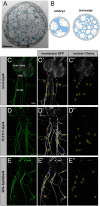
Fig. 1.
Fig. 1.
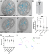
Construction of Drosophila wrapping glia Split-Gal4 driver. (A) TEM cross-section of a third instar larval nerve. Light, round profiles of larval axons (ax) are surrounded by darker (pseudo-colored cyan) wrapping glia. Subperineurial (SPG) and perineurial glia (PG; not colored) form the outer layers of the nerve. (B) Schematic of larval nerve cross-sections at embryonic and third instar stages depicting ensheathment status. Axons are depicted in white; wrapping glia coverage is shown in blue. (C-E″) Expression patterns of Gal4 lines in third instar larvae. Each Gal4 depicted is driving a membrane-bound GFP (green) and nuclear mCherry (magenta). Wrapping glia nuclei along nerves are indicated with yellow circles. (C-C″) Nrv2-Gal4 drives UAS expression exclusively in wrapping glia in the PNS (nuclei along nerves), but also in multiple types of CNS glia. (D-D″) IT.0117-Gal4 drives UAS expression in wrapping glia and a small subset of CNS neurons. (E-E″) WG-SplitGal4 (nrv2-Gal4DBD and IT.0117-Gal4VP16AD) drives UAS expression exclusively in wrapping glia.
Fig. 2.
Fig. 2.
Genetic ablation of wrapping glia impairs larval crawling behavior. (A) Expression pattern of WG-SplitGal4 driving UAS-CD8:GFP (green) and HRP counterstain (magenta). Scale bar: 50 µm. (B) Third instar larva with genetically ablated wrapping glia. Note the lack of GFP along nerves. Scale bar: 50 µm. (C) TEM of a nerve from a wrapping glia-ablated animal. Note axons in contact with each other without intervening glial membrane. Scale bar: 1 µm (D) Larval crawling behavior is impaired when wrapping glia are ablated. Unpaired t-test, P<0.0001. n=number of larvae/condition. Error bars represent s.d. (E) Representative crawling paths of control and ablated larvae.
Fig. 3.
Fig. 3.
Ddr is required for normal wrapping glia morphogenesis. (A-C) Ddr RNAi knockdown with two independent constructs; nrv2-Gal4-driven tdTomato is pseudocolored green, Futsch+ axons (magenta). (D) The Ddr genetic locus: coding region (cyan); Df(2L)BSC186; BAC clone for rescue; Ddr transcripts RG and RD (exons shown in cyan); RNAi target regions (magenta); and locations of guide RNAs used for the CRISPR-mediated mutagenesis (magenta) are depicted. (E-G) Representative images of Ddr homozygous mutants. Compared with control (E), both CRISPR mutant alleles show severe defective glia coverage in nerve cross-sections (F,G). (H) Ddr is a transmembrane receptor tyrosine kinase characterized by extracellular discoidin and discoidin-like domains and an intracellular tyrosine kinase domain. The mutant alleles generated by CRISPR-Cas9 result in truncated peptides without any full domains. (I-L) Representative images of Ddr loss-of-function and rescue experiments. (I) nrv2-Gal4, UAS-CD8:GFP/+ control. (J) Ddr41-2F/DfBSC186 (‘Ddr mutant’). This example shows a ‘moderate’ phenotype; more examples can be seen in Fig. S2A. (K) A BAC containing the Ddr locus restores normal morphology, as does expression of a 1xUAS-Ddr construct in wrapping glia (L). (M) Categorical scoring of nerve wrapping glia phenotypes. n=number of nerves scored (8-14 larvae per condition). Scale bars: 5 µm.
Fig. 4.
Fig. 4.
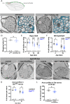
Loss of Ddr impairs axon ensheathment in larval nerves. TEM cross-sections of an abdominal nerve from third instar larva from each of the following genotypes: (A) control: nrv2-Gal4, UAS-CD8:GFP/+; (B) control: w1118; (C) Ddr41-2F/Ddr41-2F; nrv2-Gal4, UAS-CD8:GFP/+; (D) Ddr41-2F/Df(2L)BSC186; nrv2-Gal4, UAS-CD8:GFP/+. Wrapping glia are highlighted in cyan. (E) Schematic of larval fillets for TEM. Sections were collected ∼200 µm from the posterior tip of the VNC. (F) Quantification of WI. nrv2-Gal4, UAS-mCD8:GFP/+ controls (WI: 22.5%, n=26 nerves, 5 larvae); w1118 wild-type background strain (WI: 21%, n=15 nerves, 3 larvae; P=0.9546); Ddr−/− (WI: 14.8%, n=30 nerves, 4 larvae; ***P=0.0006); Ddr/Df (WI: 11.9%,n=33 nerves, 4 larvae; ****P<0.0001); one-way ANOVA with Dunnett's multiple comparisons against nrv2-Gal4. (G) Larval crawling, as measured by distance traveled per minute in Ddr mutants and controls (nrv2-Gal4, UAS-mCD8:GFP/+ in background of all conditions except w1118). One-way ANOVA with Tukey's multiple comparisons. n=number of larvae/condition. (H) Representative crawling paths. Error bars represent 95% confidence interval. ns, not significant. Scale bars: 1 µm.
Fig. 5.
Fig. 5.
Loss of Ddr impairs long-term neuronal survival without affecting wrapping in adult nerves. (A) The L1 vein lies along the anterior margin of the wing and contains ∼280 sensory neurons (schematized in green). Axons coalesce in the L1 sensory nerve and project into the CNS (arrow). The TEM sectioning window (red box) contains all sensory neuron axons within the nerve. (B) Cross-section TEM of the L1 nerve at 5 dpe. (B′) Magnified view from B with wrapping glia pseudocolored cyan. (C) Nerve morphology in Ddr mutant at 5 dpe. (C′) Magnified view from C with wrapping glia pseudocolored cyan. (D) WI quantification. Unpaired t-test, P=0.4378. (E) Number of glutamatergic GFP+ cell bodies in wings from aged repo>DdrRNAi knockdown animals compared with age-matched controls. 4 dpe P=0.9774; 14 dpe ***P=0.0007; 28 dpe **P=0.005 (two-way ANOVA with Sidak's multiple comparisons). (F) Number of glutamatergic GFP+ cell bodies in wings from aged WG-SplitGal4 >DdrRNAi knockdown animals compared with age-matched controls. 4 dpe P=0.7276; 28 dpe ****P<0.0001 (two-way ANOVA with Sidak's multiple comparisons). (G) Control nerve at 28 dpe. (H) DdrRNAi knockdown at 28 dpe. (I) Control nerve from a 28 dpe animal. (J) Representative TEM of a nerve from a 28 dpe Ddr mutant. (K) Quantification of axon profile number in control and DdrRNAi glial knockdown nerves. 4 dpe P=0.9755; 28 dpe P=0.2519 (two-way ANOVA with Sidak's multiple comparisons). (L) Quantification of axon profiles from 4 dpe and 28 dpe control and Ddr mutants shows a significant reduction in axon number. P=*0.0343 (unpaired t-test). Error bars represent 95% confidence interval. ns, not significant. Scale bars: 1 µm (B,C,G-J); 600 nm (B′,C′).
Fig. 6.
Fig. 6.
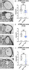
Ddr is required for normal axon caliber growth. (A-C) Axon caliber of dTSM axons as measured by the cross-sectional area in TEM images in 28 dpe DdrRNAi nerves compared with age-matched controls. **P=0.0037 (unpaired t-test). (D-F) Axon caliber of dTSM axon in 28 dpe Ddr loss-of-function nerves compared with age-matched controls. **P=0.0044 (unpaired t-test). (G-I) Axon caliber of dTSM axons in 5 dpe Ddr loss-of-function nerves compared with age-matched controls. **P=0.0073 (unpaired t-test). Error bars: 95% confidence interval. Scale bars: 1 µm.
Fig. 7.
Fig. 7.
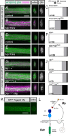
Mp genetically interacts with Ddr to promote wrapping in larval nerves. (A-C) Knockdown of Mp in wrapping glia using nrv2-Gal4 disrupts wrapping glia morphology. nrv2-Gal4-driven mCD8:GFP, green; Futsch+ axons, magenta. (D-F) Knockdown of Mp in neurons using elav-Gal4 does not affect wrapping glia morphology, visualized using the nrv2::GFP trap. (G-J) Wrapping glia morphology in Mp/+ and Ddr/+ nerves, compared with Mp/+; Ddr/+. (C,F,J) Categorical scoring of normal, moderately disrupted, and severely disrupted wrapping glia morphology. (K) A MiMiC construct that adds an in-frame GFP tag to Mp protein is localized throughout larval nerves. (L) Proposed model of Mp and Ddr interaction. A single Ddr protein is depicted in the wrapping glia membrane with extracellular discoidin (DS) domains and an intracellular kinase domain. Mp is depicted with its central collagen domain and cleavable endostatin-like (ES-like) and thrombospondin-like (Tsp-like) domains. Based on the strong phenotype observed when Mp is knocked down specifically in wrapping glia, and the lack of phenotype when knocked down in neurons, we hypothesize that wrapping glia secrete Mp, which can act as autocrine activator of Ddr to drive normal wrapping glia morphogenesis. n=number of nerves analyzed from 9-13 larvae per condition. Scale bars: 5 µm.

References

    1. Barros, C. S., Nguyen, T., Spencer, K. S. R., Nishiyama, A., Colognato, H. and Müller, U. (2009). Beta1 integrins are required for normal CNS myelination and promote AKT-dependent myelin outgrowth. Development 136, 2717-2724. 10.1242/dev.038679 - DOI - PMC - PubMed
    1. Beirowski, B., Babetto, E., Golden, J. P., Chen, Y.-J., Yang, K., Gross, R. W., Patti, G. J. and Milbrandt, J. (2014). Metabolic regulator LKB1 is crucial for Schwann cell-mediated axon maintenance. Nat. Neurosci. 17, 1351-1361. 10.1038/nn.3809 - DOI - PMC - PubMed
    1. Chen, P., Cescon, M. and Bonaldo, P. (2015). The role of collagens in peripheral nerve myelination and function. Mol. Neurobiol. 52, 216-225. 10.1007/s12035-014-8862-y - DOI - PubMed
    1. Cheng, L., Locke, C. and Davis, G. W. (2011). S6 kinase localizes to the presynaptic active zone and functions with PDK1 to control synapse development. J. Cell Biol. 194, 921-935. 10.1083/jcb.201101042 - DOI - PMC - PubMed
    1. Coutinho-Budd, J. C., Sheehan, A. E. and Freeman, M. R. (2017). The secreted neurotrophin Spätzle 3 promotes glial morphogenesis and supports neuronal survival and function. Gene Dev. 31, 2023-2038. 10.1101/gad.305888.117 - DOI - PMC - PubMed

Publication types

Substances

LinkOut - more resources