. 2023 May;72(5):1153-1167.
doi: 10.1007/s00262-022-03317-y. Epub 2022 Nov 10.

Generation of NK cells with chimeric-switch receptors to overcome PD1-mediated inhibition in cancer immunotherapy

Affiliations

Generation of NK cells with chimeric-switch receptors to overcome PD1-mediated inhibition in cancer immunotherapy

Katharina H Susek et al. Cancer Immunol Immunother. 2023 May.

Abstract

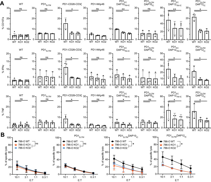
Multiple myeloma (MM) is an incurable hematological cancer, in which immune checkpoint inhibition (ICI) with monoclonal antibodies (mAbs) has failed due to uncontrollable immune responses in combination therapies and lack of efficacy in monotherapies. Although NK cell-specific checkpoint targets such as NKG2A and KIRs are currently being evaluated in clinical trials, the clinical impact of NK cells on the PD1 cascade is less well understood compared to T cells. Furthermore, while NK cells have effector activity within the TME, under continuous ligand exposure, NK cell dysfunctionality may occur due to interaction of PD1 and its ligand PD-L1. Due to above-mentioned factors, we designed novel NK cell specific PD1-based chimeric switch receptors (PD1-CSR) by employing signaling domains of DAP10, DAP12 and CD3ζ to revert NK cell inhibition and retarget ICI. PD1-CSR modified NK cells showed increased degranulation, cytokine secretion and cytotoxicity upon recognition of PD-L1+ target cells. Additionally, PD1-CSR+ NK cells infiltrated and killed tumor spheroids. While primary NK cells (pNK), expressing native PD1, showed decreased degranulation and cytokine production against PD-L1+ target cells by twofold, PD1-CSR+ pNK cells demonstrated increased activity upon PD-L1+ target cell recognition and enhanced antibody-dependent cellular cytotoxicity. PD1-CSR+ pNK cells from patients with MM increased degranulation and cytokine expression against autologous CD138+PD-L1+ malignant plasma cells. Taken together, the present results demonstrate that PD1-CSR+ NK cells enhance and sustain potent anti-tumor activity in a PD-L1+ microenvironment and thus represent a promising strategy to advance adoptive NK cell-based immunotherapies toward PD-L1+ cancers.

Keywords: Antibody-dependent cellular cytotoxicity; Chimeric switch receptor; Hematologic neoplasms; Immunotherapy; Natural killer cells; Programmed cell death 1 receptor.


Conflict of interest statement

HN is employed by Genmab and a share holder of Genmab. EA, HG and AB are consultants for Vycellix. EA and HG are shareholders of Vycellix. AKW is consultant and shareholder of VyGenBio. A patent application has been submitted, covering this work (US Provisional application no 63286205).

Figures

Fig. 1
Fig. 1
PD1-based chimeric switch receptors are stably expressed in NK-92 cells. A, B Table and vector maps depicting the design of the truncated PD1 receptor (PD1EcTM) and six chimeric switch receptors (CSR) with different signaling domains C NK-92 cells containing different PD1-CSR and sorted for positive PD1 surface staining. D PD-L1 and PD-L2 expression on NK-92 cells. E PD1, PD-L1 and PD-L2 expression on PD-L1+ Raji and PD-L1 Raji WT cells. F PD1, PD-L1 and PD-L2 expression on PD-L1+ 786-O WT and PD-L1 786-O KO cells
Fig. 2
Fig. 2
PD1-CSR+ NK-92 cells increase degranulation, cytokine production and killing of PD-L1+ target cells. A Percentage of CD107a, IFNγ and TNF by different PD1-CSR+ NK-92 cells against PD-L1+ 786-O WT and two PD-L1 786-O KO cell lines. Each data point represents the mean (± SD) of three independent experiments performed in triplicates. B Killing of PD-L1+ 786-O WT versus PD-L1 786-O KO1 and PD-L1 786-O KO2 cells by NK-92 WT, PD1EcTM+, PD1EcTMDAP10IC+ or PD1EcTMDAP12IC+ NK-92 cells. Each data point represents the mean (± SD) of three independent experiments performed in quadruplets. Statistical significance (* p < 0.05; ** p < 0.01) was determined with a two-way ANOVA test
Fig. 3
Fig. 3
PD1-CSR+ NK-92 cells increase cytotoxicity against PD-L1+ tumor spheroids. AD Killing of PD-L1+ 786-O WT versus PD-L1 786-O KO1 and PD-L1 786-O KO2 tumor spheroids by NK-92 WT, PD1EcTM+, PD1EcTMDAP10IC+ or PD1EcTMDAP12IC+ NK-92 cells. Each data point represents the mean (± SD) of six independent experiments performed in duplicates. E–G Tumor spheroids were collected, washed, dissociated and analyzed by flow cytometry. Displayed is the gating strategy (E), the percentage of 786-Odim cells per spheroid (F) and percentage of CD45+ NK-92 cells per spheroid (G). Each data point represents the mean (± SD) of three independent experiments performed in duplicates. Statistical significance (* p < 0.05; ** p < 0.01; *** p < 0.001) was determined with a two-way ANOVA test
Fig. 4
Fig. 4
PD1-CSR+ pNK cells increase degranulation and cytokine production against PD-L1+ target cells. A PD1 surface expression on untransduced or transduced CD56+ CD3 pNK cells. Each dot represents PD1 surface expression on one individual donor (n = 12). The mean of all 12 donors is displayed. B Gating strategy for degranulation assays. WT and empty vector transduced cells were gated on PD1dim population while PD1-CSR+ cells were gated on the PD1high population. CE Ratio of CD107a (C), IFNγ (D) and TNF (E) expression by different PD-CSR+ pNK cells against PD-L1+ Raji cells versus PD-L1 Raji WT cells. Each dot represents the mean of one individual donor, performed in duplicates (n = 8) The mean ± SD of all 8 donors is displayed. F–H Percentage of CD107a (F), IFNγ (G) and TNF (H) expression by different PD-CSR+ pNK cells against PD-L1+ Raji cells. Each dot represents the mean of one individual donor, performed in duplicates (n = 8) The mean ± SD of all 8 donors is displayed. I Killing of PD-L1+ Raji cells versus PD-L1 Raji WT cells by different PD-CSR+ pNK cells at an E:T of 1:1. Displayed are data from 3 independent donors with each data point representing the mean (± SD) of one experiment performed in triplicates. Statistical significance was determined with a Students t test (* p < 0.05, ** p < 0.01, ***p < 0.001, ****p < 0.0001)
Fig. 5
Fig. 5
PD1-CSR+ pNK cells increase degranulation and cytokine production against PD-L1+ autologous tumor samples. A Flow cytometry plots show the gating strategy of BM MNC (upper panel) and PBMCs (lower panel) from newly diagnosed MM patients B Percentage of PD1bright cells among CD56+CD3 pNK cells from MM donor 1 CE Percentage of CD107a (C), IFNγ (D) and TNF (E) by different PD1negative versus PD1bright PD1-CSR+ pNK cells from donor 1 against autologous BM MNC. F Percentage of PD1bright cells among CD56+CD3 pNK cells from MM donor 2 GI Percentage of CD107a (G), IFNγ (H) and TNF (I) by different PD1negative versus PD1bright PD-CSR+ pNK cells from donor 2 against autologous BM MNC. J Percentage of PD1bright cells among CD56+CD3 pNK cells from MM donor 3K–M) Percentage of CD107a (K), IFNγ (L) and TNF (M) by different PD1negative versus PD1bright PD-CSR+ pNK cells from donor 3 against autologous BM MNC. Displayed are data from each donor with each data point representing the mean (± SD) of one experiment performed in duplicates. Statistical significance was determined with a Students t test (* p < 0.05, ** p < 0.01)

References

    1. Ren D, et al. Predictive biomarkers and mechanisms underlying resistance to PD1/PD-L1 blockade cancer immunotherapy. Mol Cancer. 2020;19(1):19. doi: 10.1186/s12943-020-1144-6. - DOI - PMC - PubMed
    1. Kumpers C, et al. Immune cell infiltration of the primary tumor, Not PD-L1 status, is associated with improved response to checkpoint inhibition in metastatic Melanoma. Front Med (Lausanne) 2019;6:27. doi: 10.3389/fmed.2019.00027. - DOI - PMC - PubMed
    1. Zhu H, et al. EGFRvIII-CAR-T cells with PD-1 knockout have improved anti-glioma activity. Pathol Oncol Res. 2020;26(4):2135–2141. doi: 10.1007/s12253-019-00759-1. - DOI - PubMed
    1. Rupp LJ, et al. CRISPR/Cas9-mediated PD-1 disruption enhances anti-tumor efficacy of human chimeric antigen receptor T cells. Sci Rep. 2017;7(1):737. doi: 10.1038/s41598-017-00462-8. - DOI - PMC - PubMed
    1. McGowan E, et al. PD-1 disrupted CAR-T cells in the treatment of solid tumors: promises and challenges. Biomed Pharmacother. 2020;121:109625. doi: 10.1016/j.biopha.2019.109625. - DOI - PubMed