

Managing an open nasofrontal encephalocele after birth
- PMID: 36355194
- PMCID: PMC10006252
- DOI: 10.1007/s00381-022-05620-6
Managing an open nasofrontal encephalocele after birth
Abstract
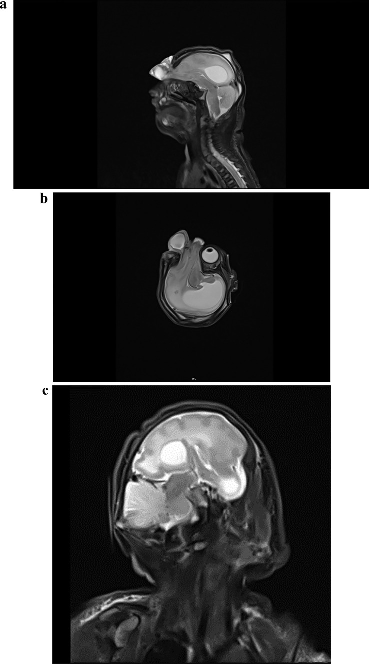
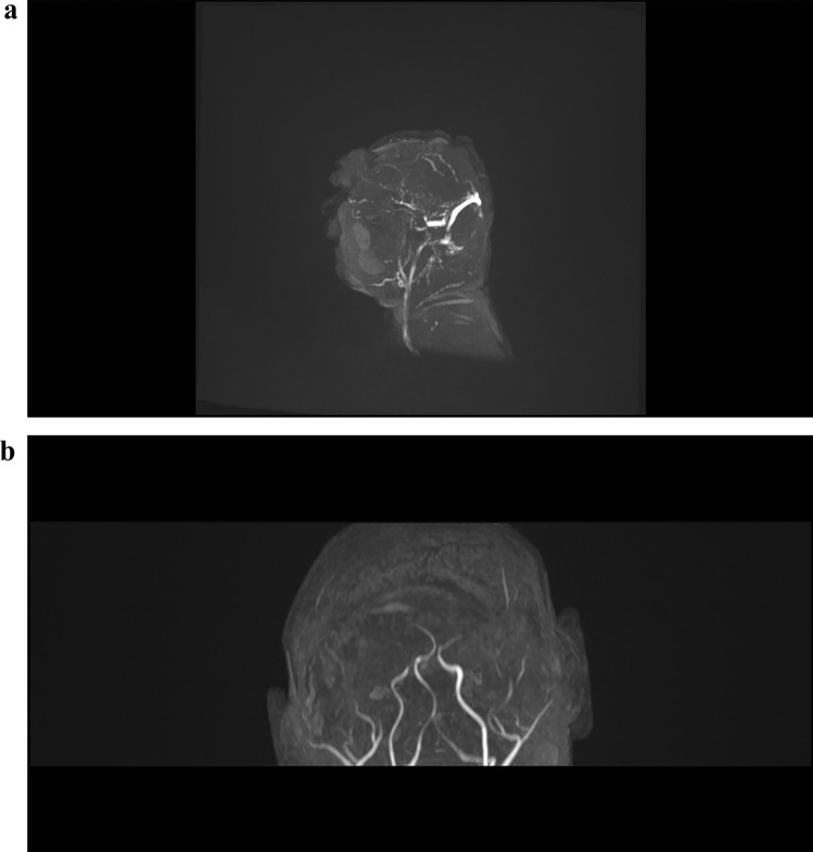
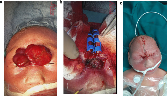
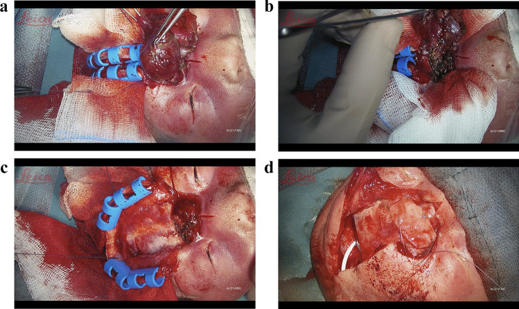
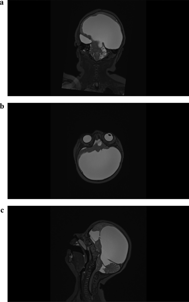
Encephaloceles are relatively uncommon in western countries. Most of the reported cases involve occipital encephaloceles. Open frontal encephaloceles comprise a rare entity. Most of them will be detected during early prenatal diagnostic, whereas the majority of the pregnancies will be terminated after the consent of the parents. Open frontal encephaloceles pose a great challenge to neurosurgeons as well as anesthesiologists, as these infants present with a microcephaly, non-physiological intracranial anatomy, and low birth weight, thus making the infant prone to excessive blood loss, hypothermia, and death. Neonates born with an incomplete cutaneous coverage are exposed to an imminent threat to life due to the risk of meningitis, necessitating surgical repair in the first days of life. We represent a rare case of an open nasofrontal encephalocele managed surgically in the first day of life. Surgery did not influence the neurological outcome of the patient.
Keywords: Case report; Encephalocele; Nasofrontal.
© 2022. The Author(s).
Conflict of interest statement
The authors declare no competing interests.
Figures
Similar articles
-
Management of an open nasofrontal encephalocele during the first day of life.Childs Nerv Syst. 2022 Jan;38(1):207-210. doi: 10.1007/s00381-021-05102-1. Epub 2021 Mar 6. Childs Nerv Syst. 2022. PMID: 33677686
-
Giant Interfrontal Encephalocele in an Infant: A Rare Entity.Pediatr Neurosurg. 2016;51(6):309-312. doi: 10.1159/000447408. Epub 2016 Aug 12. Pediatr Neurosurg. 2016. PMID: 27513987
-
Giant encephalocele: a study of 14 patients.Pediatr Neurosurg. 2011;47(6):406-11. doi: 10.1159/000338895. Epub 2012 Jul 7. Pediatr Neurosurg. 2011. PMID: 22776867
-
Frontoethmoidal encephalocele. Report of a case.Neurocirugia (Engl Ed). 2019 Mar-Apr;30(2):94-99. doi: 10.1016/j.neucir.2018.02.006. Epub 2018 Mar 31. Neurocirugia (Engl Ed). 2019. PMID: 29610064 Review. English, Spanish.
-
Hydrocephalus and occipital encephaloceles: presentation of a series and review of the literature.Childs Nerv Syst. 2021 Nov;37(11):3437-3445. doi: 10.1007/s00381-021-05312-7. Epub 2021 Aug 14. Childs Nerv Syst. 2021. PMID: 34390379 Review.
References
-
- Suwanwela C,Sukabote C, Suwanwela N (1971) Frontoethmoidal encepahlomeningocele. Surgery 69:617–625 - PubMed
-
- Andrewas BT, Meara JG (2010) Reconstruction of frontoethmoidal encephalocele defects. Atlas Oral Maxillofacial Surg Clin NAm 18:129–138 - PubMed
-
- Singh AK, Upadyaya DN (2009) Sinsipital encephaloceles. J Craniofacial Surgery 20:1851–1855 - PubMed
-
- Holmes AD, Meara JG, Kolker AR et al (2001) Frontoethmoidal encephaloceles: reconstruction and refinements. J Craniofac Surg 12(1):1–18 - PubMed
Publication types
MeSH terms
LinkOut - more resources
Full Text Sources
