. 2023:2598:87-114.
doi: 10.1007/978-1-0716-2839-3_8.

Chondrogenic Differentiation of Human-Induced Pluripotent Stem Cells

Affiliations

Chondrogenic Differentiation of Human-Induced Pluripotent Stem Cells

Amanda R Dicks et al. Methods Mol Biol. 2023.

Abstract

The generation of large quantities of genetically defined human chondrocytes remains a critical step for the development of tissue engineering strategies for cartilage regeneration and high-throughput drug screening. This protocol describes chondrogenic differentiation of human-induced pluripotent stem cells (hiPSCs), which can undergo genetic modification and the capacity for extensive cell expansion. The hiPSCs are differentiated in a stepwise manner in monolayer through the mesodermal lineage for 12 days using defined growth factors and small molecules. This is followed by 28 days of chondrogenic differentiation in a 3D pellet culture system using transforming growth factor beta 3 and specific compounds to inhibit off-target differentiation. The 6-week protocol results in hiPSC-derived cartilaginous tissue that can be characterized by histology, immunohistochemistry, and gene expression or enzymatically digested to isolate chondrocyte-like cells. Investigators can use this protocol for experiments including genetic engineering, in vitro disease modeling, or tissue engineering.

Keywords: Chondrogenesis; Human iPSCs; Stem cells; Tissue-engineered cartilage.


Conflict of interest statement

Conflicts of Interest FG is an employee of Cytex Therapeutics, Inc. The other authors declare that they have no competing interests.

Figures

Fig. 1
Fig. 1
Overview schematic of the protocol. hiPSCs undergo mesodermal differentiation in monolayer for 12 days. The cells go through the anterior primitive streak, paraxial mesoderm, early somite, sclerotome, and finally chondroprogenitor stage. Cells are then cultured in a 3D pellet culture to become chondrocytes and synthesize cartilaginous matrix. The protocol then has four options to either digest the tissue to isolate single cells or validate chondrogenesis with histology (Saf-O and IHC), biochemical assays (DMMB for sGAG and PicoGreen for dsDNA), and/or RT-qPCR
Fig. 2
Fig. 2
Phase contrast images of cells throughout mesodermal differentiation. (a) Induce cells when they are 30–40% confluent. (bf) As cells differentiate, from (b) anterior primitive streak, (c) paraxial mesoderm, (d) early somite, (e) sclerotome, to (f) chondroprogenitor, they spread and become more spindled. (g) If cells are induced at too high of a density, they may not fully differentiate and form nodules in the center of the colonies. Scale bar = 1 mm
Fig. 3
Fig. 3
Anticipated results – gene expression and matrix quantification. (a–c) Chondrogenic transcription factor SOX9 gene expression should increase early, followed by matrix genes COL2A1 and ACAN. (d–e) Relative to the expression of COL2A1, lower gene expression of fibrocartilage and hypertrophic cartilage markers COL1A1 and COL10A1, respectively, was observed. (f) sGAG/DNA ratio should increase throughout chondrogenesis, reaching 20–30 ng/ng at day 28 and over 40 ng/ng at day 42. Mean ± SEM, n = 4. *p < 0.05 compared to previous time point
Fig. 4
Fig. 4
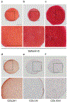
Anticipated results – histology. (a–c) Robust, homogenous safranin-O staining for sGAGs in three different cell lines. (d–f) Pellet in panel a with IHC labeling of COL2A1 (d), COL1A1 (e), and COL10A1 (f). Scale bar = 500 μm

Similar articles

Cited by

References

    1. Mansour JM (2003) Biomechanics of Cartilage. In: Kinesiology: the mechanics and pathomechanics of human movement, vol 2e, pp 66–75
    1. Guilak F (2011) Biomechanical factors in osteoarthritis. Best Pract Res Clin Rheumatol 25(6):815–823. 10.1016/j.berh.2011.11.013 - DOI - PMC - PubMed
    1. Antons J, Marascio MGM, Nohava J et al. (2018) Zone-dependent mechanical properties of human articular cartilage obtained by indentation measurements. J Mater Sci Mater Med 29(5):57. 10.1007/s10856-018-6066-0 - DOI - PubMed
    1. Sophia Fox AJ, Bedi A, Rodeo SA (2009) The basic science of articular cartilage: structure, composition, and function. Sports Health 1(6):461–468. 10.1177/1941738109350438 - DOI - PMC - PubMed
    1. Xia Y, Momot KI, Chen Z et al. (2017) Chapter 1 Introduction to Cartilage. In: Biophysics and biochemistry of Cartilage by NMR and MRI. The Royal Society of Chemistry, pp 1–43. 10.1039/9781782623663-00001 - DOI

Publication types

LinkOut - more resources