. 2022 Dec 16;50(6):1541-1553.
doi: 10.1042/BST20210240.

Laminin N-terminus (LaNt) proteins, laminins and basement membrane regulation

Affiliations

Laminin N-terminus (LaNt) proteins, laminins and basement membrane regulation

Natasha D Chavda et al. Biochem Soc Trans. .

Abstract

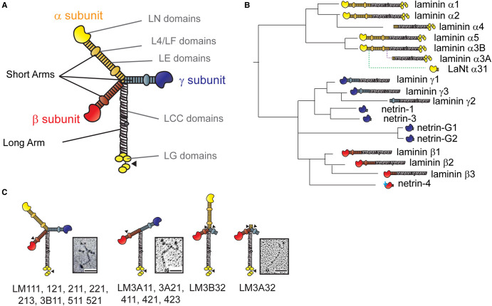
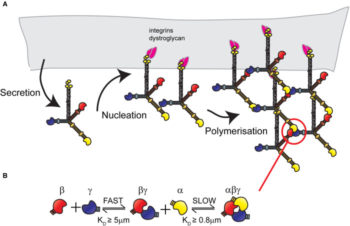
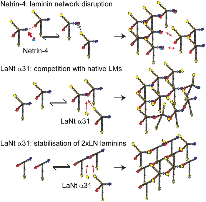
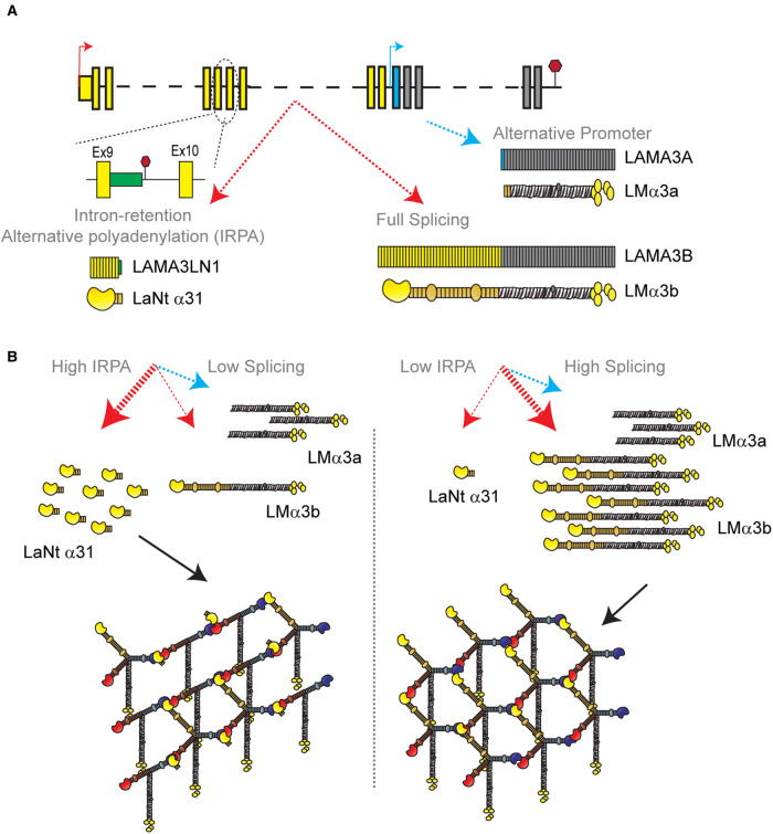
Basement membranes (BMs) are structured regions of the extracellular matrix that provide multiple functions including physical support and acting as a barrier, as a repository for nutrients and growth factors, and as biophysical signalling hubs. At the core of all BMs is the laminin (LM) family of proteins. These large heterotrimeric glycoproteins are essential for tissue integrity, and differences between LM family members represent a key nexus in dictating context and tissue-specific functions. These variations reflect genetic diversity within the family, which allows for multiple structurally and functionally distinct heterotrimers to be produced, each with different architectures and affinities for other matrix proteins and cell surface receptors. The ratios of these LM isoforms also influence the biophysical properties of a BM owing to differences in their relative ability to form polymers or networks. Intriguingly, the LM superfamily is further diversified through the related netrin family of proteins and through alternative splicing leading to the generation of non-LM short proteins known as the laminin N-terminus (LaNt) domain proteins. Both the netrins and LaNt proteins contain structural domains involved in LM-to-LM interaction and network assembly. Emerging findings indicate that one netrin and at least one LaNt protein can potently influence the structure and function of BMs, disrupting the networks, changing physical properties, and thereby influencing tissue function. These findings are altering the way that we think about LM polymerisation and, in the case of the LaNt proteins, suggest a hitherto unappreciated form of LM self-regulation.

Keywords: basement membrane; laminin; netrin.


Conflict of interest statement

The authors declare that there are no competing interests associated with the manuscript.

Figures

Figure 1.
Figure 1.. Laminin domain architecture, evolution and heterotrimer assemblies.
(A) Archetypal laminin structure. Yellow, red and blue regions indicate short arms generated from individual laminin chains, grey represents the long arm. LN — laminin N-terminus domain, LE — laminin-type epidermal growth factor-like domain, L4/LF — laminin domain IV globular domain, LCC — laminin coiled-coil domain, LG — laminin globular domain. (B) Derivation, relatedness and approximated order of appearance of laminin family members. Solid lines represent gene duplication and rearrangement events. Dotted lines indicate the evolution of an additional promoter (for LMα3A) and of intron-retention and alternative polyadenylation for LaNt α31. (C) Diagram of different laminin assemblies. Numbers represent chain composition e.g. LM111 is laminin α1β1γ1. Arrowheads indicate sites of proteolytic processing. Inset images are rotary shadowing electron microscopy images of purified LM111 (left) from [55], LM411 (middle) from [43] and LM332 (right) from [30]. Scale bars 50 mm. These images have been cropped from the original figures and are re-used in accordance with Creative Commons BY 4.0 license agreement.
Figure 2.
Figure 2.. Laminin network assembly.
(A) Network assembly involves, first, secretion of the fully trimerised laminin protein. Then, in the ‘nucleation' step, the proteins bind to cell surfaces receptors including integrins α3β1, α6β1 and α6β4 or dystroglycans. Once sufficient local concentration is achieved through receptor clustering, the ‘polymerisation' step occurs via interaction between laminin N-terminal domains. (B) Laminin ternary node formation is a two-step process involving a rapid formation of relatively unstable βγ dimers, followed by slower interaction with an α laminin N-terminal domain to form a more stable αβγ ternary node.
Figure 3.
Figure 3.. Netrin-4 and LaNt α31 predicted effects on laminin networks.
(Top) Netrin-4 binding with high affinity to laminin γ-chains can outcompete the laminin β-chains. However, α-chains cannot bind to γ/netrin-4 complexes. Therefore, netrin-4 disrupts laminin networks leading to locally increased pore size and decreased basement membrane stiffness. (Middle) LaNt α31 has an identical laminin N-terminal domain as LMα3b and therefore LaNt α31 will compete with approximately equal affinity for α-chain binding sites. This will lead to a partially disrupted network with reduced stiffness, with the level of disruption proportional to the expression level. (Bottom) In basement membranes containing T-shaped laminins, where only the β- and γ-chains contain N-terminal domains (e.g. LM411), the LaNt α31 protein may stabilise transient βγ interactions allowing the formation of stable ternary nodes. This would lead to linear arrays of the two-arm laminins. These arrays could also be cross-linked via the integration of some three-arm laminins within the local structure. The mechanical properties of the formed network would depend on the ratio of the three-arm to two-arm laminins and the local LaNt α31 concentrations.
Figure 4.
Figure 4.. LaNt α31 distribution in human tissue.
Immunohistochemistry images of formalin-fixed paraffin-embedded human larynx, pancreas, kidney and breast tissue sections obtain from US Biomax (MBN481, US Biomax, Rockville, Maryland, U.S.A.) processed with mouse monoclonal antibodies against LaNt α31 [79]. Scale bar 200 µm.
Figure 5.
Figure 5.. Intron-retention and alternative polyadenylation as a two-layer laminin regulation mechanism.
(A) Schematic of LAMA3 gene indicating three distinct transcripts and proteins produced. Red arrow — LAMA3B promoter, blue arrow — LAMA3A promoter. Yellow boxes — LAMA3B exons, grey boxes — LAMA3A and B shared exons, blue box — LAMA3A specific exon, green box — protein coding intronic sequence included in LAMA3LN1 but not present in LAMA3B transcript. (B) Potential effects of changing the intron-retention/splicing rates on protein production and laminin network assembly.

References

    1. Pozzi, A., Yurchenco, P.D. and Iozzo, R.V. (2017) The nature and biology of basement membranes. Matrix Biol. 57-58, 1–11 10.1016/j.matbio.2016.12.009 - DOI - PMC - PubMed
    1. Aumailley, M. (2013) The laminin family. Cell Adh. Migr. 7, 48–55 10.4161/cam.22826 - DOI - PMC - PubMed
    1. Hohenester, E. and Yurchenco, P.D. (2013) Laminins in basement membrane assembly. Cell Adh. Migr. 7, 56–63 10.4161/cam.21831 - DOI - PMC - PubMed
    1. Bardhan, A., Bruckner-Tuderman, L., Chapple, I.L.C., Fine, J.D., Harper, N., Has, C.et al. (2020) Epidermolysis bullosa. Nat. Rev. Dis. Primers 6, 78 10.1038/s41572-020-0210-0 - DOI - PubMed
    1. Barraza-Flores, P., Bates, C.R., Oliveira-Santos, A. and Burkin, D.J. (2020) Laminin and integrin in LAMA2-related congenital muscular dystrophy: from disease to therapeutics. Front. Mol. Neurosci. 13, 1 10.3389/fnmol.2020.00001 - DOI - PMC - PubMed

Publication types

MeSH terms