. 2022 Nov 1;63(12):16.
doi: 10.1167/iovs.63.12.16.

Formation and Fixation of the Annulus of Zinn and Relation With Extraocular Muscles: A Plastinated Histologic Study and Its Clinical Significance

Affiliations

Formation and Fixation of the Annulus of Zinn and Relation With Extraocular Muscles: A Plastinated Histologic Study and Its Clinical Significance

Chunjing Ma et al. Invest Ophthalmol Vis Sci. .

Abstract

Purpose: This study aimed to clarify the formation and fixation of the annulus of Zinn (AZ) and its relationship with the extraocular muscles by using ultrathin plastination and three-dimensional models.

Methods: Eighteen cadaveric heads (36 sides of the orbital apex) were plastinated to coronal (16 sides), sagittal (13 sides), and horizontal (5 sides) ultrathin plastination sections to be investigated at both macroscopic and microscopic levels. One cadaveric head was used for endoscopic dissection to identify anatomic landmarks.

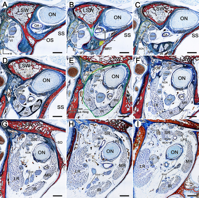
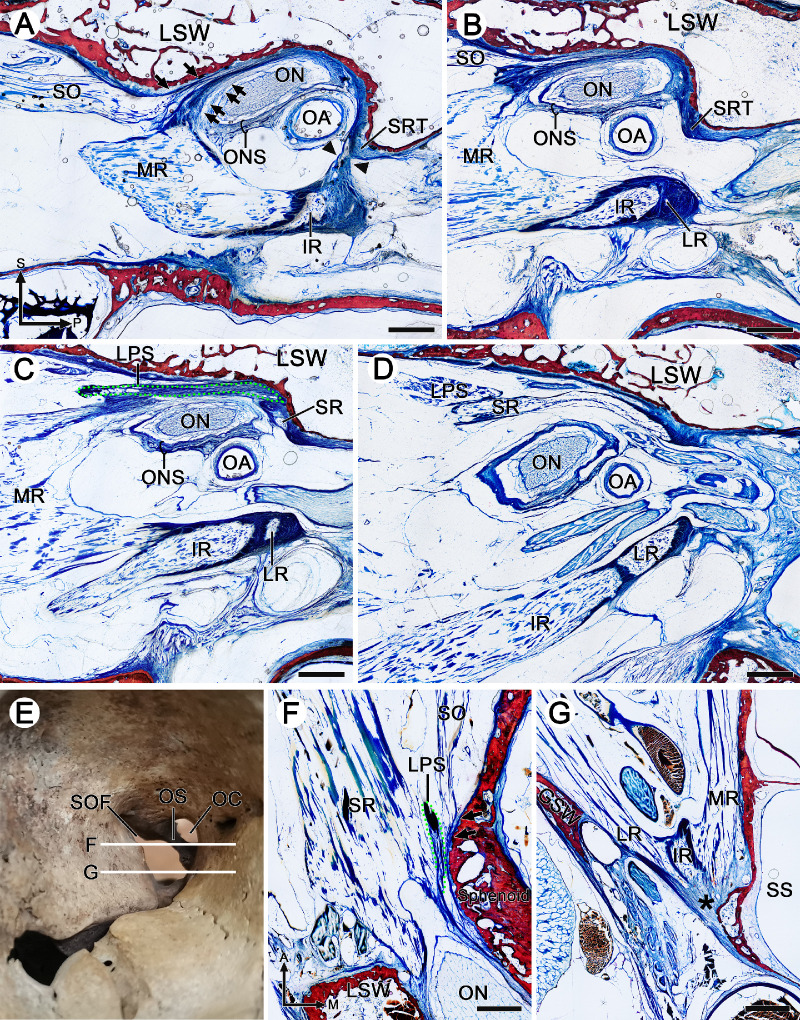
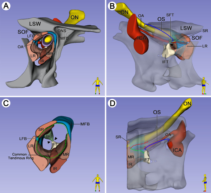
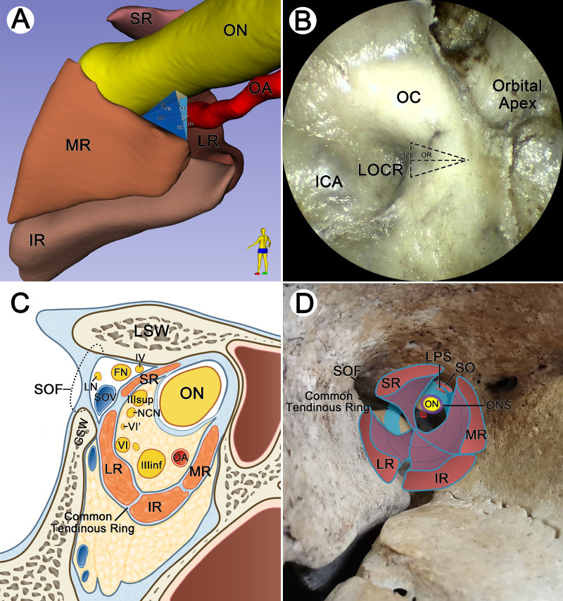
Results: There were two fibrous triangles adhered to both ends of the anterior surface of the optic strut. The superior rectus muscle originated from the superior fibrous triangle, and the lateral, inferior, and medial rectus muscles emerged from the inferior fibrous triangle. It was not until 5.46 ± 0.41 mm anterior to the optic strut that the complete tendinous ring composed of rectus muscles, optic nerve sheath, and periosteum was formed. The superior oblique and levator palpebrae superioris muscles originated from the medial fibrous band of the AZ. At the posterior of the AZ, there was a potential passage between the medial rectus muscle and the optic nerve.

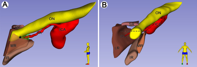
Conclusions: The fixation of the AZ was composed of the connection of the annular tendon to the optic strut posteriorly and the attachment of the complete tendinous ring to the lesser and greater wings of the sphenoid bone anteriorly. The triangular route area between the optic nerve and medial rectus muscle was located on the anterior side of the base of the optic strut.


Conflict of interest statement

Disclosure: C. Ma, None; X. Zhu, None; X. Chu, None; L. Xu, None; W. Zhang, None; S. Xu, None; L. Liang, None

Figures

Figure 1.
Figure 1.
Origin of rectus muscles, LPS and SO, and the formation of the common tendinous ring in the orbital apex. Plastinated sections from the cadaveric head of a 67-year-old male. (A) The coronal section passing through the OS. (BI) The coronal sections of 1 mm, 2 mm, 4 mm, 6 mm, 7 mm, 9 mm, 11 mm, and 14 mm anterior to the OS, respectively. (A) An annular tendon (red semicircle dotted line) was attached to the inferolateral side of the OS. The ON and OA were located on the superomedial side of the OS, and cranial nerves (CNs) IV, VI, III, FN, LN, and NCN were contained in the lateral space of the OS. Laterally, CNs IV, FN, and LN were arranged into a long strip shape (yellow ellipse dotted line in AC). (B) The SFT (green dotted line) was connected to the IFT (red dotted line). The SFT was attached to the anterior surface of the upper OS, while the IFT was mostly attached to the inferolateral surface of the lower OS. The fibers of the IR could be seen. The position of the MR (yellow triangle area) was inferred based on the section of 2 mm anterior to the OS because the muscle bundles of the MR have not yet appeared. Two black dotted lines were drawn at the lowest point of the ONS and the vertex of the MR, respectively, and the vertical distance was measured. The window distance (oblique double-headed arrow) between the ON and MR was also measured. (C) The SFT of the SR separated from the IFT and attached to the inferior margin of the LSW, and the muscle bundles of the MR, IR, and LR emerged from the IFT. Medially, the SRT fused with the superolateral side of the ONS. (D) Laterally, the LR fused with the lateral tendon of the SR to form a lateral fibrous band (white asterisk). Medially, the tendon of the MR did not tightly attach to the lateral wall of the sphenoid sinus (arrows). Note that the CN VI gave rise to an early branching (VI′). The CNs III, VI, VI ′, and NCN entered intraorbital space, while CNs IV, FN, and LN entered extraorbital space. (E) The MR fused with the inferomedial part of the ONS to form the medial fibrous band (white asterisk). The common tendinous ring (green ellipse dotted line) was formed by four rectus muscles, periosteum, and ONS. Laterally, the CNs IV and FN coursed along the superolateral aspect of the SR in triangular extraorbital space (yellow dotted line). The SOV was tightly attached to the lateral fibrous band. (F) The tendon connection between the LR and IR was separated (black asterisk). The medial fibrous bands between the SR and MR thickened (white asterisk). (G) The SO was tightly attached to the superomedial aspect of the orbital wall. (H) The LPS emerged from the medial fibrous band between the SR and MR. (I) More anteriorly, the superomedial side of the common tendinous ring separated, and the muscle bundles of the LPS coursed along the superomedial aspect of the SR. In addition, before the formation of the complete fibrous ring, there was a potential space between the MR and ON in the lateral wall of the SS, which was filled with loose adipose tissue in BE. FN, frontal nerve; GSW, greater wing of the sphenoid bone; IFT, inferior fibrous triangle; IIIinf, inferior division of the oculomotor nerve; IIIsup, superior division of the oculomotor nerve; IV, trochlear nerve; LN, lacrimal nerve; LSW, lesser wing of the sphenoid bone; M, medial; MRT, tendon of the medial rectus muscle; NCN, nasociliary nerve; OA, ophthalmic artery; ONS, optic nerve sheath; OS, optic strut; S, superior; SFT, superior fibrous triangle; SOV, superior ophthalmic vein; SRT, tendon of the superior rectus muscle; SS, sphenoid sinus; VI, abducens nerve; VI′, early branching of the abducens nerve. Bar: 2 mm.
Figure 2.
Figure 2.
Plastinated sections from cadaveric heads of a 77-year-old male (AD) and a 75-year-old male (FG). (AD) Sagittal sections of 1 mm, 2 mm, 3 mm, and 5 mm of the lateral side of the OS connected with the body of the sphenoid bone. (F, G) Corresponding to the horizontal sections in E. (A) Posteriorly, the SRT was connected to the tendon of the MR, IR, and LR (arrowheads). Anteriorly, the muscle bundles of the MR widened superiorly to fuse with the ONS. The tendon of the SO was embedded superiorly to the tendon complex (double arrows) formed by the ONS and MR and attached to the inferior margin of the LSW (arrows). (B) The connection between the SRT and the tendon of the MR, IR, and LR separated, and the muscle bundles of the MR enlarged and widened anteriorly. (C) The muscle bundles of the SR were visible, and the tendon of the LPS (green dotted line) fused with the tendon of the SR and MR but attached to the anterosuperior aspect of the SR. (D) Laterally, the LPS moved anteriorly along with the SR. (F) The tendon of the LPS (green dotted line) was visible and fused with the ONS and medial side of the SR. The attachment point of the SO was located on the orbital wall (arrows) in the anteromedial aspect of the SR and LPS. (G) The tendon of MR, IR, and LR fused posteriorly (black asterisk). A, anterior; OC, optic canal; P, posterior. Bar: 2 mm.
Figure 3.
Figure 3.
Plastinated sections used for three-dimensional reconstruction were obtained from the cadaveric head of a 76-year-old male. (A) Spatial relationship of the vital structures of the orbital apex on the three-dimensional reconstructed model. (B) The SFT was located inferior to the LSW, while the IFT was attached to the inferolateral of the OS. As the SR extended posteriorly, the SFT of the SR fused with the IFT of the LR, IR, and MR (arrowheads). (C) With the muscle bundles of the SR, LR, IR, and MR extended anteriorly, the common tendinous ring (green dot line) formed based on rectus muscles with the help of the LFB and MFB. (D) Medially, there was a potential passage between the inferior border of the ON (red dotted line) and the superior border of the MR (green dotted line) anterior to the OS. ICA, internal carotid artery; LFB, lateral fibrous bands; MFB, medial fibrous bands; SOF, superior orbital fissure.
Figure 4.
Figure 4.
Spatial relationship between ON and MR on the three-dimensional reconstructed model. Plastinated sections used for three-dimensional reconstruction were obtained from the cadaveric head of a 39-year-old male. (A) The ON coursed inferolaterally along with the ophthalmic artery. Posteriorly to the pentagram, the ON was located superiorly to the MR. There was a potential space between the ON and MR (green triangle area). (B) Anterior to the position of the pentagram in A, the ON intersected the MR (black dotted line), locating the superior portion of the MR.
Figure 5.
Figure 5.
Three-dimensional model in A from the same cadaveric head in Figure 4. Specimen in B from the cadaveric head of a 75-year-old male. (A) Diagram of measurement on the three-dimensional reconstructed model. H1 and H2 represent vertical and window distances, respectively. SH1 and SH2 are vertical and window triangular regions, respectively. OR represents the horizontal distance from the orbital opening of the optic strut to the formation of the common tendinous ring. OA-WT is the vertical distance of the OA to the window triangle. (B) The anatomic landmarks in the region of the orbital apex under endoscopic view. The anterior triangle of the optic strut was speculated (triangle dotted area). (C) The schematic diagram of Figure 1E shows the relationship between the common tendinous ring and vital cranial nerves. (D) Schematic diagram of the relationship between the rectus muscles, LPS, SO, and the common tendinous ring. LOCR, lateral optic carotid recess.

Similar articles

Cited by

References

    1. Standring S. Gray's Anatomy: The Anatomical Basis of Clinical Practice. 41st ed. London, UK: Churchill Livingstone; 2015: 668–670.
    1. Lin J, Hu W, Wu Q, Zhang J, Yan W.. An evolving perspective of endoscopic transnasal optic canal decompression for traumatic optic neuropathy in clinic. Neurosurg Rev. 2021; 44(1): 19–27. - PubMed
    1. Tu Y, Xu M, Kim AD, Wang MTM, Pan Z, Wu W.. Modified endoscopic transnasal orbital apex decompression in dysthyroid optic neuropathy. Eye Vis (Lond). 2021; 8(1): 19. - PMC - PubMed
    1. Chen B, Zhang H, Zhai Q, Li H, Wang C, Wang Y.. Traumatic optic neuropathy: a review of current studies. Neurosurg Rev. 2022; 45(3): 1895–1913. - PubMed
    1. Singh S, Curragh DS, Selva D.. Augmented endoscopic orbital apex decompression in dysthyroid optic neuropathy. Eye (Lond). 2019; 33(10): 1613–1618. - PMC - PubMed

Publication types