Case Reports
. 2023 Jan 3;133(1):e162137.
doi: 10.1172/JCI162137.

Identification of a neutrophil-specific PIK3R1 mutation facilitates targeted treatment in a patient with Sweet syndrome

Affiliations
Case Reports

Identification of a neutrophil-specific PIK3R1 mutation facilitates targeted treatment in a patient with Sweet syndrome

Shreya Bhattacharya et al. J Clin Invest. .

Abstract

BackgroundAcute febrile neutrophilic dermatosis (Sweet syndrome) is a potentially fatal multiorgan inflammatory disease characterized by fever, leukocytosis, and a rash with a neutrophilic infiltrate. The disease pathophysiology remains elusive, and current dogma suggests that Sweet syndrome is a process of reactivity to an unknown antigen. Corticosteroids and steroid-sparing agents remain frontline therapies, but refractory cases pose a clinical challenge.MethodsA 51-year-old woman with multiorgan Sweet syndrome developed serious corticosteroid-related side effects and was refractory to steroid-sparing agents. Blood counts, liver enzymes, and skin histopathology supported the diagnosis. Whole-genome sequencing, transcriptomic profiling, and cellular assays of the patient's skin and neutrophils were performed.ResultsWe identified elevated IL-1 signaling in lesional Sweet syndrome skin caused by a PIK3R1 gain-of-function mutation specifically found in neutrophils. This mutation increased neutrophil migration toward IL-1β and neutrophil respiratory burst. Targeted treatment of the patient with an IL-1 receptor 1 antagonist resulted in a dramatic therapeutic response and enabled a tapering off of corticosteroids.ConclusionDysregulated PI3K/AKT signaling is the first signaling pathway linked to Sweet syndrome and suggests that this syndrome may be caused by acquired mutations that modulate neutrophil function. Moreover, integration of molecular data across multiple levels identified a distinct subtype within a heterogeneous disease that resulted in a rational and successful clinical intervention. Future patients will benefit from efforts to identify potential mutations. The ability to directly interrogate the diseased skin allows this method to be generalizable to other inflammatory diseases and demonstrates a potential personalized medicine approach for patients with clinically challenging disease.Funding SourcesBerstein Foundation, NIH, Veterans Affairs (VA) Administration, Moseley Foundation, and H.T. Leung Foundation.

Keywords: Dermatology; Inflammation; Neutrophils; Signal transduction; Skin.


Figures
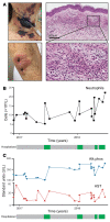
Figure 1
Figure 1. The patient with refractory Sweet syndrome required multiple hospitalizations to control the disease.
(A) Clinical photographs of the patient’s skin lesions. Panels on the right are H&E-stained images of the patient’s skin biopsy, which demonstrate a diffuse dermal neutrophilic infiltration. Scale bar: 200 μM. Original magnification, ×20 (enlarged inset). (B and C) Time course of the patient’s peripheral neutrophilia (B) and liver enzyme fluctuations (C) during her multiple clinical flares. The patient’s 4 hospitalizations for disease flares are denoted by the green boxes. Alk phos, alkaline phosphatase; AST, aspartate aminotransferase.
Figure 2
Figure 2. Refractory Sweet syndrome lesions reveal IL-1β–dominant inflammation.
(A) PCA of gene expression generated from skin dermis from the patient with refractory Sweet syndrome (n = 3, purple), other patients with Sweet syndrome (n = 7, blue), and healthy controls (n = 13, red). (B) Gene ontology categories that were most highly enriched in the transcriptome of the refractory patient with refractory disease compared with the healthy controls. (C) Increased IL1B transcript levels were detected in the refractory patient’s dermis (purple) compared with levels in healthy control dermis (blue). Individual dermis samples from patients with Sweet syndrome exhibited varied levels of IL1B transcripts. Three patients had transcript levels greater than 2.5 SDs above the average for the healthy controls (yellow box). Data represent the mean ± SEM. ***P < 0.001, by 2-tailed Student’s t test.
Figure 3
Figure 3. PIK3R1 gain-of-function mutation increases neutrophil migration toward IL-1β.
(A) Venn diagram illustrates the intersection between differentially expressed genes and neutrophil-specific gene mutations in the patient with refractory disease. Gene names are listed in the box. (B) Schematic depicting the PIK3R1 gene. The mutation location is identified by the black arrow. Below the schematic is the DNA sequence chromatogram that confirms the mutation. (C) Western blot demonstrates increased phosphorylation of AKT at S473 in overexpressed p.W335C p85 (MT) cells compared with overexpressed p85 (WT) cells. The experiment was repeated independently 3 times. (D) Cellular respiratory burst detected by DCFDA in LPS-treated WT and MT p85 cells (n = 8/group). (E) Transwell migration of WT and MT p85 cells in response to IL-1β (n = 6/group). (F) qPCR (n = 3/group) and (G) Western blot show increased IL-1R1 expression in MT p85 cells compared with WT p85 cells. The experiment was repeated independently 3 times. (H) Pharmacologic treatment with an IL-1RA blocked IL-1β–mediated Transwell migration (n = 3/group). Data represent the mean ± SEM. *P < 0.05 and **P < 0.01, and ***P < 0.001, by 2-tailed Student’s t test.
Figure 4
Figure 4. Clinical remission of refractory Sweet syndrome with an IL-1RA.
(A) Neutrophils from the patient with refractory Sweet syndrome exhibited increased AKT activation and IL-1R1 expression. The experiment repeated 2 times. (B) Photograph shows disease clearance following anakinra treatment. (C) H&E staining of post-treatment skin biopsy with an absence of neutrophilic infiltration. (D) Neutrophil and liver enzyme counts for the patient with refractory disease after initiation of anakinra treatment. When anakinra was paused, her clinical symptoms, reflected in laboratory values, returned over several days.

References

    1. Cohen PR. Sweet’s syndrome — a comprehensive review of an acute febrile neutrophilic dermatosis. Orphanet J Rare Dis. 2007;2:34. doi: 10.1186/1750-1172-2-34. - DOI - PMC - PubMed
    1. Nelson CA, et al. Neutrophilic dermatoses: pathogenesis, Sweet syndrome, neutrophilic eccrine hidradenitis, and Behçet disease. J Am Acad Dermatol. 2018;79(6):987–1006. doi: 10.1016/j.jaad.2017.11.064. - DOI - PubMed
    1. Nelson CA, et al. Sweet syndrome in patients with and without malignancy: a retrospective analysis of 83 patients from a tertiary academic referral center. J Am Acad Dermatol. 2018;78(2):303–309. doi: 10.1016/j.jaad.2017.09.013. - DOI - PubMed
    1. Heath MS, Ortega-Loayza AG. Insights into the pathogenesis of Sweet’s Syndrome. Front Immunol. 2019;10:414. doi: 10.3389/fimmu.2019.00414. - DOI - PMC - PubMed
    1. Guo H, et al. The PI3K/AKT pathway and renal cell carcinoma. J Genet Genomics. 2015;42(7):343–353. doi: 10.1016/j.jgg.2015.03.003. - DOI - PMC - PubMed

Publication types

Substances