. 2022 Oct 24;15(11):1309.
doi: 10.3390/ph15111309.

Neuroprotective Effect of Red Sea Marine Sponge Xestospongia testudinaria Extract Using In Vitro and In Vivo Diabetic Peripheral Neuropathy Models

Affiliations

Neuroprotective Effect of Red Sea Marine Sponge Xestospongia testudinaria Extract Using In Vitro and In Vivo Diabetic Peripheral Neuropathy Models

Rania Magadmi et al. Pharmaceuticals (Basel). .

Abstract

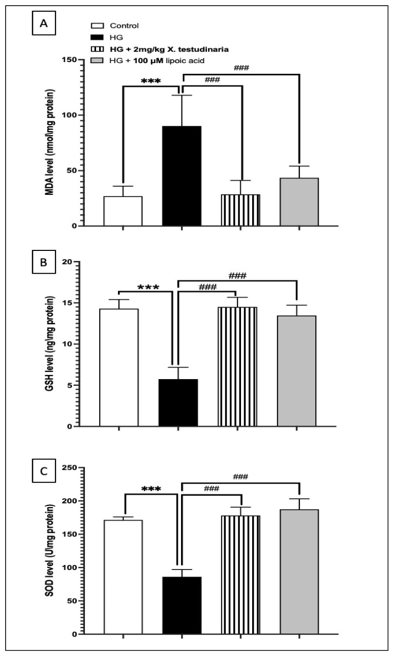
Diabetic peripheral neuropathy (DPN) is a common complication of diabetes. Oxidative stress plays an important role in the pathophysiology of DPN. Red Sea marine sponge Xestospongia testudinaria extract has a promising neuroprotective effect, presumably owing to its antioxidant and anti-inflammatory properties. Thus, this study aimed to investigate the neuroprotective effect of the sponge X. testudinaria extract on in vitro and in vivo models of DPN. Mice dorsal root ganglia (DRG) were cultured with high glucose (HG) media and used as an in vitro model of DPN. Some of the DRGs were pre-treated with 2 mg/mL of X. testudinaria. The X. testudinaria extract significantly improved the HG-induced decreased neuronal viability and the neurite length. It improved the oxidative stress biomarkers in DRG cultures. The DPN model was induced in vivo by an injection of streptozotocin at a dose of 150 mg/kg in mice. After 35 days, 0.75 mg/kg of the X. testudinaria extract improved the hot hyperalgesia and the DRG histology. Although the sponge extract did not reduce hyperglycemia, it ameliorated the oxidative stress markers and pro-inflammatory markers in the DRG. In conclusion, the current study demonstrates the neuroprotective effect of Red Sea sponge X. testudinaria extract against experimentally induced DPN through its antioxidant and anti-inflammatory mechanisms.

Keywords: Red Sea marine sponge; Xestospongia testudinaria; anti-inflammatory; antioxidant; diabetic peripheral neuropathy.


Conflict of interest statement

The authors declare no conflict of interest.

Figures

Figure 1
Figure 1
The effect of X. testudinaria extract on DRG neurons. (A) shows the effect of X. testudinaria extract on DRG viability. (B) shows the schematic tracings of the neurites from DRG culture in different groups. Neurites and nuclei were visualized with β-tubulin III and DAPI, respectively. (C) shows a quantitative analysis of total neurites length in DRG cultures. Data are presented as mean ± SEM of three independent experiments. Statistical analysis was carried out using a one-way ANOVA followed by Tukey post hoc test. **, ***: the data were statistically significant from the corresponding control group at p < 0.01 and p < 0.001, respectively. ###: the data were statistically significant from the corresponding high glucose (HG) group at p < 0.001.
Figure 2
Figure 2
The effect of X. testudinaria extract on oxidative stress biomarkers in high glucose (HG) -DRG culture: (A) malondialdehyde (MDA) level, (B) glutathione (GSH), and (C) superoxide dismutase enzymes (SOD). Data are presented as mean ± SEM for the three independent experiments. Statistical analysis was carried out using a one-way ANOVA followed by Tukey post hoc test. ***: statistical significance from the corresponding control group was set at p < 0.001. ###: statistical significance from the corresponding HG group was set at p < 0.001.
Figure 3
Figure 3
Paw withdrawal latency to hot stimuli in streptozotocin (STZ)-induced diabetic mice. Data are presented as mean ± SD for the mice (n = 6) in each group. Statistical analysis was carried out using one-way ANOVA followed by Tukey post hoc test. ****: statistical significance from the corresponding initial reading was set at p < 0.001. ns: nonsignificant.
Figure 4
Figure 4
Effect of X. testudinaria extract on histopathological changes of DRG in streptozotocin (STZ)-induced diabetes in mice using H and E: a photomicrograph of the dorsal root ganglia (DRG) of the control group (A), X. testudinaria extract-only group (B), STZ-diabetic group (C), and STZ-diabetic on X. testudinaria-treated group (D). Arrows show neurons and stars mark large-diameter nerve cells. Thick black arrows show satellite cells. H&E scale bar at 50 µm was used.
Figure 5
Figure 5
Effect of X. testudinaria extract on histopathological changes of DRG in streptozotocin (STZ)-induced diabetes in mice using reticulin stain: a photomicrograph of the dorsal root ganglia (DRG) of control group (A), X. testudinaria-only group (B), STZ-diabetic group (C), and STZ-diabetic on X. testudinaria-treated group (D). Arrows show reticular stroma and stars mark the distorted stroma, which give rise to the alveolar pattern. Reticulin stain was used and a scale bar 50 µm.
Figure 6
Figure 6
Fasting blood glucose (FBG) level in streptozotocin (STZ)-induced diabetic mice. Data are presented as mean ± SD for the mice (n = 6) in each group. Statistical analysis was carried out using a one-way ANOVA followed by Tukey post hoc test. *: statistical significance from the corresponding control group was set at p < 0.05. #: statistical significance from the corresponding week 0 was set at p < 0.05.
Figure 7
Figure 7
Effect of X. testudinaria extract on oxidative biomarkers in the dorsal root ganglia (DRG) of streptozotocin (STZ)-induced diabetic neuropathic mice: (A) malondialdehyde (MDA) level, (B) glutathione (GSH) level, and (C) superoxide dismutase enzymes (SOD) level. Data are presented as mean ± SD for mice (n = 6) in each group. Statistical analysis was carried out using a one-way ANOVA followed by Tukey post hoc test. *: statistical significance from the corresponding control group was set at p < 0.05. #: statistical significance from the corresponding STZ group was set at p < 0.05.
Figure 8
Figure 8
Effect of X. testudinaria extract on tumor necrosis factor (TNF)-α (A) and nuclear factor kappa B (NF-κB) (B) content in DRG of streptozotocin (STZ)-induced diabetic neuropathic mice. Data are presented as mean ± SD for the mice (n = 6) in each group. Statistical analysis was carried out using a one-way ANOVA followed by Tukey post hoc test. *: statistical significance from the corresponding control group was set at p < 0.05. #: statistically significance from the corresponding STZ group was set at p < 0.05.
Figure 9
Figure 9
A schematic overview of pathogenesis of diabetic neuropathy and possible mechanism (s) through which X. testudinaria extract could exert its diabetic neuroprotective effects.

Similar articles

Cited by

References

    1. Algeffari M.A. Painful Diabetic Peripheral Neuropathy among Saudi Diabetic Patients is Common but Under-recognized: Multicenter Cross-sectional study at primary health care setting. J. Fam. Community Med. 2018;25:43–47. doi: 10.4103/jfcm.JFCM_145_16. - DOI - PMC - PubMed
    1. Callaghan B.C., Cheng H.T., Stables C.L., Smith A.L., Feldman E.L. Diabetic neuropathy: Clinical manifestations and current treatments. Lancet Neurol. 2012;11:521–534. doi: 10.1016/S1474-4422(12)70065-0. - DOI - PMC - PubMed
    1. Edwards J.L., Vincent A.M., Cheng H.T., Feldman E.L. Diabetic neuropathy: Mechanisms to management. Pharmacol. Ther. 2008;120:1–34. doi: 10.1016/j.pharmthera.2008.05.005. - DOI - PMC - PubMed
    1. Sandireddy R., Yerra V.G., Areti A., Komirishetty P., Kumar A. Neuroinflammation and oxidative stress in diabetic neuropathy: Futuristic strategies based on these targets. Int. J. Endocrinol. 2014;2014:674987. doi: 10.1155/2014/674987. - DOI - PMC - PubMed
    1. Skapare E., Konrade I., Liepinsh E., Strele I., Makrecka M., Bierhaus A., Lejnieks A., Pirags V., Dambrova M. Association of reduced glyoxalase 1 activity and painful peripheral diabetic neuropathy in type 1 and 2 diabetes mellitus patients. J. Diabetes Complicat. 2013;27:262–267. doi: 10.1016/j.jdiacomp.2012.12.002. - DOI - PubMed

LinkOut - more resources