. 2022 Oct 28;15(11):1340.
doi: 10.3390/ph15111340.

Pyrazole-Based Thrombin Inhibitors with a Serine-Trapping Mechanism of Action: Synthesis and Biological Activity

Affiliations

Pyrazole-Based Thrombin Inhibitors with a Serine-Trapping Mechanism of Action: Synthesis and Biological Activity

Calvin Dunker et al. Pharmaceuticals (Basel). .

Abstract

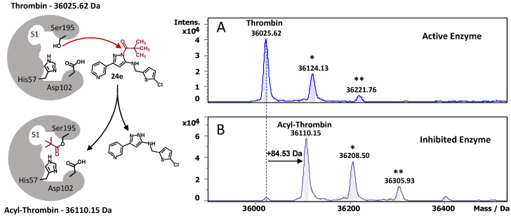
New antithrombotic drugs are needed to combat thrombosis, a dangerous pathology that causes myocardial infarction and ischemic stroke. In this respect, thrombin (FIIa) represents an important drug target. We herein report the synthesis and biological activity of a series of 1H-pyrazol-5-amine-based thrombin inhibitors with a serine-trapping mechanism of action. Among synthesized compounds, flexible acylated 1H-pyrazol-5-amines 24e, 34a, and 34b were identified as potent 16-80 nM thrombin inhibitors, which showed practically no off-targeting effect against other physiologically relevant serine proteases. To prove that synthesized compounds are covalent thrombin inhibitors, the most potent derivative 24e (FIIa IC50 = 16 nM) was studied in a mass-shift assay, where it has been shown that 24e transfers its acyl moiety (pivaloyl) to the catalytic Ser195 of thrombin. Performed herein docking studies also confirmed the covalent mechanism of thrombin inhibition by synthesized compounds. Acylated aminopyrazoles found during this study showed only limited effects on plasma coagulation in activated partial thrombin time (aPTT) and prothrombin time (PT) in vitro assays. However, such thrombin inhibitors are expected to have virtually no effect on bleeding time and can be used as a starting point for developing a safer alternative to traditional non-covalent anticoagulants.

Keywords: Ullmann reaction; anticoagulants; covalent inhibitor; dabigatran; pyrazole; pyrazolo[1,5-a]quinazolin-5(4H)-ones; pyrazolo[5,1-b]quinazolin-9(4H)-ones; thrombin; thrombosis.


Conflict of interest statement

The authors declare no conflict of interest.

Figures

Figure 1
Figure 1
Exemplary structures of small molecule thrombin (FIIa) inhibitors. Dabigatran (1) [4] is a non-covalent direct thrombin inhibitor, compounds 24 [14,15,16] are covalent serine-trapping thrombin inhibitors. Potential inhibitors 24e, 25, 34, and 10 developed in this work.
Figure 2
Figure 2
Schematic representation of thrombin’s Ser195 interaction with acylated 1H-pyrazol-5-amine 3 (top) vs. proposed interaction of thrombin with cyclic amides 10e (bottom).
Scheme 1
Scheme 1
Synthesis of acylated pyrazoles 9 and pyrazolo[5,1-b]quinazolin-9(4H)-ones 10. (a) NaH, THF, reflux under N2, 16 h, 6a 88%, 6b 75%, 6c 72%; (b) 1. HCl (1M), 5 min; 2. NH2NH2·H2O, EtOH, reflux, 16 h, 7a 75%, 7b 94%, 7c 76%; (c) 1. AcOH, molecular sieves (3Å), EtOH, r.t., 24 h; 2. NaBH4, EtOH, 0 °C to r.t., 16–24 h, 8a 88%, 8b 70%, 8c 68%, 8d 76%, 8e 78%, 8f 73%, 8g 79%, 8h 38%, 8i 43%; (d) pyridine/THF, 0 °C to r.t., 2–3 h, 9a 64%, 9b 53%, 9c 40%, 9d 81%, 9e 57%, 9f 37%, 9g 42%, 9h 54%, 9i 41%; (e) CuI, Cs2CO3, 1,10-phenanthroline, DMF, 80 °C, 0.5–3 h, 10a 82%, 10b 35%, 10c 55%, 10d 43%, 10e 90%, 10f 28%, 10g 84%, 10h 72%, 10i 58%.
Figure 3
Figure 3
X-ray crystal structure of 10a displaying the thermal ellipsoids at the 50% probability level (A); X-ray crystal structure of 19 displaying the thermal ellipsoids at the 50% probability level (B).
Scheme 2
Scheme 2
Synthesis of aminopyrazol- and aminotriazole-based cyclic derivatives 15, and 1923. (a) pyridine/THF, 0 °C to r.t., 2–4 h, 14 63%, 16, 78% 17, 74% 18 59%; (b) CuI, Cs2CO3, 1,10-phenanthroline, DMF, 80 °C, 0.5–1 h, 15 21%, 19 80%, 20 + 21 (1:1) 79%, 22 + 23 (3:7) 68%; (c) CuI, Cs2CO3, 1,10-phenanthroline, DMF, microwave irrad. at 150 °C, 1 h, 20 + 21 (7:3) 92%, 22 + 23 (3:7) 95%.
Scheme 3
Scheme 3
Synthesis of [1,2,4]triazolo[1,5-a]quinazolin-5(4H)-ones 21 and 23. (a) 260 °C, 15 min, neat 16′ 80%, 17′ 76%; (b) CuI, Cs2CO3, 1,10-phenanthroline, DMF, 80 °C, 1 h, 21 67%, 23 42%.
Scheme 4
Scheme 4
Synthesis of acylated 1H-pyrazol-5-amines 24be,gi and 25. (a) pyridine/THF, 0 °C to r.t., 2–4 h, 24b 44%, 24c 40%, 24d 56%, 24e 52%, 24g 44%, 24h 53%, 24i 30%, 25 56%.
Scheme 5
Scheme 5
Synthesis of acylated 1H-pyrazol-5-amines 26a,b and 29a,b. (a) pyridine/THF, 0 °C to r.t., 2–4 h, 26a 68%, 26b 77%; (b) HCl (1M), 5 min; (c) N2, n-BuLi, ™78 °C to r.t., THF, 16 h, 28b 59%; (d) MsOH, EtOH, reflux, 45 min, 29a 56%, 29b 97%.
Scheme 6
Scheme 6
Synthesis of acylated 4-fluoro-3-phenyl-1H-pyrazol-5-amines 34a,b. (a) N2, Ph2P(O)Cl, LiHMDS, ™78 °C to r.t., THF, 75 min, 31 17%; (b) i-PrOH, reflux, 3 h, 32 55%; (c) 1. AcOH, molecular sieves (3Å), EtOH, 0 °C to r.t., 24 h; 2. NaBH4, EtOH, 0 °C to r.t., 24 h, 33 77%; (d) pyridine/THF, 0 °C to r.t., 24 h, 34a 21%, 34b 34%.
Figure 4
Figure 4
Deconvoluted ESI(+)-MS mass spectra of native thrombin (A) and acyl-thrombin complex (B) formed after the enzyme incubation with 79 μM thrombin inhibitor 24e. The peaks of interest are labelled with the corresponding deconvoluted masses. A mass shift of 84.5 Da (B) was observed, which corresponds to the inhibitor’s acyl moiety adduct to thrombin. The schematic representation of thrombin and the covalent complexes of thrombin with the inhibitor are also shown. *—single and **—double sulfuric acid or phosphoric acid adduct [30].
Figure 5
Figure 5
The influence of compounds on plasma coagulation (in vitro). Selected acylated 1H-pyrazol-5-amines were tested at 200 μM and dabigatran (1) was tested at 2 μM. The activated partial thromboplastin time (aPTT) and prothrombin time (PT) are shown in sec. The fold increase in aPTT and PT compared to the effect of DMSO is shown under the diagram. Tests were performed at least in triplicate, and the average with standard deviation (SD) is given.
Figure 6
Figure 6
Four superimposed thrombin X-ray crystal structures (A—ribbons, B—molecular surface) with bound inhibitors exhibiting the 5-chlorothiophenyl moiety (PDB ID: 4LOY [32], 4LXB [32], 6EO8 [33], and 6YQV). In all superimposed structures, the inhibitors’ 5-chlorothiophenyl moiety resides in the S1 pocket. Calculated covalent binding conformation of inhibitor 25 (cyan stick model) in the active site of thrombin (C—close-up view and D—overall structure with molecular surface). Amino acid residues are depicted as orange or blue stick models and are numbered according to the amino acid sequence of chymotrypsinogen residue numbering. Oxygen, nitrogen, and sulfur atoms are colored in red, blue, and yellow, respectively. Substrate-binding sites are labeled (S1–S4 and S1′). Hydrogen bonds are black lines. PDB ID used for docking: 6CYM [16].

References

    1. Jackson S.P. Arterial thrombosis-insidious, unpredictable and deadly. Nat. Med. 2011;17:1423–1436. doi: 10.1038/nm.2515. - DOI - PubMed
    1. Beckman M.G., Hooper W.C., Critchley S.E., Ortel T.L. Venous thromboembolism: A public health concern. Am. J. Prev. Med. 2010;38:S495–S501. doi: 10.1016/j.amepre.2009.12.017. - DOI - PubMed
    1. Wendelboe A.M., Raskob G.E. Global Burden of Thrombosis: Epidemiologic Aspects. Circ. Res. 2016;118:1340–1347. doi: 10.1161/CIRCRESAHA.115.306841. - DOI - PubMed
    1. Wienen W., Stassen J.M., Priepke H., Ries U.J., Hauel N. In-Vitro profile and ex-vivo anticoagulant activity of the direct thrombin inhibitor dabigatran and its orally active prodrug, dabigatran etexilate. Thromb. Haemost. 2007;98:155–162. - PubMed
    1. Samama M.M., Contant G., Spiro T.E., Perzborn E., Flem L.L., Guinet C., Gourmelin Y., Rohde G., Martinoli J.L. Laboratory assessment of rivaroxaban: A review. Thromb. J. 2013;11:11. doi: 10.1186/1477-9560-11-11. - DOI - PMC - PubMed

LinkOut - more resources