. 2022 Nov 10;15(11):1381.
doi: 10.3390/ph15111381.

Preparation of Transdermal Patch Containing Selenium Nanoparticles Loaded with Doxycycline and Evaluation of Skin Wound Healing in a Rat Model

Affiliations

Preparation of Transdermal Patch Containing Selenium Nanoparticles Loaded with Doxycycline and Evaluation of Skin Wound Healing in a Rat Model

Dhiya Altememy et al. Pharmaceuticals (Basel). .

Abstract

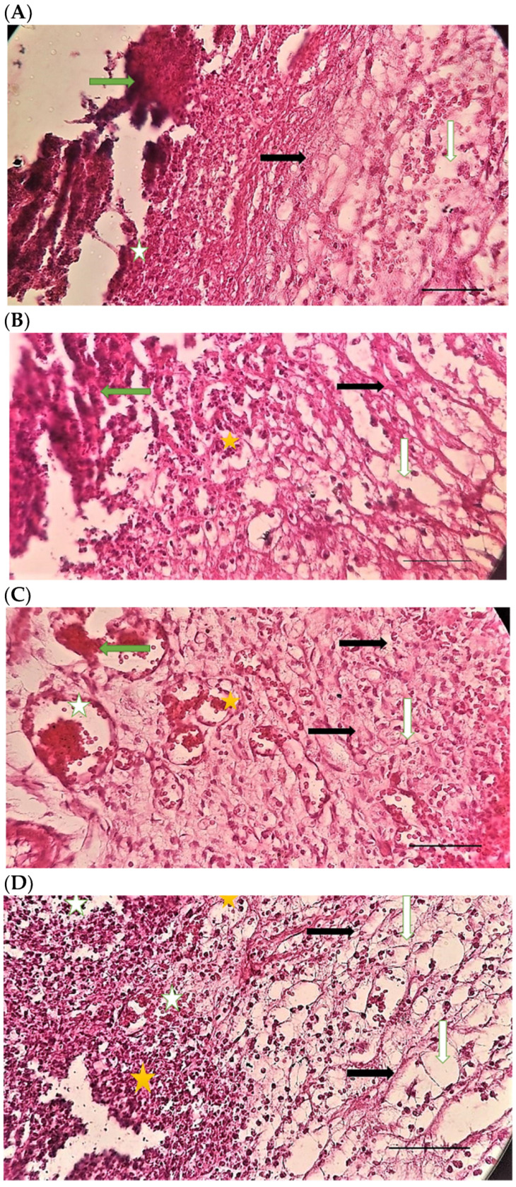
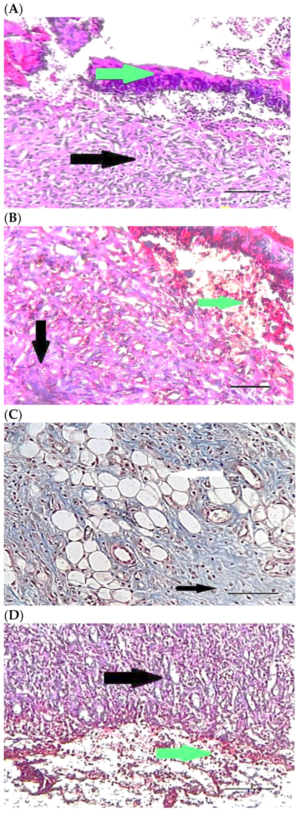
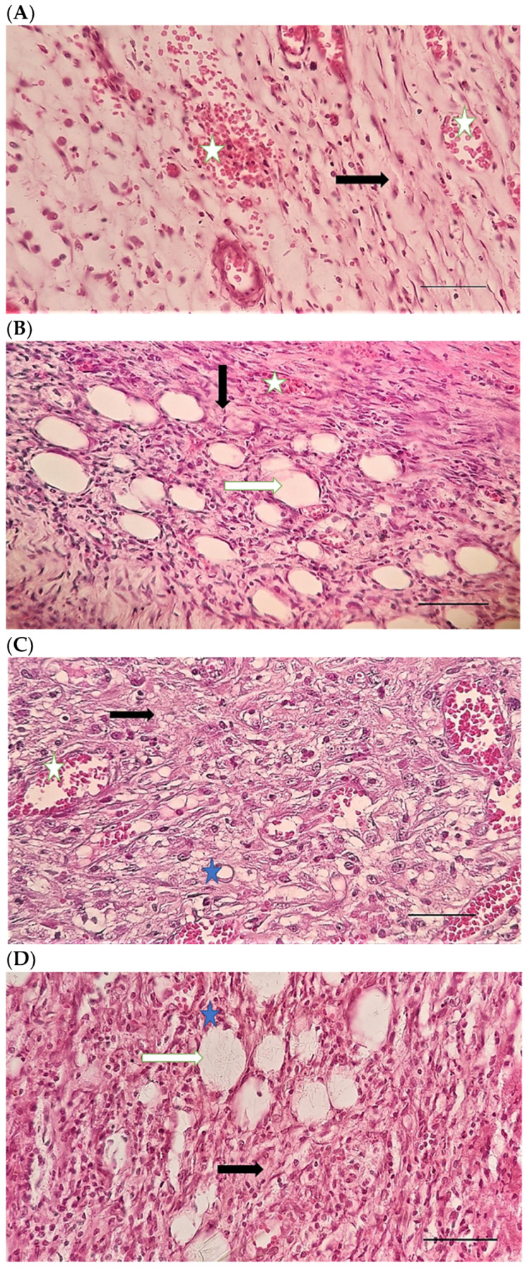
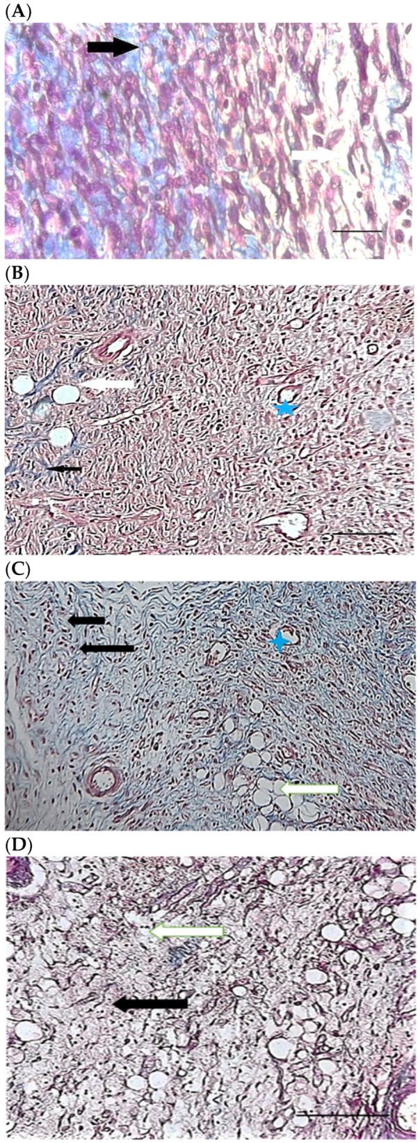
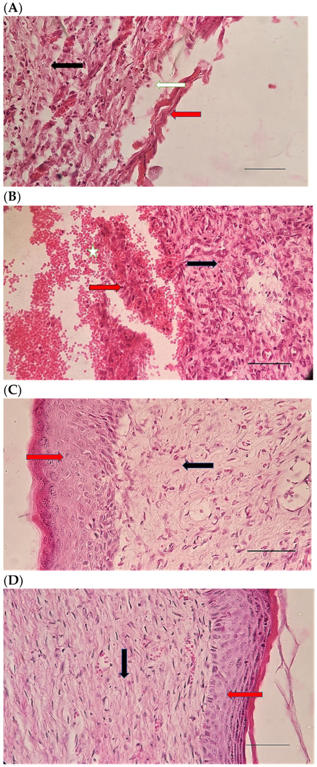
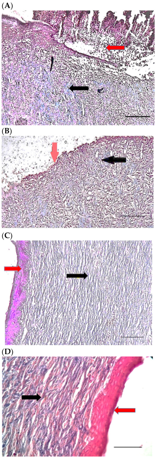
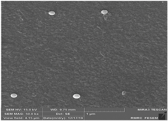
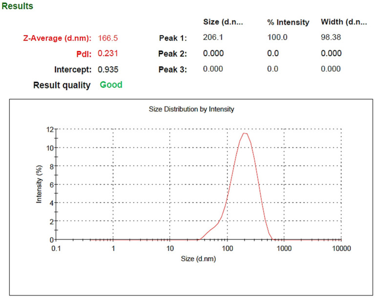
The present study aimed to prepare and evaluate a controlled-release system based on a chitosan scaffold containing selenium nanoparticles loaded with doxycycline. Its topical application in skin wound healing in rats was investigated. Therefore, 80 female rats were used and, after creating experimental skin defects on their back, were randomly divided into four equal groups: the control group without any therapeutic intervention; the second group received a chitosan transdermal patch (Ch); the third group received chitosan transdermal patch loaded with selenium nanoparticles (ChSeN), and the last group received chitosan transdermal patch containing selenium nanoparticle loaded by doxycycline (ChSeND). Morphological and structural characteristics of the synthesized patches were evaluated, and in addition to measuring the skin wound area on days 3, 7, and 21, a histopathological examination was performed. On the third day of the study, less hemorrhage and inflammation and more neo-vascularization were seen in the ChSeND group. Moreover, on day 7, less inflammation and collagen formation were recorded in the ChSeN and ChSeND groups than in the other groups. At the same time, more neo-vascularization and re-epithelialization were seen in the ChSeND group on days 7 and 21. In addition, on day 21 of the study, the most collagen formation was in this group. Examination of the wound area also showed that the lowest area belonged to the ChSeND group. The results showed that the simultaneous presence of selenium nanoparticles and doxycycline in the ChSeND group provided the best repair compared to the control, Ch and ChSeN groups.

Keywords: chitosan; doxycycline; rat; selenium nanoparticle; skin wound healing.


Conflict of interest statement

The authors declare no conflict of interest.

Figures

Figure 6
Figure 6
H&E stained microscopic sections (Bar = 100 μm) wound in rats on day 3 of the study. (A) Control group; Hemorrhage (white star), lack of collagen fibers formation (black arrow), edema (white arrow). (B) Ch group; Edema (white arrow), thin collagen fiber (black arrow), clot (green arrow), inflammation (yellow star). (C) ChSeN group; Severe hyperemia (white star), fine collagen fibers (black arrow). (D) ChSeND group; Edema (white arrow), hemorrhage (white star), fine collagen fibers (black arrow), inflammation (yellow star).
Figure 7
Figure 7
Mason trichrome stained microscopic sections (Bar = 50 μm) wound in rats on day 3 of the study. (A) Control group; Clot and hemorrhage (green arrow), fine collagen fiber (black arrow). (B) Ch group; Clot (green arrow), fine collagen fiber (black arrow), edema (white arrow). (C) ChSeN group; Fine collagen fiber (black arrow), edema (white arrow). (D) ChSeND group; Clot and hemorrhage (green arrow), fine collagen fiber (black arrow).
Figure 8
Figure 8
H&E stained microscopic sections (Bar = 100 μm) wound in rats on day 7 of the study. (A) Control group; Fine collagen fibers (black arrow), hyperemia and hemorrhage (white star). (B) Ch group; Edema (white arrow), fine collagen fibers (black arrow), hyperemia (white star). (C) ChSeN group; Fine collagen fibers (black arrow), newly formed vessels (blue star), hyperemia (white star). (D) ChSeND group; Edema (white arrow), relatively thick collagen fibers (black arrow).
Figure 9
Figure 9
Mason trichrome stained microscopic sections (Bar = 100 μm) wound in rats on day 7 of the study. (A) Control group; edema (white edema), scattered and thin collagen fibers (black arrow). (B) Ch group; Collagen fibers (black arrow), edema (white arrow), newly formed vessels (blue star). (C) ChSeN group; edema (white edema), collagen fibers (black arrow), hyperemia (blue star). (D) ChSeND group; edema (white arrow), collagen fibers (black arrow).
Figure 10
Figure 10
H&E stained microscopic sections (Bar = 100 μm) wound in rats on day 21 of the study. (A) Control group; Formation of incoherent collagen fibers (black arrow), incomplete re-epithelialization (red arrow), edema (white arrow). (B) Ch group; Fine collagen formation (black arrow), incomplete epithelial tissue formation (red arrow), hemorrhage (white star). (C) ChSeN group; Fine collagen formation (black arrow), relatively thick epithelium formation (red arrow). (D) ChSeND group; Regular epithelial tissue formation (red arrow), relatively thick collagen fibers (black arrow).
Figure 11
Figure 11
Mason trichrome stained microscopic sections (Bar = 50 μm) wound in rats on day 21 of the study. (A) Control group; Incomplete re-epithelialization (red arrow), collagen fibers (black arrow). (B) Ch group; Incomplete epithelium formation (red arrow), scattered collagen fibers (black arrow). (C) ChSeN group; Re-epithelialization (red arrow), collagen fibers (black arrow). (D) ChSeND group; Good re-epithelialization (red arrow), relatively thick collagen fibers (black arrow).
Figure 1
Figure 1
FE-SEM image of SeN. According to the picture, the size of nanoparticles is about 200 nm.
Figure 2
Figure 2
Size distribution of SeN.
Figure 3
Figure 3
Zeta potential of SeN.
Figure 4
Figure 4
FTIR spectra of SeN.
Figure 5
Figure 5
Doxycycline and SeN were released from the transdermal patch at 32 °C and pH = 7.4 (n = 3).
Figure 12
Figure 12
In vitro techniques for measurement of adhesion strength of the transdermal patch.

Similar articles

Cited by

References

    1. Saghazadeh S., Rinoldi C., Schot M., Kashaf S.S., Sharifi F., Jalilian E., Nuutila K., Giatsidis G., Mostafalua P., Derakhshandeh H., et al. Drug delivery systems and materials for wound healing applications. Adv. Drug Deliv. Rev. 2018;127:138–166. doi: 10.1016/j.addr.2018.04.008. - DOI - PMC - PubMed
    1. Boateng J.S., Matthews K.H., Stevens H.N., Eccleston G.M. Wound healing dressings and drug delivery systems: A review. J. Pharm. Sci. 2008;97:2892–2923. doi: 10.1002/jps.21210. - DOI - PubMed
    1. Dutta P., Tripathi S., Mehrotra G., Dutta J. Perspectives for chitosan based antimicrobial films in food applications. Food Chem. 2009;114:1173–1182. doi: 10.1016/j.foodchem.2008.11.047. - DOI
    1. Cerdá C., Sánchez C. Climent, B.; Vázquez, A.; Iradi, A.; Amrani, F.E.; Bediaga, A.; Sáez, G.T. Oxidative stress and DNA damage in obesity-related tumorigenesis. Adv. Exp. Med. Biol. 2014:5–17. - PubMed
    1. Singh R., Shitiz K., Singh A. Chitin and chitosan: Biopolymers for wound management. Int. Wound J. 2017;14:1276–1289. doi: 10.1111/iwj.12797. - DOI - PMC - PubMed

LinkOut - more resources