. 2022 Nov 10;17(11):e0277248.
doi: 10.1371/journal.pone.0277248. eCollection 2022.

Optimal timing of steroid initiation in response to CTLA-4 antibody in metastatic cancer: A mathematical model

Affiliations

Optimal timing of steroid initiation in response to CTLA-4 antibody in metastatic cancer: A mathematical model

Nourridine Siewe et al. PLoS One. .

Abstract

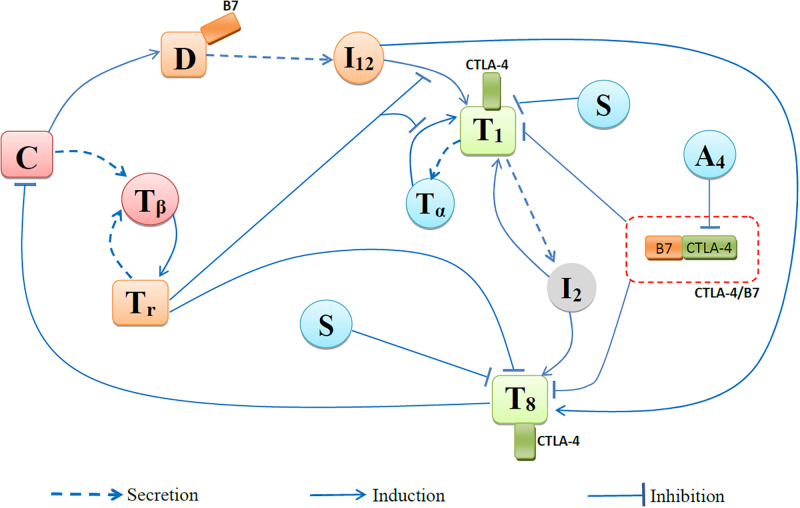
Immune checkpoint inhibitors, introduced in recent years, have revolutionized the treatment of many cancers. However, the toxicity associated with this therapy may cause severe adverse events. In the case of advanced lung cancer or metastatic melanoma, a significant number (10%) of patients treated with CTLA-4 inhibitor incur damage to the pituitary gland. In order to reduce the risk of hypophysitis and other severe adverse events, steroids may be combined with CTLA-4 inhibitor; they reduce toxicity, but they also diminish the anti-cancer effect of the immunotherapy. This trade-off between tumor reduction and the risk of severe adverse events poses the following question: What is the optimal time to initiate treatment with steroid. We address this question with a mathematical model from which we can also evaluate the comparative benefits of each schedule of steroid administration. In particular, we conclude that treatment with steroid should not begin too early, but also not very late, after immunotherapy began; more precisely, it should start as soon as tumor volume, under the effect of CTLA-4 inhibitor alone, begins to decrease. We can also compare the benefits of short term treatment of steroid at high doses to a longer term treatment with lower doses.


Conflict of interest statement

The authors have declared that no competing interests exist.

Figures

Fig 1
Fig 1. Network of cells and cytokines.
Fig 2
Fig 2. Simulation of the average densities/concentrations of the variables with/without anti-CTLA-4 and prednisone at γA4=2×10⁻⁸ g/cm3⋅d, γS = 7 × 10⁻⁹ g/cm3⋅d (low) and γS = 700 × 10−9 g/cm3⋅d (high).
All the model variables, with various drug combinations. All parameters are as in Tables 1 and 2 of Supplementary Information. A4 alone reduces tumor growth but increases TNF-α, while A4, when combined with S, reduces TNF-α but increases tumor growth. All species are in units of g/cm3.
Fig 3
Fig 3. Simulation of the average densities/concentrations of the variables with/without anti-CTLA-4 and prednisone at γA4=2×10⁻⁸ g/cm3⋅d, γS = 7 × 10⁻⁹ g/cm3⋅d (low) and γS = 700 × 10−9 g/cm3⋅d (high).
Tumor volume only in various treatment combinations. All parameters are as in Tables 1 and 2 of Supplementary Information. A4 alone reduces tumor growth but increases TNF-α, while A4, when combined with S, reduces TNF-α but increases tumor growth. The pair (*, *) represents the tumor volume and the concentration of TNF-α at day 40. All species are in units of g/cm3.
Fig 4
Fig 4. Levels of IL-12 with/without anti-CTLA-4 and prednisone at γA4=2×10⁻⁸ g/cm3⋅d, γS = 7 × 10⁻⁹ g/cm3⋅d (low) and γS = 700 × 10−9 g/cm3⋅d (high).
Fig 5
Fig 5. T cell levels with/without anti-CTLA-4 and prednisone at γA4=2×10⁻⁸ g/cm3⋅d, γS = 700 × 10−9 g/cm3⋅d (high) and γS = 7000 × 10−9 g/cm3⋅d (alone).
Density of Th1 cells (A) and density of CD8+ T cells (B).
Fig 6
Fig 6. Schedules for administering steroid with doses γS = 1, 0.5 and 0.25 mg/kg⋅d.
γA4 is a dose of anti-CTLA-4, is administered at days 1, 21 and 42.
Fig 7
Fig 7. Different protocols of treatment with prednisone and anti-CTLA-4.
(S1) − (S7): Tumor volume (left) after 10 weeks and average TNF-α (right) A4 is injected in the first day of each cycle, at γA4=3×10⁻⁶ g/cm3⋅d. S is given at various schedules (S1)–(S7) as in (23) at γS = 10−6 g/cm3⋅d (1 mg/kg⋅d), γS = 0.5 × 10⁻⁶ g/cm3⋅d (0.5 mg/kg⋅d), and γS = 0.25 × 10⁻⁶ g/cm3⋅d (0.25 mg/kg⋅d). (a) The 10-week end-time tumor volumes are displayed on the left panels and the average of TNF-α taken over the levels that exceed the no-drug case, Tα,ave, on the right panel. Tumor volume at t = 0 is 0.01 cm3.
Fig 8
Fig 8. Different protocols of treatment with prednisone and anti-CTLA-4.
(S1) − (S7): Tumor volume. A4 is injected in the first day of each cycle, at γA4=3×10⁻⁶ g/cm3⋅d. S is given at various schedules (S1)–(S7) as in (23) at γS = 10−6 g/cm3⋅d (1 mg/kg⋅d), γS = 0.5 × 10⁻⁶ g/cm3⋅d (0.5 mg/kg⋅d), and γS = 0.25 × 10⁻⁶ g/cm3⋅d (0.25 mg/kg⋅d). Tumor volume for 18 weeks. Tumor volume at t = 0 is 0.01 cm3.
Fig 9
Fig 9. Schedules for administering steroid with doses γS = 0.6, 0.6, 0.3 and 0.15 mg/kg⋅d.
γA4 is the dose of anti-CTLA-4, administered at days 1, 21 and 42.
Fig 10
Fig 10. Different protocols of treatment with prednisone and anti-CTLA-4.
(S1)-(S7) : Tumor volume (left) after 10 weeks and average TNF-α (right). A4 is injected in the first day of each cycle, at γA4=3×10⁻⁶ g/cm3⋅d. S is given at various schedules (S1)–(S7) as in (23) at γS = 0.6 × 10⁻⁶ g/cm3⋅d (0.6 mg/kg⋅d), γS = 0.3 × 10⁻⁶ g/cm3⋅d (0.3 mg/kg⋅d), and γS = 0.15 × 10⁻⁶ g/cm3⋅d (0.15 mg/kg⋅d). The 10-week end-time tumor volumes are displayed on the left panels and the average of TNF-α taken over the levels that exceed the no-drug case, Tα,ave, on the right panel. Tumor volume at t = 0 is 0.01 cm3.
Fig 11
Fig 11. Different protocols of treatment with prednisone and anti-CTLA-4.
(S1)-(S7) : Tumor volume. A4 is injected in the first day of each cycle, at γA4=3×10⁻⁶ g/cm3⋅d. S is given at various schedules (S1)–(S7) as in (23) at γS = 0.6 × 10⁻⁶ g/cm3⋅d (0.6 mg/kg⋅d), γS = 0.3 × 10⁻⁶ g/cm3⋅d (0.3 mg/kg⋅d), and γS = 0.15 × 10⁻⁶ g/cm3⋅d (0.15 mg/kg⋅d). Tumor volume for 16 weeks. Tumor volume at t = 0 is 0.01 cm3.

Similar articles

Cited by

References

    1. Wang DY, Johnson DB, Davis EJ. Toxicities Associated With PD-1/PD-L1 Blockade. Cancer J. 2018;24(1):36–40. doi: 10.1097/PPO.0000000000000296 - DOI - PMC - PubMed
    1. Winer A, Bodor JN, Borghaei H. Identifying and managing the adverse effects of immune checkpoint blockade. J Thorac Dis. 2018;10(Suppl 3):S480–S489. doi: 10.21037/jtd.2018.01.111 - DOI - PMC - PubMed
    1. Scarpati GDV, Fusciello C, Perri F, Sabbatino F, Ferrone S, Carlomagno C, et al.. Ipilimumab in the treatment of metastatic melanoma: management of adverse events. OncoTargets and therapy. 2014;7:203–209. doi: 10.2147/OTT.S57335 - DOI - PMC - PubMed
    1. Kichloo A, Albosta M, Dahiya D, Guidi JC, Aljadah M, Singh J, et al.. Systemic adverse effects and toxicities associated with immunotherapy: A review. World J Clin Oncol. 2021;12(3):150–163. doi: 10.5306/wjco.v12.i3.150 - DOI - PMC - PubMed
    1. Fucà G, Galli G, Poggi M, Lo Russo G, Proto C, Imbimbo M, et al.. Modulation of peripheral blood immune cells by early use of steroids and its association with clinical outcomes in patients with metastatic non-small cell lung cancer treated with immune checkpoint inhibitors. ESMO Open. 2019;4(1):e000457. doi: 10.1136/esmoopen-2018-000457 - DOI - PMC - PubMed

Publication types