

Machine learning based skin lesion segmentation method with novel borders and hair removal techniques
- PMID: 36355845
- PMCID: PMC9648757
- DOI: 10.1371/journal.pone.0275781
Machine learning based skin lesion segmentation method with novel borders and hair removal techniques
Erratum in
-
Correction: Machine learning based skin lesion segmentation method with novel borders and hair removal techniques.PLoS One. 2024 Dec 17;19(12):e0316115. doi: 10.1371/journal.pone.0316115. eCollection 2024. PLoS One. 2024. PMID: 39689083 Free PMC article.
Abstract
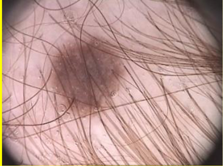
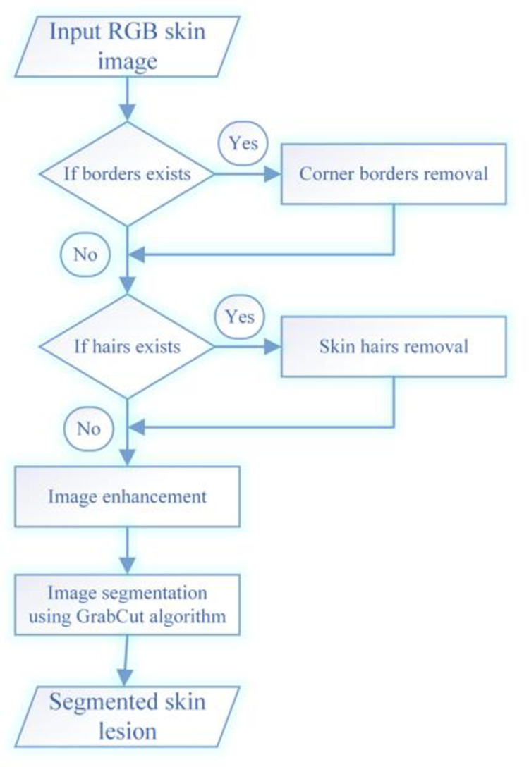
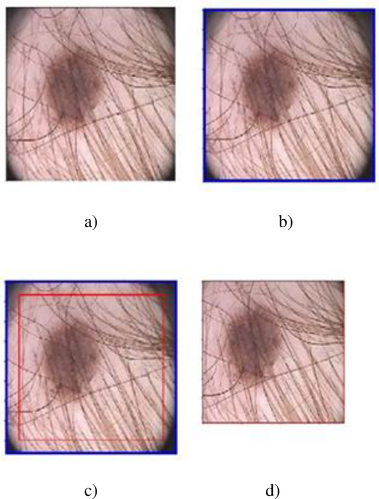
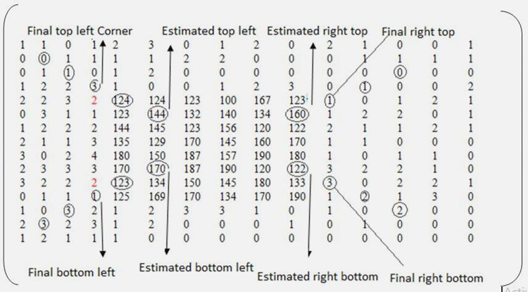
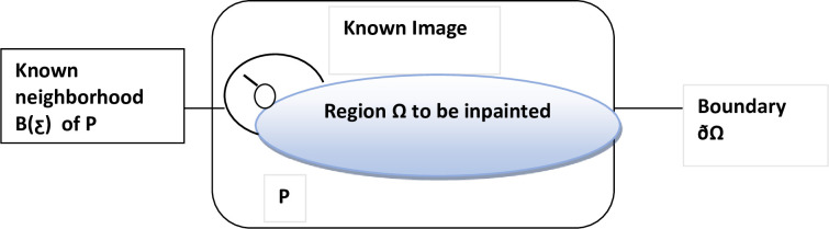
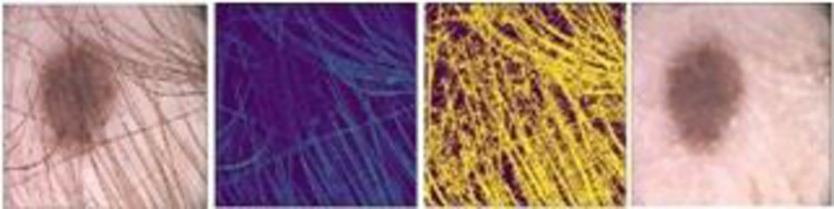
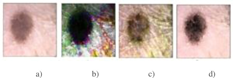
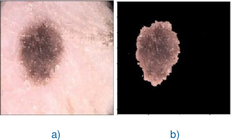
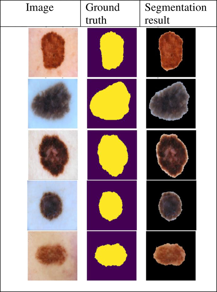
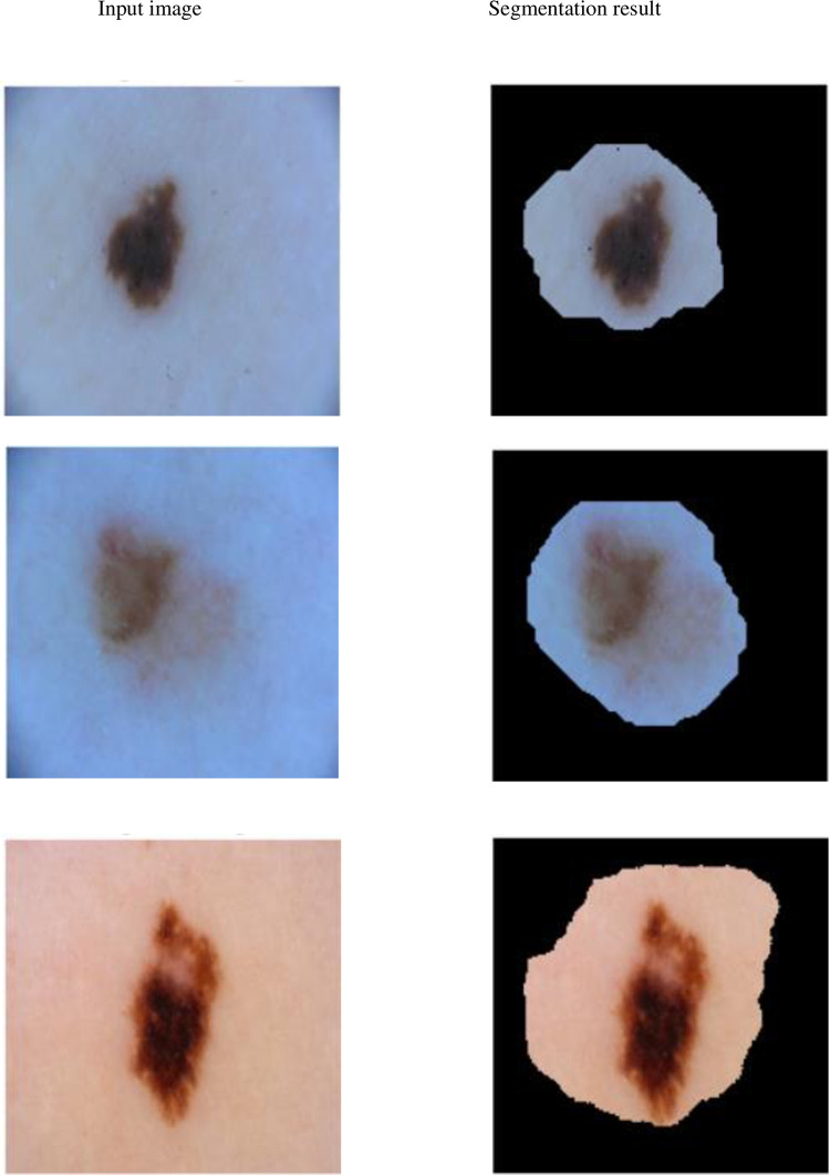
The effective segmentation of lesion(s) from dermoscopic skin images assists the Computer-Aided Diagnosis (CAD) systems in improving the diagnosing rate of skin cancer. The results of the existing skin lesion segmentation techniques are not up to the mark for dermoscopic images with artifacts like varying size corner borders with color similar to lesion(s) and/or hairs having low contrast with surrounding background. To improve the results of the existing skin lesion segmentation techniques for such kinds of dermoscopic images, an effective skin lesion segmentation method is proposed in this research work. The proposed method searches for the presence of corner borders in the given dermoscopc image and removes them if found otherwise it starts searching for the presence of hairs on it and eliminate them if present. Next, it enhances the resultant image using state-of-the-art image enhancement method and segments lesion from it using machine learning technique namely, GrabCut method. The proposed method was tested on PH2 and ISIC 2018 datasets containing 200 images each and its accuracy was measured with two evaluation metrics, i.e., Jaccard index, and Dice index. The evaluation results show that our proposed skin lesion segmentation method obtained Jaccard Index of 0.77, 0.80 and Dice index of 0.87, 0.82 values on PH2, and ISIC2018 datasets, respectively, which are better than state-of-the-art skin lesion segmentation techniques.
Copyright: © 2022 Rehman et al. This is an open access article distributed under the terms of the Creative Commons Attribution License, which permits unrestricted use, distribution, and reproduction in any medium, provided the original author and source are credited.
Conflict of interest statement
The authors have declared that no competing interests exist
Figures

Similar articles
-
ChimeraNet: U-Net for Hair Detection in Dermoscopic Skin Lesion Images.J Digit Imaging. 2023 Apr;36(2):526-535. doi: 10.1007/s10278-022-00740-6. Epub 2022 Nov 16. J Digit Imaging. 2023. PMID: 36385676 Free PMC article.
-
Efficient skin lesion segmentation using separable-Unet with stochastic weight averaging.Comput Methods Programs Biomed. 2019 Sep;178:289-301. doi: 10.1016/j.cmpb.2019.07.005. Epub 2019 Jul 8. Comput Methods Programs Biomed. 2019. PMID: 31416556
-
LAMA: Lesion-Aware Mixup Augmentation for Skin Lesion Segmentation.J Imaging Inform Med. 2024 Aug;37(4):1812-1823. doi: 10.1007/s10278-024-01000-5. Epub 2024 Feb 26. J Imaging Inform Med. 2024. PMID: 38409610 Free PMC article.
-
Deep Learning Approaches Towards Skin Lesion Segmentation and Classification from Dermoscopic Images - A Review.Curr Med Imaging. 2020;16(5):513-533. doi: 10.2174/1573405615666190129120449. Curr Med Imaging. 2020. PMID: 32484086 Review.
-
ACCPG-Net: A skin lesion segmentation network with Adaptive Channel-Context-Aware Pyramid Attention and Global Feature Fusion.Comput Biol Med. 2023 Mar;154:106580. doi: 10.1016/j.compbiomed.2023.106580. Epub 2023 Jan 25. Comput Biol Med. 2023. PMID: 36716686 Review.
Cited by
-
Correction: Machine learning based skin lesion segmentation method with novel borders and hair removal techniques.PLoS One. 2024 Dec 17;19(12):e0316115. doi: 10.1371/journal.pone.0316115. eCollection 2024. PLoS One. 2024. PMID: 39689083 Free PMC article.
-
Automatic Assessment of AK Stage Based on Dermatoscopic and HFUS Imaging-A Preliminary Study.J Clin Med. 2024 Dec 10;13(24):7499. doi: 10.3390/jcm13247499. J Clin Med. 2024. PMID: 39768422 Free PMC article.
-
Grid-Based Structural and Dimensional Skin Cancer Classification with Self-Featured Optimized Explainable Deep Convolutional Neural Networks.Int J Mol Sci. 2024 Jan 26;25(3):1546. doi: 10.3390/ijms25031546. Int J Mol Sci. 2024. PMID: 38338828 Free PMC article.
References
-
- Ge Z., Demyanov S., Chakravorty R., Bowling A., and Garnavi R., “Skin Disease Recognition Using Deep Saliency Features and Multimodal Learning of Dermoscopy and Clinical Images,” 2017, pp. 250–258. doi: 10.1007/978-3-319-66179-7_29 - DOI
-
- “Melanoma skin cancer report. The Global Coalition for Melanoma Patient Advocacy,” https://melanomapatients.org.au/wp-content/uploads/2020/04/2020-campaign...., 2020.
-
- Fakrul I. T., “Automatic Skin Lesion Segmentation Using GrabCut in HSV Colour Space.,” Computer Vision and Pattern Recognition, 2018.
Publication types
MeSH terms
LinkOut - more resources
Full Text Sources
Medical
Miscellaneous
