Comparison between Colorimetric In Situ Hybridization, Histopathology, and Immunohistochemistry for the Diagnosis of New World Cutaneous Leishmaniasis in Human Skin Samples
- PMID: 36355886
- PMCID: PMC9695648
- DOI: 10.3390/tropicalmed7110344
Comparison between Colorimetric In Situ Hybridization, Histopathology, and Immunohistochemistry for the Diagnosis of New World Cutaneous Leishmaniasis in Human Skin Samples
Abstract
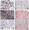
New world cutaneous leishmaniasis (NWCL) is an anthropozoonosis caused by different species of the protozoan Leishmania. Colorimetric in situ hybridization (CISH) was shown to satisfactorily detect amastigote forms of Leishmania spp. in animal tissues, yet it was not tested for the diagnosis of human NWCL. The aim of this study was to compare CISH, histopathology (HP), and immunohistochemistry (IHC) techniques to diagnose NWCL in human cutaneous lesions. The sample comprised fifty formalin-fixed, paraffin-embedded skin biopsy specimens from patients with NWCL caused by L. (V.) braziliensis. These specimens were analyzed by CISH, using a generic probe for Leishmania, IHC, and HP to assess the sensitivity of these methods by using a parasitological culture as a standard reference. Additional specimens from three patients diagnosed with cutaneous mycoses were also included to evaluate cross-reactions between CISH and IHC. The sensitivities of IHC, CISH, and HP for detecting amastigotes was 66%, 54%, and 50%, respectively. IHC, unlike CISH, cross-reacted with different species of fungi. Together, these results demonstrate that CISH may be a complementary assay for the detection of amastigote in the laboratorial diagnosis routine of human NWCL caused by L. (V.) braziliensis.
Keywords: Leishmania braziliensis; diagnosis; immunoperoxidase; in situ hybridization; tegumentary leishmaniasis.
Conflict of interest statement
The authors declare no conflict of interest.
Figures


References
-
- Lainson R. Espécies neotropicais de Leishmania: Uma breve revisão histórica sobre sua descoberta, ecologia e taxonomia. Rev. Panamazonica Saude. 2010;1:13–32. doi: 10.5123/S2176-62232010000200002. - DOI
-
- Brazil Ministério da Saúde . Manual de Vigilância da Leishmaniose Tegumentar Americana. 2nd ed. Ministério da Saúde; Brasília, Brazil: 2017.
-
- Pan American Health Organization Leishmaniasis. [(accessed on 26 July 2022)]. Available online: https://www.paho.org/en/topics/leishmaniasis.
-
- Pena H.P., Belo V.S., Xavier-Junior J.C.C., Teixeira-Neto R.G., Melo S.N., Pereira D.A., Fontes I.C., Santos I.M., Lopes V.V., Tafuri W.L., et al. Accuracy of diagnostic tests for American tegumentary leishmaniasis: A systematic literature review with meta-analyses. Trop. Med. Int. Health. 2020;10:168–1181. doi: 10.1111/tmi.13465. - DOI - PubMed
Grants and funding
LinkOut - more resources
Full Text Sources
Research Materials
Miscellaneous

